259
Revista da Sociedade Brasileira de Medicina Tropical 35(3): 259-262, mai-jun, 2002.
RELATO DE CASO
Identification of
Leishmania chagasi
from skin in
Leishmania/
HIV co-infection: a case report
Identificação de
Leishmania chagasi
na pele em co-infecção
Leishmania
/HIV: relato de caso
Marcela Orsini1, 2, Marcelo Silva3, Zélia Maria Profeta da Luz1, Jolandt Disch1, Octávio Fernandes4,
Dalton Moreira5, Antonio Carlos Martins Guedes6 and Ana Rabello1
Abstract A case of HIV/Leishmania co-infection presenting both visceral and cutaneous manifestations is reported. Leishmania infection was confirmed by conventional methods (parasitological approach and serology) and by PCR. Leishmania chagasi isolated from the skin lesion was characterized by enzyme electrophoresis and by restriction fragment length polymorphism of the internal transcribed spacer of the ribosomal gene.
Key-words: Leishmania. HIV.
Resumo É descrito um caso de co-infecção Leishmania/HIV com manifestações cutâneas e visceral. Infecção pela leishmania foi confirmada através de métodos convencionais (parasitológicos e sorológicos) e através da PCR. A espécie Leishmania chagasi isolada da pele foi caracterizada por eletroforese enzimática e por polimorfismo de fragmento obtido por enzima de restrição.
Palavras-chaves:Leishmania. HIV.
1. Laboratório de Pesquisas Clínicas do Centro de Pesquisas René Rachou da Fundação Oswaldo Cruz, Belo Horizonte, MG. 2. Curso de Pós Graduação em Medicina Tropical da Universidade Federal de Minas Gerais, Belo Horizonte, MG. 3. Clínica Integrada de Infectologia do Hospital Santo Ivo, Belo Horizonte, MG. 4. Departamento de Imunologia e Medicina Tropical do Instituto Oswaldo Cruz da Fundação Oswaldo Cruz, Rio de Janeiro, RJ. 5. Centro de Referência e Treinamento em Doenças Infecto-Parasitárias Orestes Diniz da Universidade Federal de Minas Gerais, Belo Horizonte, MG. 6. Faculdade de Medicina da Universidade Federal de Minas Gerais, Belo Horizonte, MG.
Endereço para correspondência: Dra. Marcela Orsini. Laboratório de Pesquisas Clínicas/CPqRR/FIOCRUZ. Av. Augusto de Lima 1715, 30190-002 Belo Horizonte, MG, Brazil.
Tel: 55 31 3295-3566, Fax: 55 31 3295-3115. E-mail: [email protected]
Recebido para publicação em 21/12/2000.
Co-infection by Leishmania spp and HIV has been considered an emerging disease in several parts of the world11.In Brazil, more than 200,000 cases of
leishmaniasis have been notified over the last decade1.
Between 1980 and 1991, the Brazilian Ministry of Health recorded an annual mean of 27,000 new cases of cutaneous (CL) and muco-cutaneous leishmaniasis (ML) and 2,800 new cases of visceral infection. These figures in the annual reports have increased during the past three years reaching 35000 cases of CL and 3,500 patients with visceral leishmaniasis (VL)3. In Minas
Gerais state, records have shown 1569 cases of VL (population of 15 million people), 30% (445) of which were from the metropolitan region.
Growth of the Brazilian population, acceleration of urbanization processes, man-made environmental changes and rural-urban migration contributed to the increased and modified geographic spread of
Leishmania spp parasite. Such that, nowadays many people living in dense urban zones live in contact with the causative agent. During the past decade, many urban outbreaks of VL caused by Leishmania (Leishmania)chagasi have been reported in Brazil7 10.
260
Orsini M et al
According to estimates of the World Health Organization (WHO), 2-9% of the South European AIDS patients develop VL. Gradoni et al5 observed a
500 times higher prevalence of Leishmania spp infection among HIV infected subjects in Sicily.
In Brazil, out of 61 actively searched Leishmania/ HIV co-infected cases, 64% were CL or ML and 36% presented the visceral form of the disease. In only
n i n e p a t i e n t s t h e L e i s h m a n i a s p e c i e s w a s c h a r a c t e r i z e d a n d i n a l l c a s e s b e l o n g e d t o the V i a n n i a s u b g e n u s (A. Rabello : personal communication, 1999).
This paper reports a fatal case of Leishmania/HIV co-infection presenting both cutaneous and visceral manifestations that were caused by Leishmania (Leishmania) chagasi.
CASE REPORT In December 1995, after successive episodes of
sinusitis and pneumonia, HIV infection was diagnosed in a 43-year-old patient from Belo Horizonte, Minas Gerais State, Brazil. HIV serology was performed by ELISA and confirmed by Western Blot. At that time, the patient presented a good healthy condition and clinical manifestation of seborrheic dermatitis.
In January 1996, with 87 CD4 positive lymphocyte/ mm3 and viral load of 73,000 RNA copies/ml, the patient
received zidovudine, didanosine and trimethoprim-sulfamethoxazole. Clinical conditions remained unchanged until March 1997, when the patient presented fever (38°C), weight loss (7.5kg) and splenomegaly. Investigation of the putative etiology of the fever of unknown origin was not successful. X-ray and computerized tomography of the thorax and abdominal ultrasonography were normal, as well as the otorhinolaryngology exam. Urine and blood laboratory analysis also did not reveal any abnormalities. Serology for CMV infection, Epstein-Barr virus infection, syphilis, brucellosis and toxoplasmosis were negative. Microbiologic culture of urine and faeces and investigation of Mycobacterium sp were negative. Direct examination of the bone marrow showed no pathologic abnormalities. Anti-retroviral therapy was changed to stavudine and lamivudine which induced clinical improvement with reduction of the spleen size and regression of the fever leading to the patient’s return to normal daily activities.
In January 1998, the patient presented diarrhea and
Shigella sp and Cryptosporidium sp were identified in the feces. Treatment (floxacillin for 15 days) was considered successful. One month later, there was a progressive worsening of symptoms, with alopecia, caquexia and peripheral neuritis.
In August 1998, the patient presented pancytopenia (platelets: 34,000/ml, leukocytes: 1,200/ml, granulocytes:
700/ml, hemoglobin: 8.1%) and alterations in several biochemical parameters (aspartate aminotransferase: 71 mg%, alanine aminotransferase: 40mg%, alkaline phosphatase: 150mg%, prothrombin activity: 85%) were observed. In December, 1998, cutaneous and oral-pharyngeal lesions suggestive of herpes zoster, appeared. Fluconazole and acyclovir acheived total recovery of the oral-pharyngeal lesions and partial recovery of the cutaneous lesions was observed after 14 days of treatment. However, the cutaneous hyperkeratosis lesions did not heal completely and a biopsy was taken in June 1999. Although initial histopathological diagnosis of keratoacanthoma was not confirmed, during a microscopic revision of the slides
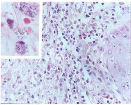
Leishmania spp amastigotes were found in the interior of macrophages in a mild lymphohistiocytary inflammatory dermal infiltrate (Figure 1). In August, the patient presented new cutaneous lesions located in the upper and lower limbs and hepatosplenomegaly. A skin biopsy from the new lesions was performed and
Leishmania spp was detected by imprint and culture. Hemoculture and PCR using DNA extracted from peripheral mononuclear cells or skin with a set of primers directed towards the minicircle conserved region, were positive. Parasites isolated from the biopsy specimen were submitted to in vitro culture and DNA was extracted. Electrophoresis of parasite enzymes identified L. chagasi
as the causative agent. Restriction fragment length polymorphism of amplicons corresponding to the internal transcribed spacers of the ribosomal gene confirmed this finding (Figure 2).
Treatment with meglumine antimoniate (10mg/kg for 40 days) was initiated and lead to partial improvement of the skin lesions, regression of the hepatosplenomegaly and negative hemoculture. In November the patient was hospitalized, presenting fever, progressive dyspnea, caquexia and disseminated cutaneous lesions. Supportive measures were implemented, but nevertheless the patient died on December 7th.
DISCUSSION Both AIDS and leishmaniasis are highly prevalent in
Brazil and the number of cases of co-infections is growing9 In the immune competent patient CL is
commonly characterized by a single ulcer that responds satisfactorily to treatment. Cure is assured for 95% of the patients for whom treatment has been applied in time6. Cure and resistance to infection demands an
adequate cellular immune response of the Th1 type, with production of gamma-interferon and IL-12. The infection leads to disease and its complications when a cellular immune response of the type Th2 is elicited9. Similarly
261
Revista da Sociedade Brasileira de Medicina Tropical 35: 259-262, mai-jun, 2002
Figure 1 - Lymphohistiocytary inflammatory infiltrate in the dermal surface with amastigotes in the interior of macrophages (HE, 400X). On the left corner, macrophages with amastigotes (HE, 1000X).
Figure 2 - Profiles of Ecor-I amplified KDNA of Leishmania
s p p : L a n e 1 : 1 0 0 b p l a d d e r . L a n e 2 : L . l a i n s o n i. Lane 3: L . g u y a n e n s i s. L a n e 4 : L . c h a g a s i ( p a t i e n t ) . L a n e 5 : L. guyanensis. Lane 6: L. mexicana. Lane 7: L. chagasi.
The patient here described presented cutaneous and visceral manifestation. Initial confirmation of infection by Leishmania spp was performed by microscopic examination of the skin biopsy. Cutaneous manifestations of L. chagasi in HIV infected patients has been observed by several other authors2 4 8.
In this patient, the diagnosis of leishmaniasis was not anticipated. Spleen enlargement was attributed to specific reaction to HIV and/or drugs and Leishmania
262
therefore delayed leading to aggravation of the patient’s situation. The described case illustrates the severity of an association of Leishmania spp infection and HIV and
underlines the need for inclusion of Leishmania spp evaluation in AIDS patients in regions where leishmaniasis occurs.
REFERENCES
1. Alvar J, Canavate C, Gutierrez-Solar B, Jimenez M, Laguna F, Lopez-Velz R, Molina R, Moreno J. Leishmania and Human Immunodeficiency Virus coinfection: the first 10 years. Clinical of Microbiology Reviews 298-318, 1997.
2. Dauden E, Fernadez-Penaz P, Rios L. Leishmaniasis presenting as a dermatomyositis-like syndrome in AIDS. Journal of American Academy of Dermatology 35, 316-319, 1996.
3. Fundação Nacional de Saúde, Informe Epidemiológico do Sistema Unico de Saúde, Ministério da Saúde, Brasília - Brasil, 2000.
4. Gonzalo HC, Arazo GP, Alvarez CG, Alonso CF, Jorda RP, Aguirre EJM. Leishmaniasis visceral e infección por el HIV: dos nuevos casos. Annals of Medicine Interna 8: 208-209, 1991.
5. Gradoni L, Scalone A, Gramiccia M, Troiani M. Epidemiological surveillance of leishmaniasis in HIV-1-infected individuals in Italy. AIDS 10:785-791, 1996.
6. Marzochi MCA. Leishmanioses no Brasil: as leishmanioses
tegumentares. Jornal Brasileiro de Medicina, 63: 82-99, 1992.
7. Passos VMA, Falcão, AL, Mazorchi MCA, Gontijo CMF, Dias ES, Barbosa-Santos EGO, Guerra HL, Katz N. Epidemiological
a s p e c t s o f A m e r i c a n C u t a n e o u s L e i s h m a n i a s i s i n a periurban area of the metropolitan region of Belo Horizonte, Minas Gerais, Brazil, Memórias do Instituto Oswaldo Cruz 88: 103-110, 1993.
8. Postigo C, Liamas R, Zarco C, Rubio R, Pulido F, Costa
J R , I g l e s i a s L . C u t a n e o u s l e s i o n s i n p a t i e n t s w i t h v i s c e r a l l e i s h m a n i a s i s a n d H I V i n f e c t i o n . J o u r n a l o f Infection 35: 265-268, 1997.
9. Rabello A, Andrade MO, Dish J. Co-infecção Leishmania/
HIV. Revista da Sociedade Brasileira de Medicina Tropical 31 (supl III): 81, 1998.
10. Vasconcelos IAB, Vasconcelos AW, Momen H, Grimaldi Jr G, Alencar JE. Epidemiological studies on american leishmaniasis in Ceará State, Brazil molecular characterization of the
Leishmania isolates. Annals of Tropical Medicine and Parasitology 82: 547-554, 1988.
11. World Healh Organization. Division of Control of Tropical Diseases. Epidemiological analyses of retrospective cases of
Leishmania/HIV co-infection. World Health Organization/LEISH/ 96.39.1996.