MODELS, BIOLOGICAL
Histological study of rat ovaries cryopreserved by vitriication or slow freezing and
reimplanted in the early or late postmenopausal stage¹
João Marcos de Meneses e SilvaI, Luiz Gonzaga Porto PinheiroII, José Alberto Dias LeiteIII, Lígia Helena Ferreira MeloIV, Franciele Osmarini LunardiV, Rômulo Cesar Costa Barbosa FilhoVI, Cindy Vitalino MendonçaVII
DOI: http://dx.doi.org/10.1590/S0102-86502014000500003
IFellow PhD degree, Postgraduate Program in Medical and Surgical Sciences, Department of Surgery, School of Medicine, Federal University of Ceara
(UFC), Fortaleza-CE, Brazil. Design of the study, technical procedures, analysis and interpretation of data, manuscript writing, critical revision.
IIPhD, Full Professor, Department of Surgery, School of Medicine, UFC, Fortaleza-CE, Brazil. Scientiic and intellectual content of the study, critical
revision.
IIIPhD, Full Professor, Department of Surgery, School of Medicine, UFC, Fortaleza-CE, Brazil. Scientiic and intellectual content of the study. IVMD, Department of Radiology, General Army Hospital of Fortaleza, Fortaleza-CE, Brazil. Technical procedures; acquisition, analysis and
interpretation of data.
VMaster Veterinarian, Veterinary Medicine, Veterinary School, State University of Ceara (UECE), Fortaleza-CE, Brazil. Technical procedures; scientiic
and intellectual content of the study.
VIResident, Department of Surgery, Hospital Nossa Senhora da Conceição, Fortaleza-CE, Brazil. Acquisition of data, technical procedures. VIIGraduate student, School of Medicine, UFC, Fortaleza-CE, Brazil. Technical procedures, English language.
ABSTRACT
PURPOSE: To compare two rat ovary cryopreservation techniques (vitriication vs. slow freezing) and two postmenopausal stages (early vs. late) with regard to graft take.
METHODS: Thirty-three Wistar rats were submitted to bilateral oophorectomy. One ovary was submitted to histological analysis while the other was cryopreserved by slow freezing or vitriication. The cryopreserved ovary was thawed and reimplanted in the greater omentum one week (early menopause) or one month (late menopause) after oophorectomy. One month after ovary reimplantation, the graft take was evaluated macroscopically and histologically.
RESULTS: Six of the animals were used ascontrols and seven died. The histological indings of 20 animals included atretic follicles (n=4), primordial follicles (n=2), and corpus luteum with primordial follicles (n=3). No ovarian tissue was found in 11 animals. Vitriication resulted in a higher graft take rate than slow freezing (50% vs. 38.5%), but the difference was not statistically signiicant. However, the graft take rate was 9.3 times higher in the early than in the late postmenopausal stage (61.5% vs. 14.3%) (p=0.043). CONCLUSION: Vitriication was superior to slow freezing as ovarian cryopreservation technique, and grafting was signiicantly more successful when the ovary was reimplanted in the late postmenopausal stage.
Introduction
With the advent of new therapies against chronic degenerative diseases, especially neoplasia, the life expectancy of many young patients has been extended. Ovarian function may be lost prematurely as a result of treatment for neoplasia (depending on the age of patient), the chemotherapy regimen and the irradiation levels used. In addition, even though ovulation resumes after oncological treatment, fertility may have been compromised. For such patients, the maintenance of ovarian function is often a priority, with direct implications on future quality of life¹. Thus, many women seek medical care in an attempt to postpone the menopause and its complications and maintain ovarian function as long as possible.
The menopause can be a very distressing and debilitating period, with a range of accompanying manifestations, including vasomotor symptoms, insomnia, headache, genito-urinary disorders, luctuations in libido, bone-joint changes and osteoporosis².
In a recently published review, chances of spontaneous gestation were estimated at 28% and 5% for women under 20 and over 25 years of age, respectively, following treatment for cancer with radiotherapy and chemotherapy³.
Cryopreservation is an experimental fertility preservation technique involving the surgical removal and cryopreservation of the ovaries of mostly young women prior to chemotherapy in order to avoid exposure to systemic gonadotoxic compounds. Following treatment, provided the patient is believed to be disease-free, the ovaries may be reimplanted. Though still in the experimental stage, cryopreservation is considered a promising alternative for the recovery of fertility4-5.
The recovery of ovarian function by way of reimplantation of cryopreserved tissue has been successfully accomplished in both animals and humans. However, due to the small number of patients submitted to this procedure so far, the technique has not been fully standardized6-7.
Several animal models have been designed to evaluate compounds capable of improving the techniques and outcomes of cryopreservation. Over time, a number of different compounds have been used in the cryopreservation of reproductive cells. Thus, several studies were conducted in the 1990s evaluating the cryoprotective eficiency of DMSO, ethylene glycol, propylene glycol and even glycerol (used since 1949)7.
Initially, most research was focused on the cryopreservation of spermatozoa. Already in 1951, the irst calf
was born using frozen bull sperm. The new techniques facilitated selection and reproduction of domestic animals. Eventually, cryopreservation techniques were adapted for use in human organs, at irst for oocytes and follicles, then for ovaries and embryos8-9.
In 1954, for the irst time, a child was born as the result of insemination with semen stored for six weeks in dry ice. Ten years later, in 1964, a child was born which had been conceived with semen stored in liquid nitrogen for ive months. More recently, with the advent in 2004 of proper cryopreservation techniques, a child was engendered using semen frozen for 21 years, making the duration of the seminal phase of human life practically unlimited10-11.
The evolution of techniques of cryopreservation of female germ cells has been remarkable. The irst studies on ovarian tissue cryopreservation were performed in rodents more than half a century ago, and results have been very encouraging since. Current achievements include the maintenance of the morphology and viability of the ovarian follicle structure and the restoration of endocrine function after cryopreservation and transplantation of ovarian tissue.
After orthotopic transplantation of previously frozen and thawed whole ovary, Parrott12 described the irst birth: a single offspring in mice. Using a similar protocol, other researchers obtained mouse pregnancy rates between 33% and 73%and 3-4 births per litter. Yet others reported promising results (23 births) after orthotopic transplantation of frozen/thawed half ovaries, demonstrating that ovarian tissue retrieved post-mortem was viable even after a period of storage at low temperatures13.
In 2004, Donnez and colleagues published an important study on human reproductive medicine particularly relevant to young women facing the prospect of gonadotoxic treatment. One of their patients experienced restoration of her endocrine and gametogenic functions and conceived 11 months after orthotopic autotransplantation of ovarian fragments6.
The study showed that women treated for and cured of cancer can bear healthy children without the use of assisted reproduction techniques such as in vitro fertilization4,7.More recently, other researchers demonstrated that it is possible to maintain endocrine and gametogenic activity in the graft for an extended period. One of their patients was submitted to ovarian tissue transplantation after treatment for cancer14.
volume, structure and function, especially related to plasma membrane permeability and luidity and its implications for cell metabolism. The main complication is cryoinjury which involves one or more successive or independent events: formation of
intracellular and extracellular ice crystals, chemical toxicity of
cryoprotectors, osmotic shock and fracture damage15.
Proper training in the techniques of cryopreservation and
autotransplantation in animals is indispensable for surgeons who
wish to offer this treatment option for patients in whom ovarian
function may be compromised by radio and chemotherapy.
Methods
The experimental protocols were reviewed and approved
by the Ethics Committee for Animal Research of the Federal University of Ceara and iled under entry number 79/2012.
The study used thirty-three-month-old female Wistar rats
(RattusNovergicus) weighing approximately 200g. The animals were
housed in individual cages in a controlled environment (circadian cycle, 22 ± 2ºC, constant humidity, food and water ad libitum).
Ovarian tissue
Following excision, the ovaries (~2 mm3) were dissected
free of fat and mesentery. One of the ovaries was submitted to
histological analysis; the other was cryopreserved.
Design of the experiment
Stage I: Thirty-three Wistar rats were submitted to
bilateral oophorectomy. One ovary was immediately submitted to
histological analysis, while the other was cryopreserved by slow freezing (n=10) or vitriication (n=23)7,16 .Two animals from the slow
freezing group and ive animals from the vitriication group died. The controls included six animals from the vitriication group and two from the slow-freezing group. The latter two animals provided suficient material for control and experimental procedures.
Stage II: The cryopreserved ovary was thawed and
reimplanted in the greater omentum one week after oophorectomy (early menopause) (Group A, n=7; 3 vitriied and 4 slow-frozen) or one month after oophorectomy (late menopause) (Group B, n=13; 9 vitriied and 4 slow-frozen).
Stage III: One month after ovary reimplantation, the animals
were euthanized, the graft was excised and graft take was evaluated.
Vitriication and warming procedures
Whole ovaries were initially equilibrated in HEPES containing 10% v/v ethylene glycol and 10% v/v DMSO for 20 min at room temperature and then immersed for 3 min in a vitriication solution containing 17% v/v ethylene glycol, 17% v/v DMSO and 0.75 M sucrose in HEPES. The ovaries were then transferred individually with minimal vitriication solution to the surface of a metal cube loating in liquid nitrogen. The vitriied ovaries were kept in liquid nitrogen (-196°C) for 30-60 min. Using cooled forceps, the samples were placed in cooled cryovials containing liquid nitrogen, according to a protocol adapted from Wang et al.16 Prior to reimplantation, the vials containing the samples were removed from liquid nitrogen storage and exposed to room temperature for one minute. Then each sample was submitted to three 5-min baths in a solution containing minimal essential medium + 10% fetal calf serum and decreasing concentrations of sucrose (0.5, 0.25 and 0.0 M), using a protocol adapted from Lunardiet al.7.
Slow freezing
The procedure was carried out according to the protocol described by Gosden et al.17. Ovarian strips were placed in 2-mL cryovials illed with a cryoprotective solution containing modiied phosphate-buffered saline (DPBS) medium, 1.5 mmol/LDMSO and 20% human serum albumin (HSA). Using a programmable freezer (Custom BioGenic Systems Model 2100 Controlled Rate Freezing System), the cryovials were i) cooled from 0°C to −8°C at −2°C/min; ii) cooled to −40°C at −0.3°C/min; (iii) cooled to −150°C at −30°C/ min, and (iv) immediately transferred to liquid nitrogen for storage at −196°C. To thaw the ovarian cortical tissue, the cryovials were removed from the dewarand kept at room temperature for 2 min, followed by immersion in a water bath at 37°C for 2 min under gentle shaking. The contents of the cryovials were quickly emptied into culture dishes with Leibovitz L-15 medium and washed three times with fresh medium to remove residual cryoprotector before further processing.
Histology
in both early (one week) and late (one month) postmenopausal rats18.
Statistical analysis
The study variables were dichotomous (presence/absence). Using the software GraphPadPrism 5.0, indings were submitted to Fisher´s exact test and expressed as absolute frequencies and percentages. The level of statistical signiicance was set at 5% (p<0.05).
Results
Slow freezing
Two of the 10 rats which received an ovarian graft cryopreserved by slow freezing died (mortality: 20%). The graft was evaluated for viability prior to reimplantation in two (25%) of the eight surviving animals. In one of these (50%), primordial follicles and corpus luteum were observed, matching indings in the literature (42%). Upon euthanasia, no ovarian graft was found in ive (62.5%) animals. In the remaining three animals (38.5%), the graft had taken. One of these (12.8%) presented atretic follicles while 2 (25.6%) presented primordial follicles and corpus luteum (Table 1). All three animals with successful grafting belonged to Group B (late postmenopausal stage).
Vitriication
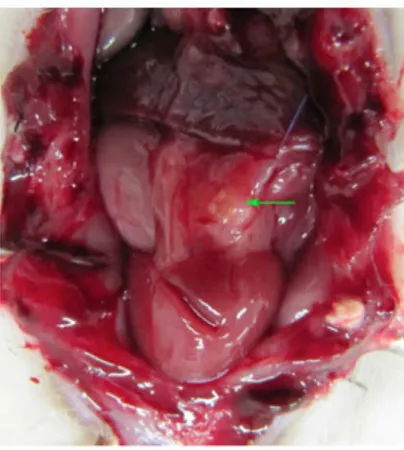
Five of the 23 rats which received an ovarian graft cryopreserved by vitriication died (mortality: 15.1%). Six randomly selected grafts evaluated for viability (but not implanted) presented primordial follicles and corpus luteum (5/6; 83.3%) and atretic and primordial follicles (1/6; 16.7%). The remaining 12 grafts were implanted. Upon euthanasia, no ovarian graft was found in six (50%) animals, just inlammatory/scar tissue and/or ibrosis. In the other six animals (50%), the graft had taken (Figure 1).
FIGURE 1 -Abdominal cavity of female Wistar rat after laparotomy showing ovary (arrow) reimplanted in the omentum. The color of the tissue and the absence of macroscopic evidence of necrosis are suggestive of good graft take.
TABLE1 -Histological indings following reimplantation of ovarian tissue in 20 female Wistar rats.
Legend: CL = corpus luteum; AF = atretic follicles; PF = primordial follicles.
Ovary cryopreservation and reimplantation
ANIMAL Postmenopausal stage Histological indings following reimplantation Cryopreservation technique
Early Late Vitriication Slow freezing
1 X PF + CL X
2 X AF X
3 X AF + PF X
4 X AF X
5 X Absence of ovarian tissue X
6 X AF X
7 X Absence of ovarian tissue X
8 X Absence of ovarian tissue X
9 X Absence of ovarian tissue X
10 X Absence of ovarian tissue X
11 X AF + PF X
12 X Absence of ovarian tissue X
13 X Absence of ovarian tissue X
14 x Absence of ovarian tissue X
15 x Absence of ovarian tissue X
16 x AF X
17 x Absence of ovarian tissue X
18 X Absence of ovarian tissue X
19 X PF + CL X
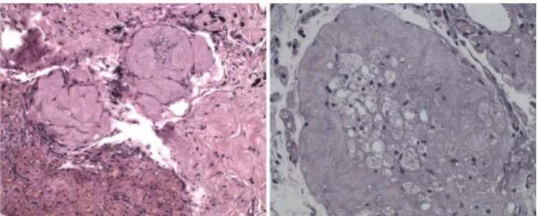
Three of these (25%) presented atretic follicles, 2 (16.7%) presented both atretic and primordial follicles, and one (8.3%) presented primordial follicles and corpus luteum (Figures 2 and 3).
Of the 20 animals in the inal sample, seven belonged to Group A (early postmenopause) and 13 belonged to Group B (late menopause). In the latter group, nine ovaries were vitriied, followed by ive (55.5%) successful reimplantations, while four ovaries were slow-frozen, followed by only one successful reimplantation (25%). Nevertheless, the difference between the techniques did not reach statistical signiicance (p=0.559).In Group A, three ovaries were vitriied, followed by one (33.3%) successful reimplantation, while four ovaries were slow-frozen, with no successful reimplantation (0%). Again, the difference was not signiicant (p=0.429). However, when the two postmenopausal stages (early vs. late) were compared with regard to graft take, a statistically signiicant difference emerged: the graft take rate was 9.3 times higher in late postmenopausal rats (61.5%) than in early postmenopausal rats (14.3%) (p=0.043).
Discussion
Despite being a promising technique, ovarian tissue cryopreservation has only resulted in about 20 human live births so far. Much research is needed to improve success rates for both slow-frozen and vitriied tissue and to ensure the health of children born from such treatments. In one study, for example, 27 patients received similar treatments, eight had evidence of antral follicle growth and oocytes were retrieved, but only one gave birth19.
In the current study, we used a rat model to compare two cryopreservation techniques (slow freezing vs. vitriication) and to evaluate the inluence of postmenopausal status (early vs. late) on graft take. Our indings raise questions about the ideal moment of ovary reimplantation in relation to hormonal metabolism. In other words, the time of absence of ovarian function appears to determine how the organism reacts to reimplantation.
Vitriication resulted in a higher graft take rate than slow freezing (50% vs. 38.5%) and follicles were present in all specimens. The success rate of slow freezing was similar to the rates reported in the literature: in vitro studies suggest that vitriication of ovarian tissue may be an improvement over the 70% oocyte viability loss from slow freezing7.
During the 2009 World Congress on Fertility Preservation in Belgium, a list was given of the advantages and disadvantages of vitriication as applied to human ovary cryopreservation. The advantages included i) excellent survival of ovarian stroma and blood vessels, ii) the technique is quick to perform, iii) no sophisticated equipment is needed, iv) the technique is clinically feasible, and v) excellent survival in tissue culture. On the other hand, the disadvantages included i) no live births reported until
FIGURE 2 -Atretic follicles after reimplantation (Magniication: x200
and x400).
FIGURE 3 -Primordial follicles and corpus luteum after reimplantation
(Magniication: x200 and x400).
Most (5/6) of the animals with successful grafting belonged to Group B (late postmenopausal stage) (Group B=83.3% vs. Group A=16.7%), but the difference between the groups with regard to take rate was not statistically signiicant (p=0.080).
Slow freezing vs. vitriication
Upon thawing, the ovarian tissue was preserved in 50% of slow-frozen specimens versus 100% of vitriied specimens; however, the difference was not statistically signiicant (p=0.400). Vitriication resulted in a higher graft take rate than slow freezing (50% vs. 37.5%), but the difference was not statistically signiicant (p=0.400).
Postmenopausal stage
2009, ii) the technique requires a considerable level of training, iii) the technique is very sensitive to concentrations, osmolarities and swiftness of procedure, and iv) ovarian tissues may be damaged by toxicity if the surgeon is not properly trained. However, recent studies describing human births following ovarian cryopreservation and reimplantation, along with the indings of the current study, indicate that the technique has been mastered despite its complexity. The subject of rat postmenopausal status has been little discussed in the literature. In our experiment, a signiicant difference was observed between early and late postmenopausal rats (Groups A and B) with regard to graft take rates. This inding may be interpreted in light of two important studies, one by López-Grueso et al.20 and one by Ahmed et al.18.
The irst study looked at oxidative stress and estrogen replacement therapy (ERT) at different stages of the menopause (early vs. late). Early, but not late, onset of ERT was found to prevent an ovariectomy-associated increase in mitochondrial hydrogen peroxide levels, oxidative damage to lipids and proteins, and a decrease in glutathione peroxidase and catalase activity in rats. The authors concluded that, possibly, the ER
α/β ratio is increased by ovariectomy and reduced by immediate estrogen replacement. On positron emission tomography, the former decreased brain glucose uptake in vivo while the latter was beneicial when administered immediately after deprivation. Ovariectomy decreases GLUT-1 and 3 glucose transporters in the brain, and only early onset estrogen administration prevents it. Plasma from rats treated with estrogen immediately after ovariectomy show similar metabolomics proiles as controls. The authors provided a molecular basis for the recommendation of early onset ERT and explained its lack of effectiveness if a signiicant time period elapses after ovariectomy and probably after the onset of menopause20. The inding of a better physiological response to ERT during early, rather than late, menopause contradicts the indings of the present study.
The second study, which evaluated mammary gland response to ERT in rodents, found the treatment to be more effective in late postmenopausal animals. The authors examined the effects of ERT on mammary gland morphology, cell proliferation and progesterone receptor regulation in early postmenopause (one week post-ovariectomy) and late postmenopause (ive weeks post-ovariectomy). The study explored the effects of ERT on the early vs. late postmenopausal mammary gland and demonstrated that late ovariectomy-induced menopause was associated with improved proliferative response to estrogen, as shown morphologically by pronounced duct end hyperplasia. This enhanced mitogenic response to estrogen
was localized to the duct end epithelium (4-fold increase) and adjacent stromal cells (6.5-fold increase). Long-term estrogen treatment produced a lower steady-state proliferative response. However, both epithelial and stromal cell proliferations were 2-fold higher when long-term estrogen treatment was started at 5 weeks post-ovariectomy vs. 1 week post-ovariectomy. In other words, an apparent permanent enhanced proliferative response to estrogen was observed in late postmenopausal animals. In contrast, no morphological response and minimal epithelial and stromal cell proliferation were observed after any length of estrogen treatment in early postmenopausal animals18.
Our results suggest that, despite claims of improved effects in relation to oxidative stress during early postmenopausal stages, ovarian graft take rates are higher in the late postmenopausal stage than in the early postmenopausal stage. This is supported by the enhanced mammary gland response to ERT observed in late postmenopausal rodents.
Vitriication was shown to be superior to slow freezing as ovarian cryopreservation technique, and grafting was signiicantly more successful when the ovary was reimplanted in the late postmenopausal stage. These results may be helpful in future studies, especially when determining the ideal time of onset of ERT in women.
Conclusions
Both vitriication and slow freezing were feasible though the former yielded better results when the time of reimplantation was taken into account. Regardless of the time of cryopreservation, rat ovary reimplantation is more likely to be successful in the late postmenopausal stage.
References
1. Teinturier C, Hartmann O, Valteau-Couanet D, Benhamou
E, Bougneres PF. Ovarian function after autologous bone marrow
transplantation in childhood: High-dose busulfan is a major cause of ovarian failure. Bone Marrow Transplant. 1998;22:989-94. 2. Binkley N, Krueger D. Combination therapy for osteoporosis:
considerations and controversy. Curr Osteporos Rep. 2005;3:150-4. 3. Lobo RA. Potential options for preservation of fertility in women. N
Engl J Med. 2005;353:64-73.
4. Lee SJ, Schover LR, Partridge AH Patrizio P, Wallace HW, Hagerty
K, Beck LN, Brennan VL, Oktay K. American Society of Clinical
Oncology recommendations on fertility preservation in cancer
patients. J ClinOncol. 2006;24:2917-31.
5. Oktay K, Karlikaya G. Ovarian function after transplantation
of frozen, banked autologous ovarian tissue. N Engl J Med.
2000;342:1919.
7. Lunardi FO, Araújo VR, Faustino LR, Carvalho AA, Gonçalves RFB, Bass CS, BAO SN, OlaziaName KP, Campello CC,
De Figueiredo JR, Ribeiro Rodrigues APl. Morphologic, viability
and ultrastructural analysis of vitriied sheep preantralfollicles en closed in ovarían tissue.Small Rumin Res. 2012;107:121-30 8. Polge C. Fertilizing capacity of bull spermatozoa after freezing at
degrees C. Nature. 1952;169(4302):626-7.
9. Stewart DL. Storage of bull spermatozoa at low temperatures. Vet
Rec.1951;63:65–6.
10. Bunge RG, Keettel WC, Sherman JK. Clinical use of frozen semen: report of four cases. Fertil Steril. 1954;5(6):520-9.
11. Horne G, Atkinson AD, Pease EH, Logue JP, Brison DR, Lieberman BA.Live birth with sperm cryopreserved for 21 years prior to cancer treatment: case report. Hum Reprod. 2004;19:1448-9.
12. Parrot DMY. The fertility of mice with orthopic ovarian grafts derived from frozen tissue. J Reprod Fertil. 1960;1:230-41. 13. Mukaida T, Nakamura S, Tomiyama T, Wada S, Kasai M, Takahashi
K. Successful birth after transfer of vitriied human blastocysts
with use of a cryoloop containerless technique. Fertil Steril.
2001;76(3):618-20.
14. Ernst E, Bergholdt S, Jorgensen JS, Andersen CY. The irst woman
to give birth to two children following transplantation of frozen/
thawed ovarian tissue. Hum Reprod. 2010;25:1280-1.
15. Kasai M, Ito K, Edashige K. Morphological appearance of the
cryopreserved mouse blastocyst as a tool to identify the type of
cryoinjury. Hum Reprod. 2002;17(7):1863-74.
16. Wang XQ, Catt S, Pangestu M, Temple-Smith P. Live offspring from vitriied blastocysts derived from fresh and cryopreserved ovarian tissue grafts of adult mice. Reproduction. 2009;138:527-35. 17. Gosden RG, Baird DT, Wade JC, Webb R. Restoration of fertility
to oophorectomized sheep by ovarian autografts stored at −196 degrees C. Hum Reprod. 1994;9:597–603.
18. Raafat AM, Hofseth LJ, Li S, Bennett JM,Haslam SZ.A mouse
model to study the effects of hormone replacement therapy on normal mammary gland during menopause: enhanced proliferative response to estrogen in late postmenopausal
mice. Endocrinology.1999;140(6):2570-80.
19. Kawamura K, Cheng Y, Suzuki N, Deguchi M, SatoY, Takae S, Ho C, Kawamura N, Tamura M, Hashimoto S, Sugishita Y, Morimoto
Y, Hosoi Y, Yoshioka N, Ishizuka B, Hsueh AJ. Hippo signaling disruption and Akt stimulation of ovarian follicles for infertility
treatment. Proc Natl Acad Sci USA. 2013;110 (43):17474-9 20. López-Grueso R, Gambini J, Mohamed K, Monleón D, Díaz A, Alami
M, Bonet CB, Viña J. Early, butnot late-onset estrogen replacement therapy prevents oxidative stress and metabolic alterations caused
by ovariectomy. Antioxid Redox Signal. 2014;20(2):236-46.
Correspondence:
Luiz Gonzaga Porto Pinheiro Faculdade de Medicina-UFC Departamento de Cirurgia
Rua Professor Costa Mendes 1608, 3° andar, Bloco Didático 60.430-140 Fortaleza - CE Brasil
Tel.: (55 85)3366-8062 Fax: (55 85)3366-8065
Received: Dec 17, 2013 Review: Feb 21, 2014 Accepted: March 18, 2014 Conlict of interest: none
Financial source: none