Inhibitory effects of a standardized extract of
Justicia
pectoralis
in an experimental rat model of airway
hyper-responsiveness
Carlos T. M. Mouraa, Francisco J. Batista-Limaa, Teresinha S. Britoa, Alfredo A. V. Silvaa,
Luan C. Ferreiraa, Cassia R. Roquea, Karoline S. Arag~aoa, Alexandre Havta, Francisco N. Fonsecab, Luzia K. A. M. Lealband Pedro J. C. Magalh~aesa
aDepartment of Physiology and Pharmacology, School of Medicine andbDepartment of Pharmacy, School of Pharmacy, Odontology and Nursing,
Federal University of Ceara, Fortaleza, Brazil
Keywords
biological evaluation of natural products; tissue and cellular pharmacology
Correspondence
Pedro J. C. Magalh~aes, Departamento de Fisiologia e Farmacologia, Faculdade de Medicina, Universidade Federal do Ceara, R. Cel. Nunes de Melo 1127, Rodolfo Teofilo, 60430-270 Fortaleza, CE, Brazil.
E-mail: pjcmagal@ufc.br
Received September 7, 2016 Accepted December 10, 2016
doi: 10.1111/jphp.12689
Abstract
Objective Justicia pectoralisis a plant useful for the treatment of respiratory dis-eases. Here, we studied the antiasthmatic properties of a standardized extract of
J. pectoralis(Jp).
Methods Ovalbumin (OVA)-sensitized rats were actively challenged with saline or OVA to study airway hyper-responsiveness after oral treatment with saline or Jp. The ability of Jp to inhibit hyper-reactivity was evaluated in isolated trachea mounted in isolated organ bath chamber.
Key findings Using KCl or carbachol as contractile agents, tracheal rings of OVA-challenged rats contracted with higher magnitude than trachea of rats chal-lenged with saline. Such hyper-responsive phenotype of OVA-chalchal-lenged tissues decreased with Jp administration. In Ca+-free medium, Jp or its major con-stituent coumarin inhibited preferentially the contractions induced by Ca2+ addi-tion in tissues of OVA-challenged rats stimulated with KCl or acetylcholine. In tissues depleted of their internal Ca+stores in the presence of thapsigargin, Jp inhibited the contraction induced by capacitative Ca2+entry. By gavage, Jp abol-ished the increase caused by challenge with OVA on the levels of IL-1band
TNF-ain the bronchoalveolar fluid and also impaired the changes in gene expression of canonical transient receptor proteins.
Conclusions Jp has antiasthmatic properties in an experimental model that reproduces tracheal hyper-reactivity.
Introduction
Justicia pectoralis Jacq. var. stenophylla Leonard (Acan-thaceae) is a herb largely used in folk medicine of South and Central America to treat respiratory complaints.[1]Named popularly as ‘chamba’ in the Brazilian north-east, this spe-cies is useful as expectorant and is in the list of plants poten-tially useful for phytotherapy purposes according to the Brazilian Health Surveillance Agency (Anvisa).[2] A few studies reinforce the notion thatJ. pectoraliscan be effective in the respiratory system,[1,3]and a recent preliminary study revealed potential antiasthmatic properties of an aqueous extract obtained from J. pectoralis by its ability to inhibit histamine-induced contraction on guinea-pig trachea.[4]
In the city of Fortaleza, Brazil, pharmacists involved with a project named Farmacias Vivas (Living Pharmacies) maintain species ofJ. pectoralisin cultivated gardens under low-light conditions, to produce traditional remedies for respiratory diseases.[5]De Vrieset al.[6]firstly revealed that extracts of J. pectoralis yielded after purification several coumarin compounds. From the local species of J. pec-toralis cultivated in Fortaleza, Lino et al.[7] reported the occurrence of coumarin and 7-hydroxycoumarin (umbellif-erone) as major constituents, besides small amounts of acetylated coumaric acid, acetylated melilotic acid and
Plants possessing coumarin as a major constituent share antinociceptive, anti-inflammatory and bronchodilator properties.[1,8,9]Regarding pharmacological actions on the respiratory system, Limet al.[10] reported that the extract from roots of Angelica decursiva and its coumarin con-stituents possess inhibitory actions against airway inflam-mation. In ovalbumin (OVA)-sensitized asthmatic mice treated with umbelliferone extracted from the aerial parts of Typha domingensis Pers., Vasconcelos et al.[11] found reduced number of eosinophils in bronchoalveolar lavage fluids and decreased mucus production and lung inflam-mation.Amburana cearensis (Allem~ao) A.C. Smith, a tree native from the Brazilian north-east rich in coumarin and amburoside A, has bronchodilator properties on air-ways.[12]Bronchodilator activity was also demonstrated to hydroalcoholic extract ofJ. pectoralis.[1]
A variety of studies demonstrated potential efficacy of
J. pectoralison respiratory system, and to the best of our knowledge, this is the first pharmacological evidence of its effects using an experimental model of asthma. Thus, the present approach evaluated whether a standardized extract of this plant for coumarin and umbelliferone can prevent the hyper-responsive phenotype caused by the antigen challenge on sensitized animals. For this purpose, OVA-sensitized rats developed airway hyper-responsive-ness induced by challenge with OVA to evaluate the effi-cacy of the acute treatment with the standardized extract of J. pectoralis. Additionally, the direct effects of the standardized extract of J. pectoralis and coumarin were tested on rat tracheal rings, and changes in gene expres-sion of canonical transient receptor proteins (TRPC) were analysed in animals gavaged with saline or with the extract ofJ. pectoralis.
Materials and Methods
Botanical material
Aerial parts of J. pectoralis were collected from cultivated specimens grown at the Phytotherapy Core (NUFITO) of the Pharmaceutical Care Center (NUASF), Fortaleza, Bra-zil. Dr Edson P. Nunes, Department of Biology, confirmed the plant’s identity by means of a voucher specimen (num-ber #16.079) deposited in the Herbarium Prisco Bezerra, Federal University of Ceara. The raw material was washed and dried in an air-forced oven (35°C, 24 h); afterwards, the plant drug was ground and sieved and the moderately coarse fraction was collected for the extraction.
Preparation of the extract of J. pectoralis
Extract was prepared by maceration followed by percola-tion. The plant drug was macerated in 20% ethanol
during 24 h. The extract was filtered and maintained in air-forced oven at 35 °C to remove ethanol and concen-trate the product (final ratio drug : extract 1 : 7.5, w/v). Solid residues corresponded to 3.8% (w : v), and the extract was maintained under refrigeration at 4 °C until the day of experiment.
Standardized extract
The concentration of coumarin and umbelliferone in the hydroalcoholic extract of J. pectoralis was determined in triplicate by high-performance liquid chromatography– photodiode array (HPLC-PDA, chromatographic system Alliance; Waters, Milford, MA, USA). Chromatographic conditions for HPLC were as follows: C18 column (2509 4.6 mm, 5lm, CX-terra; Waters), gradient elu-tion (organic phase: ACN : MeOH : THF 5.8 : 2.2 : 2.0, v/v/v; buffer phase: phosphoric acid 0.45% and triethy-lamine 0.24% in ultrapure water), flow 1.8 ml/min and detection at 323 nm. The extract was diluted 1 : 5 (v/v) in the organic phase, filtered (0.22-lm membrane) and analysed. The presence of coumarin and umbelliferone in the extract of J. pectoralis was confirmed according to Ven^ancio et al.[13] by comparing the retention time (Rt) of external standards (Rt umbelliferone =4.8 min; Rt coumarin=5.9 min; HPLC chromatograms are shown in Figure 1). Content of the two markers was estimated through calibration curves ranging from 21.0 to 359.4
lg/ml for coumarin and 2.7 to 36.8lg/ml for umbellif-erone. Finally, the content of coumarin and umbellifer-one in the extract corresponded to 1.49 and 0.17 mg/ml, respectively.
Animals
Male Wistar rats (250–350 g) were obtained from the vivarium of the Department of Physiology and Pharmacol-ogy, School of Medicine, Federal University of Ceara. The rats were housed under standard conditions in 12-h light/ 12-h dark cycles with free access to food and water. All ani-mals were handled in accordance with the ethical principles of the Guide for the Care and Use of Laboratory Animals (National Academies Press, Washington D.C., USA; 2011). Our institutional ethics committee approved the study as submitted by Dr L.K. Leal (#068/07).
Antigen sensitization
Administration of the standardized extract and antigen challenge
Antigen challenge was performed 14 days after antigen sen-sitization. Rats were challenged with the sensitizing antigen (OVA) in the course of two subsequent periods of 15 min. In brief, rats were transferred to acrylic cages (20930921 cm) and OVA was nebulized in solutions of 1 mg/ml in the first 15-min period, followed by 5 mg/ml in the second period using an ultrasonic nebulizer (Res-piraMax; NS Industria de Aparelhos Medicos, S~ao Paulo, Brazil). In a separate group, following the gavage with the standardized extract or with saline as appropriate, sensi-tized animals inhaled 0.9% NaCl in a single session of 30 min to simulate antigen challenge. Rats were euthanized 12 h later the antigen challenge with an intraperitoneal injection of sodium pentobarbital (50 mg/kg, i.p.). Forty minutes before euthanasia, rats received by gavage a single dose of the standardized extract (380 mg/kg) or saline (1 ml/100 g body weight). After euthanasia, the trachea was immediately excised to evaluate smooth muscle con-tractility. After tracheal tissue removal, bronchoalveolar
fluid was collected to cytokine-level analysis. Fluid collec-tion was performed by two subsequent flushes with 5 ml of warmed (37 °C) saline introduced by means of a cannula into the remaining portion of trachea just above its bifurca-tion. Analyses on samples of lung parenchyma evaluated gene expression of transient receptor potential cation chan-nels, subfamily C (TRPC).
Experiments with isolated trachea
After surgical excision, the trachea was carefully cut into 3–4 rings that were individually transferred to organ bath containing 5 ml Krebs–Henseleit solution maintained at 36.5°C under pH 7.4 and bubbling with 5% CO2 in O2.
The initial tension applied to the trachea was 1 g, which was adjusted every 15 min following a change of the Krebs–Henseleit solution. Krebs–Henseleit solution was prepared with distilled water and had the following compo-sition (in mM): NaCl 118.0, KCl 4.7, CaCl22.5, MgSO41.2,
NaHCO3 25.0, KH2PO4 1.2 and glucose 10.0. Isometric
contractions were recorded using force transducers
Coumarin Umbelliferone
(a)
(b)
Figure 1 Chromatogram generated by the high-performance liquid chromatography–photodiode array system for solution ofJusticia pectoralis
(MLT0201/RAD; AD Instruments, Dunedin, New Zealand), and changes in tension were recorded on a data acquisition system (Power Lab 8/30, ML870B60/C-V; AD Instruments). Tissue viability was tested by adding 60 mM K+, while
tracheal response was recorded. When two successive contractions of similar magnitude were observed, tracheal rings were stimulated with increasing concentrations of KCl (10–120 mM) or carbachol (CCh; 0.01–100lM) to obtain concentration–effect relationships.
To evaluate the direct effects of the standardized extract of J. pectoralis on contractility, tracheal rings of sensitized rats challenged with either saline or OVA were stimulated under Ca2+-free conditions with 60 mM KCl
or acetylcholine (ACh; 100lM). The stimulus was
applied in the absence or in the presence of a single con-centration of the standardized extract of J. pectoralis
(1 mg/ml) or its main constituent coumarin (300lM)
and CaCl2 (0.01–10 mM) was then added to the perfu-sion medium, and the resulting concentration–effect curve was recorded. Such protocol was conducted to evaluate the inhibitory effects of the standardized extract of J. pectoralis or coumarin on contractions putatively induced by transmembrane Ca2+influx.
Analysis of TNF-aand IL-1blevels in bronchoalveolar lavage fluid
Cytokine levels were determined in bronchoalveolar lavage fluid collected after rat euthanasia 12 h after challenge with antigen or saline. Bronchoalveolar lavage fluid was cen-trifuged at 400g (10 min) and supernatant stored at 70°C until the moment of assay. The measurement of TNF-a and IL-1b concentrations in the bronchoalveolar lavage fluid followed enzyme-linked immunosorbent assays (ELISAs) according to the instruction found in the kits (R&D Systems Inc., Minneapolis, MN, USA).
Reverse transcriptase reaction
Following lung dissection, slices from the right lung were snap-frozen in liquid nitrogen and stored at 80°C for molecular studies. To evaluate the gene expression of members of TRPC subfamily, RNA sam-ples (1lg) were incubated with 1ll oligo(dT)20 primer (50lM) and 1ll of a mixture of nucleotides (dNTP
mix, 10 mM). This reaction was warmed to 65°C for
5 min to denature the RNA and to anneal the oligo(dT) 20 primer at the end of the poly(A) mRNA tail. After cooling the reaction on ice for at least 1 min, the solu-tion was supplemented with 2ll cDNA synthesis buffer (109), 4ll MgCl2 (25 mM), 2 ll 0.1 M dithiothreitol,
1ll RNAseOUTTM
solution (40 U/ll; Invitrogen, Waltham, MA, USA) and 1ll SuperScriptTM
III RT
(200 U/ml). The tube was transferred to a thermal cycler initially for 50 min at 50 °C, procedure followed by a 5-min period of war5-ming to 85 °C and immediate cooling to 4 °C. Finally, 1ll RNase H was added to the synthe-sized cDNA and the preparation was incubated for 20 min at 37°C. The synthesized cDNA was kept in a freezer at 20°C until its amplification by real-time polymerase chain reaction (PCR).
Real-time PCR (qPCR)
This study evaluated gene expression of TRPC1, TRPC4,
TRPC5andTRPC6using an iQ5 Real-Time PCR Detection System (Bio-Rad, Hercules, CA, USA). Phospholipase A2
(tyrosine 3-monooxygenase/tryptophan 5-monooxygenase activation protein zeta polypeptide) was used as the refer-ence gene (YHWAZ). DNA primers were based on mRNA sequences available in the National Center for Biotechnol-ogy Information (http://www.ncbi.nlm.nih.gov).
Real-time PCR assays were performed at recom-mended optimal concentrations of reagents for such kind of assay. The PCR conditions followed an initial denatu-ration period of 3 min at 95°C and 40 cycles for gene amplification. Each cycle consisted of an initial denatura-tion phase of 30 s at 95 °C, followed by an annealing phase of 30 s at 60 °C and an extension phase of 45 s at 72°C. The samples were then subjected to an extension step of 3 min at 72 °C. To test the specificity of the amplifications, a melting curve after each reaction was constructed, in which the reaction temperature was sub-sequently increased by 1°C every 20 s, beginning at the annealing temperature of a given primer and ending at 95°C. Throughout the curve construction process, the changes in fluorescence were measured, and the data obtained with the iQ5 Optical System software (version 2.0; Bio-Rad) were based on the values of the threshold cycle, in which the observed fluorescence is 10-fold higher than the basal fluorescence for each PCR assay. Gene expression was obtained by applying the mathe-matical method of Pfaffl.[14]
Statistical analysis
Results
The standardized extract ofJ. pectoralis decreased tracheal hyper-responsiveness caused by antigen challenge with
ovalbumin
Figure 2 shows concentration–response curves for KCl (10–120 mM) or CCh (0.01–100lM) in rat tracheal rings. All tissues contracted in a concentration-dependent man-ner (P< 0.001, ANOVA), but those obtained from animals previously challenged with OVA revealed increased respon-siveness for either KCl (panel a) or CCh (panel d) in com-parison with saline-challenged tissues. In fact, the highest magnitude of contraction (Emax) in OVA-challenged
prepa-rations was 1.620.08 g when the contractile stimulus was 120 mMKCl (n=6; b) and 2.500.18 g (n =8; d) in response to 100lMCCh (Table 1). Such values were
sig-nificantly higher (P<0.05, two-way ANOVA and Holm– Sidak) than those recorded in tissues of saline-challenged animals (1.000.09 g for KCl; n= 14; panel a; 1.320.15 g for CCh; n =11; panel d). Table 1 shows that EC50 value for KCl was significantly smaller after anti-gen challenge with OVA (P<0.05, Mann–Whitney).
Tracheal preparations of rats challenged with saline (Figure 2, panels b and e) that were gavaged with the stan-dardized extract of J. pectoralis (Jp) produced concentra-tion–effect curves with magnitude similar (for KCl,n= 8, panel b) or smaller (for CCh,n= 8, panel e;P<0.05,
two-way ANOVA and Holm–Sidak test) than tracheal rings of rats gavaged with saline. Tracheal rings of animals treated with the extract of J. pectoralis and subjected to OVA revealed decreased magnitude of the concentration–effect curve for KCl (n = 4, panel c) and CCh (n = 4, panel f). Treatment with the extract of J. pectoralis significantly increased the EC50 for KCl, but not for CCh, in OVA-chal-lenged tissues (Table 1).
In-vitro effects of the standardized extract ofJ. pectoralisand its major constituent coumarin on contractions caused by Ca2+
influx induced by KCl or ACh
To evaluate the direct effects of the standardized extract ofJ. pectoralisand its major constituent coumarin on the contractility of rat trachea, a series of experiments was conducted with tracheal rings of animals previously sub-jected to challenge with OVA or saline. Under Ca2+-free conditions, tracheal rings were stimulated with either KCl (60 mM) or ACh (100lM) in the absence (n = 14
for KCl in a; n = 12 for ACh in d) or in the presence of the extract of J. pectoralis (1.1 mg/ml) or coumarin (300lM). Afterwards, the addition of increasing
concen-trations of CaCl2 produced the concentration–effect curves shown in Figure 3. Maximal effect induced by Ca2+addition in preparations stimulated with KCl (pan-els a–c) or ACh (panels d–f) was significantly higher (P<0.05, two-way ANOVA and Holm–Sidak test) in
Figure 2 Effects of the standardized extract ofJusticia pectoralison the concentration–effect curve of KCl or carbachol in tracheal rings from
saline- or ovalbumin-challenged rats. Contractile responsiveness of rat trachea in response to cumulative addition of KCl (10–120 mM; panels a–c)
or carbachol (0.01–100lM; panels d–f) was determined in preparations obtained from ovalbumin-sensitized rats challenged with saline (n= 14 in
a;n= 11 in d) or ovalbumin (OVA;n= 6 in a;n= 8 in d). The concentration–effect curves were generated in tissues of animals gavaged with
tracheal rings of OVA- than in saline-challenged rats. Such hyper-responsiveness was observed even in those preparations maintained in the presence of the extract of
J. pectoralis (panels b and e, respectively) or coumarin (panels c and f, respectively). While the contractions in OVA-challenged tissues were significantly smaller (P<0.05, Holm–Sidak test) in the presence of J. pec-toralis (b) or coumarin (c) under KCl stimulus in com-parison with untreated tissues (a), maximal contractions under stimulation with ACh was achieved even in the presence of J. pectoralis or coumarin, but just at higher Ca2+concentrations (3 and 10 mM).
In-vitro effects of the standardized extract ofJ. pectoralison contractions induced by Ca2+influx after Ca2+ store depletion with CCh and thapsigargin
To evaluate the effects of the standardized extract ofJ. pec-toralis on contractions induced by Ca2+ influx after Ca2+ store depletion, tracheal rings of sensitized rats subjected to challenge with OVA or saline were repeatedly stimulated to contract with 1lMCCh. Such contractions were stimulated
for at least three subsequent times with tracheal rings main-tained in Ca2+-free solution (in the presence of 1lM
Table 1 Values ofEmaxand EC50 for KCl and CCh in tracheal rings of rats subjected to challenge with saline or OVA
Emax(g) EC50
KCl CCh KCl (mM) CCh (lM)
Saline 1.000.09 (n=14) 1.320.14 (n=11) 60.9 [53.2–69.6] 0.22 [0.16–0.30]
Saline+Jp 0.810.09 (n=8) 0.990.08a(n=8) 57.4 [50.8–64.9] 0.19 [0.11–0.34]
OVA 1.620.08a(n
=6) 2.500.18a(n
=8) 47.4 [41.4–54.3]
a 0.33 [0.22
–0.51]
OVA+Jp 1.320.16b(n
=4) 1.890.32b(n
=4) 62.1 [52.2–73.8]
b 0.48 [0.28
–0.80]
OVA, ovalbumin; CCh, carbachol. Values of maximal effects (Emax) and EC50 in concentration–effect curves induced by the cumulative addition of
KCl or CCh as shown in Figure 2. The experiments were conducted in isolated preparations of rat trachea obtained from rats challenged with saline or OVA that were treated by gavage with saline or with the standardized extract ofJusticia pectoralis(Jp; 380 mg/kg). Values ofEmaxare
meanSEM, whereas EC50 values are geometric mean and 95% confidence interval.aP<0.05 vs saline;bP<0.05 vs OVA.
Figure 3 Effects of the standardized extract ofJusticia pectoralisor its main constituent coumarin on the concentration–effect curves induced
by Ca2+ on rat tracheal smooth muscle maintained in the presence of KCl or acetylcholine under Ca2+-free conditions. Isolated trachea was
obtained from ovalbumin-sensitized rats challenged with saline or ovalbumin. They were initially stimulated in Ca2+-free medium with either
60 mMKCl (panels a–c) or 100lMacetylcholine (panels d–f) in the absence (a or d) or in the presence of the extract ofJ. pectoralis(Jp; 1.1 mg/
ml; b and e) or coumarin (300lM; c and f). Time exposure to the extract ofJ. pectoralisor coumarin before Ca2+addition was min. Number of
thapsigargin), which resulted in disappearance of the CCh-induced contractions. On removal of CCh from the extracel-lular medium and readmission of normal Ca2+(2.5 mM) in
the physiological solution, a contraction was observed and such phenomenon was conducted in preparations main-tained in the absence or in the presence of the extract of
J. pectoralis (1.1 mg/ml). Figure 4 shows that contractions induced by Ca2+ readmission were significantly higher (P<0.05, Holm–Sidak test) in tracheal rings of OVA-chal-lenged (0.720.06 g;n =8) than in saline-challenged tis-sues (0.43 0.07 g;n= 8). The presence of the extract of
J. pectoralis in the perfusion medium significantly reduced (P<0.05, Holm–Sidak test) the contraction to 0.420.07 g (n=8) in OVA-challenged tracheal rings and to 0.22 0.03 g (n=8) in tissues of animals challenged with saline.
Pretreatment of sensitized rats with the extract ofJ. pectoralisreduced IL-1band TNF-ain the broncoalveolar fluid after challenge with OVA
Figure 5 shows that the values of IL-1b and TNF-a were significantly increased after antigen challenge with OVA (521.3105.7 [n= 6] and 1998.7541.8 [n =10] pg/ ml, respectively) in comparison with the bronchoalveolar fluid obtained from saline-challenged rats (76.218.4
[n=10] and 166.622.7 [n =14] pg/ml, respectively;
P<0.05, Holm–Sidak). In rats gavaged with the extract ofJ. pectoralis (380 mg/kg), the levels of IL-1band
TNF-a in the bronchoalveolar fluid of OVA-challenged rats (n = 10 and n = 9, respectively) did not differ signifi-cantly in comparison with the respective values acquired after challenge with saline (n = 10 and n = 14, respec-tively, P> 0.05).
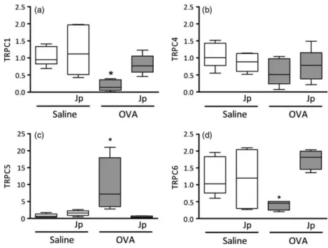
RT-PCR analysis of theTRPCgene expression in rat lung tissue
The expression of genes related to theTRPCsubfamily was investigated in tissue samples obtained from rat lung par-enchyma. Figure 6 reveals that all genes evaluated (TRPC1, TRPC4, TRPC5andTRPC6) were expressed in lung tissues. The expression of a given gene changed according to the challenge with the sensitizing antigen. While TRPC1 and
TRPC6 showed a significantly reduced expression after OVA challenge, TRPC5 significantly increased its mRNA levels (P<0.05). TRPC4 remained at values not signifi-cantly different from the values of saline-challenged ani-mals. Treatment of animals by gavage with the extract of
J. pectoralis (380 mg/kg) abolished all changes caused by OVA challenge.
Discussion
The present study shows that a standardized extract of
J. pectoralishas inhibitory effects on tracheal smooth mus-cle of rats subjected to challenge with OVA in an allergen model that reproduces many features of clinical asthma such as bronchial hyper-reactivity.[15]Administered by gav-age to sensitized rats after challenge with saline or OVA, the extract of J. pectoralis decreased the exacerbated responsiveness of rat trachea caused by the challenge with the sensitizing antigen. The present findings reinforce the notion that the extract of J. pectoralis possesses potential antiasthmatic properties.
Although the time of administration can be considered short, a few authors evaluated the pharmacological effects of the extract ofJ. pectoralisin other experimental models using 60 min as time of administration with positive results.[7,12,13] Importantly, clinically relevant information is available indicating that compounds administered orally in the therapy of asthma can be effective with a short onset of action (<30 min) in patients possessing moderate or severe asthma.[16,17] Thus, this study simu-lated experimentally a condition clinically relevant in terms of acute treatment of bronchospasm and reinforces that the active principle of J. pectoralis has properties of potential interest to pharmacological approaches on the respiratory system.
Figure 4 Effects of the standardized extract ofJusticia pectoralison contractions induced by Ca2+influx after Ca2+store depletion. Tissues were maintained in Ca2+-free medium and repeatedly stimulated with
carbachol (1lM) in the presence of thapsigargin (1lM). Once con-firmed the Ca2+store depletion by the absence of carbachol -induced
contractile response, carbachol was removed from the physiological medium and contractions were induced by Ca2+ restoration in the
absence or presence of the extract ofJ. pectoralis(Jp; 1.1 mg/ml) in tracheal rings obtained from saline- or ovalbumin-challenged animals. Number of experiments:n= 8 for all groups. Data are meanSEM. *P<0.05 compared to saline group;#P
Administered orally, the inhibitory effects of the extract of J. pectoralis on rat airways are likely due to its anti-inflammatory actions and the maintenance of the levels of pro-inflammatory cytokines in bronchoalveolar fluid of OVA-challenged rats at values that did not differ from those obtained with saline-challenged animals sup-ports such hypothesis. Increased amounts of IL-1b and TNF-a in bronchoalveolar lavage fluid are involved in the development of airway hyper-reactivity by affecting the contractile response of airway smooth muscle cells to a variety of stimuli like ACh or KCl.[18–20] The present approach confirmed that the animals subjected to chal-lenge with OVA had higher levels of IL-1band TNF-ain bronchoalveolar fluid and revealed tracheal
hyper-responsiveness under ACh, CCh or KCl stimuli. Leal
et al.[1]reported anti-inflammatory actions for the extract of J. pectoralis against carrageenan-induced paw oedema, but to our knowledge, there is hitherto no study testing the extract of J. pectoralison the respiratory system under inflammatory conditions.
Experiments carried out with the addition of the extract to the bath reveal that J. pectoralis can also exert direct effects on tracheal rings. Although the hyper-responsive phenotype remained observable in OVA-challenged tra-cheal tissues maintained in the continuous presence of the extract of J. pectoralis, hyper-responsiveness was evident only at higher Ca2+concentrations in preparations stimu-lated with ACh or KCl, findings that indicate antispasmodic actions for the extract. Cameron et al.[4]recently reported inhibitory properties of a crude extract of J. pectoralison guinea-pig tracheal contractility. In isolated tissues, the extract ofJ. pectoralisreduced the magnitude of histamine-induced contractions. Part of these antispasmodic actions may be attributable to coumarin, the major constituent of the extract ofJ. pectoralis. Myorelaxant and antispasmodic effects of coumarin and related compounds on rat airway smooth muscle contracted with cholinergic agonists or KCl were already reported.[1,21,22]
Recruitment of inflammatory cytokines like IL-1b and TNF-a further enhances contraction in airway smooth muscle cells by facilitate multiple Ca2+ signalling path-ways, which may be mediated by muscarinic G-protein-coupled receptors or by a non-receptor-dependent stimu-lus such as KCl.[19,23]The experiments in Ca2+-free med-ium (Figure 3) aimed to evaluate the contractile profile of tracheal preparations stimulated to induce Ca2+ influx by receptor-operated or voltage-operated Ca2+ channels. Activated by ACh, muscarinic receptors induce influx of extracellular Ca2+ across the cell plasma membrane majorly by receptor-operated pathways.[24] In contrast, the best-documented KCl-elicited route of Ca2+ entry into airway smooth muscle cells is via dihydropyridine-sensitive voltage-operated Ca2+ channels.[25] We have
already showed that antigen challenge with OVA increases contractile response in isolated trachea of allergic rats by a supposedly greater ionic Ca2+ channel involvement.[26] In the present study, the hyper-responsive behaviour of OVA-challenged tracheal rings is compatible with an increased influx of receptor- or voltage-operated path-ways, both putatively inhibited by the extract of J. pec-toralis and coumarin, which clearly shifted to the right the Ca2+-induced concentration–effect curve in OVA-challenged preparations maintained in the presence of either ACh or KCl, respectively.
In this study, contractions induced by Ca2+influx across plasma membrane mediated by store-operated Ca2+ chan-nels were evaluated in preparations treated with Figure 5 Effects of the standardized extract ofJusticia pectoralison
thapsigargin, a sarco/endoplasmic reticulum Ca2+-ATPase
inhibitor.[27] In tracheal rings maintained in extracellular medium without Ca2+, repeated cholinergic stimulations depleted internal Ca2+stores, a phenomenon confirmed by a gradual decline in the magnitude of tracheal contractions. After removal of the cholinergic stimulus from the extracel-lular medium, the addition of Ca2+to the physiological solution caused a contraction compatible with an expected augment in cytosolic Ca2+by store-operated pathways.[28] Higher contractions in response to extracellular Ca2+ restoration in OVA-challenged tissues suggest a role for the capacitative Ca2+ entry in the development of the hyper-reactive phenotype of airways.[29] Of note was the inhibitory effect induced by the extract of J. pectoralis, which significantly decreased the contraction induced by Ca2+in saline- or OVA-challenged tracheal rings in com-parison with their respective controls.
Gene expression of proteins belonging to the canonical transient receptor potential (TRPC) family were susceptible to antigen challenge in lung tissues. While gene expression of TRPC1 and TRPC6 decreased, TRPC5 increased and
TRPC4was refractory to the antigen challenge. Upregula-tion ofTRPC1expression was reported in rat trachea and proliferating airway smooth muscle cells, while TRPC1
downregulation diminished store-depletion-induced Ca2+ influx and airway hyper-responsiveness in the presence of inflammatory mediators.[28,30] In mice challenged with OVA, downregulation of TRPC6 resulted in increased
agonist-induced contractility of tracheal rings,[31] findings
in line with the present results obtained with rat airways. The expression of TRPC4 was unchanged by the antigen challenge, which argues against the involvement of this pro-tein on the hyper-responsive phenotype on lung tissues. In contrast, the increase in gene expression of TRPC5 was noticeable after the antigen challenge, findings that confer to this protein a potential role in the hyper-responsiveness caused by the present experimental model of asthma.
It is noteworthy that the treatment of saline-challenged animals with the extract ofJ. pectoralisdid not change the gene expression of all TRPC proteins evaluated in this study. However, lung tissues of OVA-challenged animals treated with the extract ofJ. pectoralisrevealed gene expres-sion without significant differences in comparison with sal-ine-treated tissues, suggesting that regulation of TRPC protein expression can be a target of the antiasthmatic properties of the extract of J. pectoralis. We hypothesize that such actions could be derived from its abilities in blunting the increased levels of inflammatory cytokines in airways.
Conclusion
is probable that its beneficial effects are due to its anti-inflammatory effects.
Declarations
Conflict of interest
The Author(s) declare(s) that they have no conflict of inter-ests to disclose.
Funding
This work is part of a thesis submitted by C.T.M. Moura in partial fulfilment of the requirements for the degree of Doctor in Pharmacology at Federal University of Ceara. It was supported by scholarships from CAPES, FUNCAP, and grants from CNPq (INCT-IBISAB, 573928/2008-8).
References
1. Leal LK et al. Antinociceptive, anti-inflammatory and bronchodilator activities of Brazilian medicinal plants containing coumarin: a comparative study. J Ethnopharmacol 2000; 70: 151–159.
2. Brasil. From the Formulario de Fitoterapicos da Farmacopeia Brasi-leira. Ag^encia Nacional de Vigil^ancia Sanitaria (ANVISA), 2011. Available from: http://www.anvisa.gov.br/hot site/farmacopeiabrasileira/conteudo/ Formulario_de_Fitoterapicos_da_Far macopeia_Brasileira.pdf (accessed on 03 September 2016).
3. Correa GM, Alcantara AF. Chemical constituents and biological activities of species of Justicia – a review. Rev Bras Farmacogn2012; 22: 220–238. 4. Cameron Cet al.Preliminary
investi-gations of the anti-asthmatic proper-ties of the aqueous extract ofJusticia pectoralis(fresh cut).West Indian Med J2015; 64: 320–324.
5. Matos FJA. Farmacias Vivas: Sistema de Utilizacß~ao de Plantas Medicinais Projetado para Pequenas Comunidades, 2nd edn. Fortaleza: EUFC, 1994. 6. De Vries JXet al.Constituents of
Jus-ticia pectoralis Jacq. 2. Gas chro-matography/mass spectrometry of simple coumarins, 3-phenylpropionic acids and their hydroxy and methoxy derivatives. Biomed Environ Mass Spectrom1988; 15: 413–417.
7. Lino CS et al. Analgesic and anti-inflammatory activities ofJusticia pec-toralisJacq. and its constituents: cou-marin and umbelliferone. Phytother Res1997; 11: 211–215.
8. Kim JSet al.Chemical constituents of the root ofDystaenia takeshimanaand their anti-inflammatory activity. Arch Pharm Res2006; 29: 617–623. 9. Islam MN et al. Mechanism of
anti-inflammatory activity of umbellifer-one 6-carboxylic acid isolated from Angelica decursiva. J Ethnopharmacol 2012; 144: 175–181.
10. Lim HJ et al. Inhibition of airway inflammation by the roots ofAngelica decursiva and its constituent, colum-bianadin.J Ethnopharmacol2014; 155: 1353–1361.
11. Vasconcelos JFet al.Effects of umbel-liferone in a murine model of allergic airway inflammation.Eur J Pharmacol 2009; 609: 126–131.
12. Leal LK et al. Anti-inflammatory and smooth muscle relaxant activities of the hydroalcoholic extract and chemi-cal constituents from Amburana cearensis A C Smith. Phytother Res 2003; 17: 335–340.
13. Ven^ancio ET et al. Anxiolytic-like effects of standardized extract of Justi-cia pectoralis (SEJP) in mice: involve-ment of GABA/benzodiazepine in receptor. Phytother Res2011; 25: 444– 450.
14. Pfaffl MW. A new mathematical model for relative quantification in real-time RT-PCR. Nucleic Acids Res 2001; 29: e45.
15. Kucharewicz I et al. Experimental asthma in rats. Pharmacol Rep 2008; 60: 783–788.
16. Scalabrin DM, Naspitz CK. Efficacy and side effects of salbutamol in acute asthma in children: comparison of oral route and two different nebulizer systems.J Asthma1993; 30: 51–59.
17. Vatrella A et al. Bronchodilating effects of salmeterol, theophylline and their combination in patients with moderate to severe asthma. Pulm Pharmacol Ther2005; 18: 89–92. 18. Reynolds AMet al.Cytokines enhance
airway smooth muscle contractility in response to acetylcholine and neu-rokinin A.Respirology2000; 5: 153–160. 19. Chen H et al.TNF-[alpha] modulates murine tracheal rings responsiveness to G-protein-coupled receptor ago-nists and KCl. J Appl Physiol (1985) 2003; 95: 864–872.
20. Makwana R et al. TNF-a-induces airway hyperresponsiveness to cholin-ergic stimulation in guinea pig air-ways. Br J Pharmacol 2012; 165: 1978–1991.
21. Pavlovic Iet al.Chloroform extract of underground parts of Ferula heuffelii: secondary metabolites and spasmolytic activity. Chem Biodivers 2014; 11: 1417–1427.
22. Sanchez-Recillas A et al. Semisynthe-sis, ex vivo evaluation, and SAR stud-ies of coumarin derivatives as potential antiasthmatic drugs. Eur J Med Chem2014; 77: 400–408. 23. Pelaia Get al. Molecular mechanisms
underlying airway smooth muscle contraction and proliferation: implica-tions for asthma. Respir Med 2008; 102: 1173–1181.
24. Bolton TB. Mechanisms of action of transmitters, and other substances on smooth muscle. Physiol Rev 1979; 59: 606–718.
26. Moura CTet al.Increased responsive-ness to 5-hydroxytryptamine after antigenic challenge is inhibited by nifedipine and niflumic acid in rat trachea in vitro. Clin Exp Pharmacol Physiol2005; 32: 1119–1123.
27. Lytton J et al. Thapsigargin inhibits the sarcoplasmic or endoplasmic retic-ulum Ca-ATPase family of calcium pumps.J Biol Chem1991; 266: 17067– 17071.
28. Sweeney M et al. Role of capacitative Ca2+ entry in bronchial contraction and remodeling.J Appl Physiol (1985) 2002; 92: 1594–1602.
29. Spinelli AM et al. Airway smooth muscle STIM1 and Orai1 are upregu-lated in asthmatic mice and mediate PDGF-activated SOCE, CRAC cur-rents, proliferation, and migration. Pflugers Arch2012; 464: 481–492.
30. Sousa CTet al.Sildenafil decreases rat tracheal hyperresponsiveness to carba-chol and changes canonical transient receptor potential gene expression after antigen challenge. Braz J Med Biol Res2011; 44: 562–572.


![Figure 5 shows that the values of IL-1b and TNF-a were significantly increased after antigen challenge with OVA (521.3 105.7 [n = 6] and 1998.7 541.8 [n = 10] pg/](https://thumb-eu.123doks.com/thumbv2/123dok_br/15309064.550351/7.892.105.441.120.382/figure-shows-values-tnf-significantly-increased-antigen-challenge.webp)