*Correspondence: Dong Liu. Department of Pharmacy, Tongji Hospital, Tongji
Medical College, Huazhong University of Science and Technology, 1095 Jiefang Avenue, 430030, Wuhan, China. E-mail: [email protected]
A
vol. 50, n. 3, jul./sep., 2014 http://dx.doi.org/10.1590/S1984-82502014000300019
Reduced bioavailability of cyclosporine A in rats by mung bean
seed coat extract
Xiping Li
1, Ping Gao
2, Chengliang Zhang
1, Tao Wu
1, Yanjiao Xu
1, Dong Liu
*,11Department of Pharmacy, Tongji Hospital, Tongji Medical College, Huazhong University of Science and Technology, Wuhan, Hubei, China, 2Department of Pharmacy, Wuhan Children’s Hospital, Wuhan, Hubei, China
Mung bean seed coat (MBSC) is a healthcare product in Asian countries. The aim of this study was to investigate the effect of an MBSC ethanol extract on the bioavailability of cyclosporine A (CsA) in rats. Rats were orally dosed with CsA alone or in combination with MBSC ethanol extracts (500 mg/kg, p.o.). The blood levels of CsA were assayed by liquid chromatography with an electrospray ionization source and tandem mass spectrometry (LC-MS/MS). The everted rat intestinal sac technique was used
to determine the inluence of MBSC on the absorption of CsA. The results reveal that combined CsA
intake with MBSC decreased the Cmax, AUC0-t, t1/2z and MRT0-t values of CsA by 24.96%, 47.28%, 34.73%
and 23.58%, respectively (P<0.05), and signiicantly raised the CL/F by 51.97% (P<0.01). The in vitro
results demonstrated that signiicantly less CsA was absorbed (P<0.05). The overall results indicate that after being concomitantly ingested, MBSC reduced the bioavailability of CsA, at least partially, in the absorption phase.
Uniterms: Mung bean/seed coat/health care use. Mung bean/seed coat/ethanolic extract/properties. Cyclosporine A/bioavailability.
O tegumento da semente de feijão-mungo (MBSC) é um produto para tratamento de saúde em países asiáticos. O objetivo deste estudo foi investigar o efeito de extrato etanólico de MBSC na biodisponibilidade da ciclosporina A (CVsA) em ratos. Administrou-se aos ratos CsA sozinha ou em associação com extrato etanólico de MBSC (500 mg/kg, p.o.), por via oral. Os níveis sanguíneos de
CSA foram determinados por cromatograia a líquido com ionização por electrospray, associada à espectrometria de massas (LC-MS/MS). Utilizou-se a técnica de inversão do saco intestinal de rato para
determinar a inluência do MBSC na absorção de CsA. Os resultados revelaram que a ingestão combinada
de CsA e MBSC diminuiu os valores de Cmax, AUC0-t, t1/2z e MRT0-t de CsA em 24%, 47,28%, 34,73%
e 23,58%, respectivamente (P<0.05), e aumentou, signiicativamente, CL/F em 51,79% (P<0.05). Os
resultados in vitro demostraram que, signiicativamente, menos CsA foi absorvida (P<0.05). Os resultados
totais indicaram que após ser concomitantemente ingerida, a MBSC reduziu, ao menos parcialmente, a biodisponibilidade de CsA, na fase de absorção.
Unitermos: Feijão-mungo/tegumento da semente/uso para a saúde. Feijão-mungo/tegumento da semente/
extrato etanólico/propriedades. Ciclosporina A/biodisponibilidade.
INTRODUCTION
In daily practice, drugs are often taken simultaneously with foods or nutriments. However, food-drug interactions
(FDIs), with commonly overlooked hidden risks,
unintentionally result in therapeutic failure or increased
toxicity of drugs, thus adversely affecting patient care, contributing to morbidity and prolonging treatment or hospitalization stay (Bushra, Slam, Khan, 2011).
Therefore, identifying FDIs, understanding the relevant
mechanisms, and evaluating and managing the risks
are clinically signiicant before diagnosis and treatment
which are prone to inducing acute cellular rejection, lead to hepatotoxicity, nephrotoxicity and neurotoxicity (Kuypers, 2008). Previously, Yang (2002) reported six patients who had received a renal transplantation and suffered from graft rejection after concomitantly drinking mung bean soup. The trough concentrations of CsA decreased from (390.00±112.21) ng/mL before mung bean food intake to (287.33±94.21) ng/mL (P < 0.001) after.
Mung bean (Vigna radiatae L.) seed coat (MBSC) is a traditional Chinese medicine targeting several diseases, generally by reducing fever and removing toxic substances (Yao et al., 2008). Mung bean is a ubiquitous food because
the embryo is abundant in nutrients such as starch, iber
and protein, while most bioactive phytochemicals are contained in the coats (Cao et al., 2011; Khan, Jacobsen, Eggum, 2006). Besides, the pharmacological effects of MBSC, such as antitumor (Soucek et al., 2006), antidiabetic (Peng et al., 2008), anti-inflammatory (Prabhakar et al., 1981), antimicrobial (Randhir, Lin, Shetty, 2004) and antioxidant (Soucek et al., 2006), have also been spotlighted. Hence, the present study mostly focused on the bioactivity of MBSC.
The purpose of this study was to verify the pharmacokinetic effects of mung bean seed coat (MBSC) on CsA. We designed a parallel experiment by pretreating rats with MBSC ethanol extract or vehicle solution for 7 consecutive days before the administration of CsA to investigate the resultant food-drug pharmacokinetic
interaction. Furthermore, whether intestinal absorption
was functionally modulated by MBSC was tested by everted gut sac studies.
MATERIAL AND METHODS
Chemicals and reagents
Dried MBSC was purchased from Hui-Rui Chinese Medicine Science Co., Ltd. (Bozhou, China). CsA (purity>99%) and tacrolimus (Internal standard, purity>99%) were obtained from the China Pharmacy Biological Products Examination Institute (Beijing, China). CsA was donated by Zhongmei Huadong Pharmaceutical Co., Ltd. (Hangzhou, China). HPLC-grade
methanol and acetonitrile were obtained from the Fisher Scientiic Company (Emerson, USA). Other chemicals
and reagents were all analytically pure (Jinfeng Chemical
Factory, Tianjin, China).
Experimental animals
Male Sprague-Dawley rats, aged 8-9 weeks old,
were purchased from the Experimental Animal Center of Tongji Medical College (Huazhong University of Science and Technology, China). The experimental rats were maintained at the Experimental Animal Center of Tongji
Medical College under speciic pathogen-free conditions.
The rats were housed in stainless steel cages and kept at a controlled temperature (25 ± 2 °C) and ambient humidity (50% to 75%). Light was maintained following a 12 h dark-light cycle. All of the rats were continuously provided with a chow diet and tap water throughout the experiment. The experiments were carried out according to the National Institutes of Health Guide for the Care and Use of Laboratory Animals approved by the Animal Ethics Committee of Tongji Medical College, Huazhong University of Science and Technology.
Preparation of the aqueous extract of MBSC
MBSC (500 g) was pre-immersed in 50% ethanol
aqueous solution (solid: liquid, 1:10) and then reluxed at
80 °C for 150 min and extracted three times. The combined extracts were filtered through gauze, to which was then added appropriate amounts of diatomaceous earth. This was stirred at ambient temperature for 2-3 min, and then allowed to stand for 5 min. After the removal of macromolecular compounds by adsorption, the solution was distilled under reduced pressure, and the combined dry residue was extracted with petroleum ether (500 mL) to remove lipid-soluble constituents. The aqueous phase was then collected, freeze-dried as the aqueous extract of MBSC and stored.
Overall pharmacokinetics study
Twelve rats (8-9 weeks old) were randomly divided into two groups, which were administered cyclosporine A (CsA) alone or in combination with MBSC extract respectively. Once daily for six consecutive days, the rats in the combined administration group were orally given 500 mg/kg MBSC extract (dissolved in 0.5% CMC-Na), and those in the CsA group were orally administered with equal volumes of 0.5% CMC-Na solution. All rats had free access to food and water. On the seventh day, the rats were treated with vehicle or MBSC extract. The rats were fasted for no less than 12 h before intragastric administration, but retained free access to water. Meanwhile, the two groups were orally given 10 mg/kg CsA (dissolved in olive oil) after 30 min of the above administration procedure. Blood (0.3 mL) was collected from the carotid artery at 0, 0.83, 1.5, 3, 5, 6, 8, 12, 24 and 36 h after administration. Whole blood was stored at -80 °C. After the experiment, the rats
Everted intestinal sac study
Twelve rats (8-9 weeks old) were randomly divided into a control group and an MBSC extract pretreatment group, in which the rats were orally administered with 0.5% CMC-Na solution and MBSC extract solution (dissolved in 0.5% CMC-Na) respectively once a day for six continuous days. On the seventh day, the rats were fasted for no less than 12 h before experiment with free water access. Thirty minutes after the corresponding pretreatment, the rats were
ixed after ether anesthesia. Everted sacs were prepared
by slightly modifying a procedure described previously (Sakamoto et al., 2006). The abdomens were incised open along the abdominal midline to carefully peel the intestinal canal off the mesentery. Then, 10 cm of the duodenum, jejunum, ileum and colon were disconnected, put into 37 °C K-R buffer (133 mM NaCl, 4.75 mM KCl, 3.33 mM CaCl2, 2.67 mM NaH2PO4, 0.02 mM MgCl2, 16.31 mM NaHCO3, and 8.75 mM C6H12O6, pH 7.0-7.2) and washed until the exhaustion of intestinal contents, after which the mesentery and fat on the surface of intestinal segments were cautiously removed. The rats were sacrificed by cervical dislocation under anesthesia. After being ligated onto a self-prepared plastic sleeve with one end, the intestinal canal was carefully everted and rinsed with K-R buffer, and then the other end was also ligated into a capsular shape. Blank K-R buffer (1 mL) was added to the intestinal sac, which was then put into a water bath already containing K-R buffer, magnetically stirred at 37 °C under a 95% O2/5% CO2 atmosphere. After 5 min of equilibrium, the original K-R buffer was removed from the water bath, into which was then added 250 mL of K-R liquid containing 5 μg/mL CsA. The solution (200 μL) in the intestinal sac was sampled after 15, 30, 45, 60, 75 and 90 min of incubation, and same volume of blank K-R buffer was added simultaneously. The absorptive solution was stored at -80 °C until analysis.
Lactate dehydrogenase (LDH) release of everted gut sacs
LDH is an intracellular enzyme, detected following damage to cell membranes, and has been used as a biochemical marker of intestinal wall damage (Brown
et al., 2002; Swenson, Milisen, Curatolo, 1994). The
feature was determined by means of LDH release tests, as previously reported (Rong et al., 2013).
Analytical methods
All analyses were conducted on a Shimadzu LC
system equipped with two LC-20AD pumps, an SIL-20ACHT autosampler, an SCL-10Avp control system, a DGU-20A3 on-line degasser and a CTO-20AC column oven (Chiyoda-Ku, Japan). Separation was performed on a Dimonsil C18 column (150 mm × 2.1 mm i.d., 5 mm, Dikma, China) equipped with a Phenomenex guard column (5.0 mm × 2.0 mm i.d., Phonomenon, Guangzhou, China). The mobile phase consisted of methanol and 0.1% formic acid (10:90, v/v). The temperature was maintained at 65 oC for the column and 15 oC for the autosampler. The flow rate was 0.3 mL/min. Mass spectrometric analyses were conducted on an API 3,200 LC–MS-MS
system (Applied Biosystems, Foster City, USA) equipped
with an electrospray ionization source (ESI) in triple-quadrupole mode. The curtain gas and collision activated dissociation were 20 and 5 psi, respectively. The other working parameters were set as follows: spray voltage, 5,000 V; source temperature, 450 oC; GAS1, 60 psi and GAS2, 45 psi. The declustering potential, entrance potential, collision energy and collision cell exit potential were optimized, respectively, as 77, 14, 20 and 22 V for CsA; these values were 112, 34 and 70 and 8.0 V for sirolimus (IS). LC–ESI-MS-MS was performed in positive ionization mode with multiple reaction monitoring (MRM) of the transitions m/z ([M+Na]+) 1225.8→m/z 1225.8 for CsA and ([M+NH4]+) m/z 821.9→m/z 409.4 for tacrolimus. Data acquisition and analysis were controlled using Analyst 1.5 software (Applied Biosystems).
Sample preparation
In this study, all samples were detected by liquid chromatography–electrospray ionization tandem mass spectrometry (LC-ESI-MS/MS). Rat whole blood (200 μL) or the absorptive solution from the gut sac was placed in a 10 mL glass centrifuge tube, to which was then added 50 μL of 5 M NH4AC to break the cells. The resultant solution was mixed for 1 min and allowed to stand for 10 min, to which was then added 20 μL of tacrolimus solution (1.012 μg/mL). This was the mixed, extracted with 3 mL of ether for 5 min, and centrifuged at 3,500 rpm for 5 min. The upper organic layer was collected and dried under a nitrogen stream at 40 °C. The dried residue was redissolved in 100 μL of the mobile phase, transferred to an EP tube, and centrifuged at 12,000 rpm for 5 min, from which 10 μL of the supernatant was collected for analysis.
Data transformation
the main pharmacokinetic parameters (Li et al., 2013; Zhang et al., 2012). Peak concentration (Cmax) and time-to-peak (tmax) were measured, area under the plasma concentration-time curve (AUC) was calculated by the trapezoidal rule, and the half-life of elimination (t1/2) was calculated by 0.693/ke (ke refers to the terminal elimination rate constant derived from the slope of terminal straight line of logarithmic plasma concentration-time curve). The
apparent clearance rate of oral administration (CL/F) was
calculated by dose/AUC0-t. All the other parameters were calculated based on the non-compartmental model, and the mean residence time (MRT) of drug molecules was calculated as 1.44t1/2.
The rate of drug transport was usually expressed as the apparent permeability coefficient (Papp). It was calculated from the following equation:
Papp=(dQ/dt)/(C0×A)
where dQ/dt is the steady-state appearance rate on the acceptor solution, A is the surface area of the intestinal sac and C0 is the initial concentration inside the sac.
STATISTICAL ANALYSIS
The experimental data were expressed as mean ± SD or mean, and were analyzed by SPSS 16.0 (Li, et al., 2013; Zhang et al., 2012). The main parameters of each group were subjected to one-way analysis variance (ANOVA) and Student’s t-test, with P<0.05 being statistically
signiicantly different.
RESULTS AND DISCUSSION
Method validation
Briely, the seven-point calibration curve for CsA (0,
50, 100, 200, 400, 600 and 1200 ng/mL) was constructed by plotting the peak area ratio of CsA-IS against the real concentration of the calibration standards in rat plasma and K-R buffer. Inter-day and intra-day repeatability were assessed with QC samples (50, 200 and 1000 ng/mL of CsA in rat whole blood or absorptive solution of gut sac). The accuracy and precision were defined by the
percentage of relative standard deviation (RSD) of ive
standards at five different concentrations analyzed on the same day. Stability was expressed by the relative error between the initial and tested concentration of QC samples under different sample preparatory conditions, such as short-term, long-term, freeze-thaw cycle and post-preparation stability. The results indicate that the lower
limit of quantification was 50 ng/mL with a precision (RSD) less than 9.01% and a accuracy ranging from 94.5% to 107.76%. The recovery of CsA was no less than 87.3%
with a coeficient of variation less than 3.2%. Short-term,
long-term and three freeze-thaw stability studies indicated that analytes were stable under the above conditions. Ion suppression and enhancement from plasma and K-R buffer matrix were negligible under the present conditions.
LDH release in the everted gut sac model
The results reveal that there were no significant differences in LDH activity at 30, 60, 90 and 120 min, while a significant difference was found at 180 min, suggesting that the everted gut sacs began to lose viability after 120 min. It was concluded that the everted gut sacs maintained their viability during the experimental period (90 min) and consequently this model was suitable for testing drug transport.
Effects of MBSC on the pharmacokinetics of CsA in rats
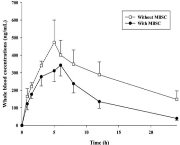
The whole blood concentration-time curves of CsA after oral dosing with CsA alone and in combination with
MBSC are illustrated in Figure 1. The pharmacokinetic
parameters of CsA are shown in Table I. Pretreating the
rats with MBSC signiicantly reduced the Cmax, AUC0-t, t1/2z
and MRT of CsA by 24.96% (P<0.05), 47.28% (P<0.01), 34.73% (P<0.05)and 23.58% (P<0.01), respectively, and
signiicantly elevated CL/F by 51.97% (P<0.01).
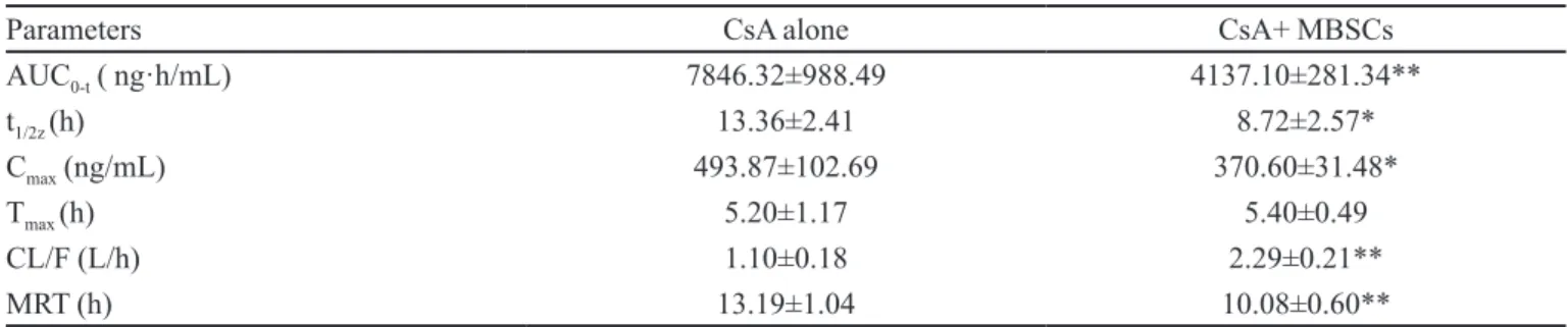
TABLE I - Effect of treatment with herbal extract on the pharmacokinetic parameters of CsA
Parameters CsA alone CsA+ MBSCs
AUC0-t ( ng·h/mL) 7846.32±988.49 4137.10±281.34**
t1/2z (h) 13.36±2.41 8.72±2.57*
Cmax (ng/mL) 493.87±102.69 370.60±31.48*
Tmax (h) 5.20±1.17 5.40±0.49
CL/F (L/h) 1.10±0.18 2.29±0.21**
MRT (h) 13.19±1.04 10.08±0.60**
Each point represents the mean±S.D. (n=6). * p<0.05, ** p<0.01, signiicantly different compared with CsA alone
FIGURE 2 - Average transport of CsA (ng) from the mucosal to serosal surface across duodenum, jejunum, ileum and colon with or without MBSC extract pretreatment (500 mg/kg, i.g.). Each point represents the mean±SD. (n=3).
Effects of MBSC on CsA absorption in intestinal gut sacs
Figure 2 shows the effects of MBSC treatment on
the absorption of CsA in the duodenum, jejunum, ileum and colon. Table II shows the Papp of CsA in each intestinal
segment in the everted gut sac study. The results demonstrate that the rank order of Papp of CsA in each intestinal segment
was as follows: ileum > duodenum ≈ jejunum > colon. The
doses of MBSC decreased the Papp of CsA in most intestinal segments, except for the duodenum. Altogether, the resultsreveal that pretreatment with 500 mg/kg MBSC
signiicantly decreased the Papp of CsA in the jejunum, ileum
and colon (P < 0.05).
In this study, combined intake of CsA with MBSC markedly decreased the AUC0-t (P<0.01), Cmax (P<0.05), t1/2z (P<0.05) and MRT (P<0.01) of CsA, and remarkably
increased the CL/F (P<0.01), demonstrating signiicantly
reduced bioavailability of CsA. The semi-log profiles (data not shown) indicate that MBSC seemed to inhibit the intestinal absorption and enhanced the intestinal/ liver elimination of CsA. It is acknowledged that combined intake of CsA with foods and beverages can affect the rate and extent of drug absorption (Chiang et al., 2006), probably by impacting intestinal physiological factors and transport (Boullata, Hudson, 2012). Our
everted gut sac study indicates that MBSC signiicantly
decreased the accumulative absorption of CsA in each intestinal segment, suggesting that reduced CsA bioavailability occurs, at least partially, at the absorption site. Given that accelerated transit of a drug through the gastrointestinal tract reduces its absorption, MBSC might shorten drug transit time like ginger does (Platel, Srinivasan, 2001). In the meantime, CsA is metabolized in the intestine/liver by CYP3A4, and the parent drug is subjected to efflux by P-gp in the intestinal apical membrane (Pal, Mitra, 2006). Dürr reported that St John’s wort lowers the blood concentration of CsA by inducing CYP3A4 and intestinal P-gp in humans (Dürr et al., 2000). In addition, Yang reported that the ingestion of ginkgo and onion decreases the bioavailability of CsA via inducing CYP3A in rats (Yang et al., 2006). Moreover, Chiang also found that ginger reduces the blood concentration of CsA by the induction of CYP3A4 (Chiang et al., 2006). However, whether MBSC can accelerate the metabolism of CsA by inducing intestinal or liver CYP3A in rats, or increase the efflux of CsA by inducing intestinal P-gp expression or activity remain unknown. The underlying
mechanisms should be further clariied in future.
CONCLUSIONS
The present study was conducted to verify the interaction between MBSC and CsA. The results of this study demonstrate that combined treatment with CsA and
MBSC could signiicantly reduce the oral bioavailability
of CsA, at least partially by inhibiting absorption, suggesting that combined use of MBSC with CsA should be closely monitored for potential food-drug interactions.
ACKNOWLEDGMENTS
This research was supported by theFundamental
Research Funds for the Central Universities (HUST:
2012QN182).
REFERENCES
BUSHRA, R.; SLAM, N.; KHAN, A.Y. Food-drug interactions. Oman. Med. J., v.26, n.2, p.77-83, 2011.
BOULLATA, J.I.; HUDSON, L.M. Drug-nutrient interactions:
a broad view with implications for practice. J. Acad. Nutr.
Diet, v.112, n.4, p.506-517, 2012.
BROWN, J.R.; COLLETT, J.H.; ATTWOOD, D.; LEY, R.W.; SIMS, E.E. Inluence of monocaprin on the permeability of a diacidic drug BTA-243 across Caco-2 cell monolayers and
everted gut sacs. Int. J. Pharm., v.245, n.1, p.133-142, 2002.
CAO, D.; LI, H.; YI, J.; ZHANG, J.; CHE, H.; CAO, J., YANG, L.; ZHU, C.; JIANG, W. Antioxidant properties of the mung bean lavonoids on alleviating heat stress. PLoS One, v.6, n.6, p.1, 2011.
CHIANG, H.M.; CHAO, P.D.; HSIU, S.L.; WEN, K.C.; TSAI, S.Y.; HOU, Y.C. Ginger signiicantly decreased the oral
bioavailability of cyclosporine in rats. Am. J. Chin. Med.,
v.34, n.5, p.845-855, 2006.
TABLE II – Apparent permeability of CsA in each intestinal segment in everted gut sac study
CsA alone
(Papp, 10-4 cm/s)
CsA+MBSCs
(Papp, 10-4 cm/s)
Duodenum 10.83±1.14 9.42 ±2.17
Jejunum 9.01±1.03 7.26 ±3.04*
Ileum 13.29 ±5.21 8.57 ±2.75*
Colon 6.71±1.55 4.87± 0.97*
DÜRR, D.; STIEGER, B.; KULLAK-UBLICK, G.A.; RENTSCH, K.M.; STEINERT, H.C.; MEIER, P.J.; FATTINGER, K. St John’s Wort induces intestinal P-glycoprotein/MDR1 and intestinaland hepatic CYP3A4. Clin. Pharmacol. Ther., v.68, n.6, p.598-604, 2000.
DREWE, J.; BEGLINGER, C.; KSSEL, T. The absorption site
of cyclosporin in the human gastrointestinal tract. Br. J.
Clin. Pharmacol., v.33, n.1, p.39-43, 1992.
HUANG, Q.; XU, J.; BEI, Y.Y.; LIU, Y.; TANG, J.Z.; ZHANG, X.N. Studies on mechanism to promote intestinal absorption
of cyclosporine A & formulation factors. Anti-Infect.
Pharm., v.7, n.4, p.246-250, 2010.
K U Y P E R S , D . R . I n f l u e n c e o f i n t e r a c t i o n s b e t w e e n immunosuppressive drugs on therapeutic drug monitoring. Ann. Transplant., v.13, n.3, p.11-18, 2008.
KHAN, M.A.; JACOBSEN, I.; EGGUM, B.O. Nutritive value
of some improved varieties of legumes. J. Sci. Food Agric.,
v.30, n.4, p.395-400, 2006.
LI, X.P.; ZHANG, C.L.; GAO, P.; GAO, J.; LIU, D. Effects of andrographolide on the pharmacokinetics of aminophylline
and doxofylline in rats. Drug Res. (Stuttg), v.63, n.5,
p.258-262, 2013.
PENG, X.; ZHENG, Z.; CHENG, K.W.; SHAN, F.; REN, G.X.; WANG, M. Inhibitory effect of mung bean extract and its costituents vitexin and isovitexin on the formation of
advanced glycation endproducts. Food Chem., v.106, n.2,
p.475-481, 2008.
PRABHAKAR, M.C.; BANO, H.; HUMAR, I.; PRABHAKAR, M.C.; BANO, H.; KUMAR, I.; SHAMSI, M.A.; KHAN,
S.Y. Pharmacological investigations on vitexin. Planta
Med., v.43, n.4, p.396-403, 1981.
PLATEL, K.; SRINIVASAN, K. Studies on the inluence of
dietary spices on food transit time in experimental rats. Nutr.
Res., v.21, n.9, p.1309-1314, 2001.
PAL, D.; MITRA, A.K. MDR- and CYP3A4-mediated
drug-herbal interactions. Life Sci., v.78, n.18, p.2131-2145, 2006.
RANDHIR, R.; LIN, Y.T., SHETTY, K.F. Stimulation of phenolics, antioxidant and antimicrobial activities in dark germinated mung bean sprouts in response to peptide and
phytochemical elicitors. Process Biochem., v.39, n.5,
p.637-646, 2004.
RONG, Z.H.; XU, Y.J.; ZHANG, C.L.; XIANG, D.C.; LI, X.P.; LIU, D. Evaluation of intestinal absorption of amtolmetin guacyl in rats: Breast cancer resistant protein as a primary
barrier of oral bioavailability. Life Sci., v.92, n.3, p.
245-251, 2013.
SAKAMOTO, S.; SUZUKI, H.; KUSUHARA, H.; SUGIYAMA, Y. Eflux mechanism of taurocholate across the rat intestinal
basolateral membrane. Mol. Pharm., v.3, n.3, p.275-281,
2006.
SOUCEK, J.; SKVOR, J.; POUCKOVA, P.; MATOUSEK, J.; SLAViK, T.; MATOUSEK, J. Mung bean sprout (phaseolus aureus) nuclease and its biological and antitumor effects. Neoplasma, v.53, n.5, p.402-409, 2006.
SWENSON, E.S.; MILISEN, W.B.; CURATOLO, W. Intestinal permeability enhancement: eficacy, acute local toxicity,
and reversibility. Pharm. Res., v.11, n.8, p.1132-1142, 1994.
YANG, C.Y.; CHAO, P.D.; HOU, Y.C.; TSAI, S.Y.; WEN, K.C.; HSIU, S.L. Marked decrease of cyclosporin bioavailability caused by coadministration of ginkgo and onion in rats. Food Chem. Toxicol., v.44, n.9, p.1572-1578, 2006.
YANG, Z.H.; ZHANG, Z.; LIU, N.B. JIONG, Y.J. Inluence of mung bean food on blood CsA concentration in renal
transplantation patients. China Pharm. J., v.37, n.3,
p.229-230, 2002.
YAO, Y.; CHEN, F.; WANG, M.; WANG, J.; REN, G. Antidiabetic activity of mung bean extracts in diabetic
KK-Ay mice. J. Agric. Food Chem., v.56, n.19,
p.8869-8873, 2008.
ZHANG, C.L.; GAO, P.; YIN, W.F.; XU, Y.J.; XIANG, D.C.; LIU, D. Dexamethasone regulates differential expression of carboxylesterase 1 and carboxylesterase 2 through
activation of nuclear receptors. J. Huazhong Univ. Sci.
Technolog. Med. Sci., v.32, n.6, p.798-805, 2012.
Received for publication on 10th September 2013