Vol. 7, No. 3, 2004 Preparation and Characterization of FeAg Granular Alloys 513
Materials Research, Vol. 7, No. 3, 513-516, 2004. © 2004
*e-mail: [email protected]
Article presented at the XV CBECIMAT, Natal - RN, November/2002
Preparation and Characterization of FeAg Granular Alloys
J. M. Soaresa*, J. H. de Araújoa, F. A. O. Cabrala, J. A. P. da Costaa, J. M. Sasakib
aDepartamento de Física Teórica e Experimental, Centro de Ciências Exatas e da Terra,
Universidade Federal do Rio Grande do Norte, 59072-970 Natal - RN
bDepartamento de Física, Universidade Federal do Ceará,
60455-970 Fortaleza - CE
Received: October 03, 2003; Revised: March 21, 2004
Fe10Ag90 granular alloys have been fabricated, using a sol-gel method, for a range of nitric acid concentrations in the start solution. The samples have been characterized by X-ray diffraction and magnetization measurements. The average Fe particle sizes derived from X-ray diffraction are in the range 24-29 nm, indicating a large variation. The coercivity obtained from the hysteresis curve is two orders of magnitude larger that of pure iron in all the samples. Moreover, the hysteresis curves do not saturate, even in fields of up to 1 T. These observations indicate that the samples contain both superparamagnetic and blocked particles. A comparison between the coercive field and the average particle diameter, determined by the Sherrer’s formula, is displayed in all acid concentrations.
Keywords: sol-gel, granular alloy, ferromagnetism
1. Introduction
Granular magnetic materials composed of magnetic nanoparticles embedded in a nonmagnetic matrix show in-teresting physical properties, such as superparamagnetism (SPM), giant magnetoresistance and giant
magneto-impedance1-3. Moreover, they have important
technologi-cal applications in magnetic recording, in optitechnologi-cal devices and in sensors4-7. Such materials have been produced as films
by sputtering, as ribbons by melt-spinning, and in powder form by sol-gel methods8,9 and mechanical alloying10.
The magnetic grain size of these materials varies from a few Å to hundreds of Å, with the grains randomly distrib-uted in a nonmagnetic metallic matrix. When these alloys are prepared by melt-spinning or by sputtering methods, the mean grain size is very small (a few Å) displaying a predominantly SPM behavior. However, granular alloys produced by other techniques such as sol-gel and mechani-cal alloying contain both SPM and blocked (BL) parti-cles10,11,14,15, displaying a more complex magnetic behavior.
Magnetization and Mössbauer Spectroscopy measure-ments have been used to study magnetic properties of the granular alloys FeCu and FeAg in powder form8,10. The
av-erage sizes of magnetic particles were estimated by the co-ercive field.
In this work a series of Fe10Ag90 granular alloys has been produced by a sol-gel method and characterized by X-ray diffraction, Mössbauer spectroscopy and magnetization. We have observed that in all samples there is a size distribution of both SPM and BL particles with a strong influence of changes in acid concentration.
2. Experimental
Fe10Ag90 granular alloys were produced by a sol-gel method8,9. The start solution was prepared from an aqueous
solution of the nitrates with five different nitric acid con-centrations: 0.25, 0.50, 0.75, 1.00 and 1.25 ml, respectively, in 300 ml of mixture. The powder obtained was reduced in a hydrogen atmosphere for 45 min at a temperature of 400 °C.
The crystalline structure of the sample was investigated by conventional X-ray diffraction using a Rigaku diffractometer with Mo-Kα radiation. The magnetization and
hysteresis curves were measured in a vibrating sample magnetometer with a maximum magnetic field of 1 T. The Mössbauer spectrum was obtained in a conventional con-stant acceleration spectrometer with a 57Co source in a
514 Soares et al. Materials Research
3. Results and Discussion
Figure 1 shows the X-ray diffractometry patterns of Fe10Ag90 granular alloy reduced at temperature of 400 °C. It is shows that the iron in the Fe10Ag90 is b.c.c. structure and the silver is f.c.c. structure; there are also some low intensity reflections from Fe3O4. Assuming that the Fe in the sample is indeed distributed as small particles, the aver-age particle diameter should be related to the Fe Bragg peak widths by Scherrer’s formula, Dm= (0.9λ)/[δ(2θ)cosθ], where λ is the Mo wavelength, δ(2θ) is the Fe(211) peak width, and θ the scattering angle16. Figure 2 shows details
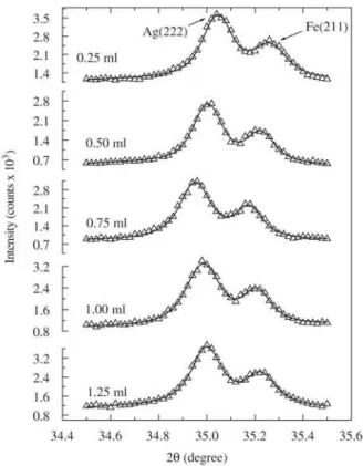
of the spectral regions around the Ag (222) and Fe (211) Bragg peaks. Applying Scherrer’s formula, we obtain the average Fe particle sizes Dm from the (211) line width for different nitric acid concentrations Vac, as shown in Table 1. We observe that Dm increases with Vac up to a concentration of 0.75 ml followed by a decrease.
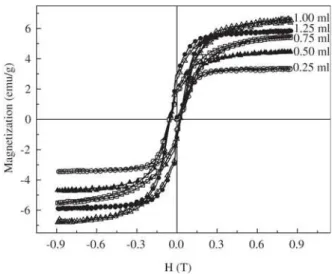
Figure 3 shows the hysteresis curves at room temperature obtained for Fe10Ag90 granular alloy samples with different nitric acid concentrations. None of the samples reach satura-tion in fields up to 1 T, showing the presence of SPM parti-cles. The coercive field, determined from the hysteresis curves, ranges from 354 to 430 Oe, which is much higher than that of pure iron, indicating fine single-domain particles.
Figure 4 shows the Mössbauer spectrum of the Fe10Ag90 (0.5 ml) sample. We can observe that the spectrum consists
Figure 1. X-ray diffractogram of Fe10Ag90 granular alloy (0.75 ml).
Figure 2. X-ray diffractogram of the Ag (222) and Fe (211) peaks of Fe10Ag90 granular alloy, in different nitric acid concentrations. The solid line was the fitting obtained using two Pseudo-Voigt functions. Table 1. Average Fe particles sizes Dm in the Fe10Ag90 granular
alloy in different nitric acid concentrations Vac.
Vac(ml) 0.25 0.50 0.75 1.00 1.25
Dm (nm) 24.5 28.0 28.6 26.2 25.3
of two components, a singlet, characteristic of an SPM phase, and a sextet corresponding to the blocked particles in the ferromagnetic α-Fe. Observe that we were able to see the presence of SPM particles, even though the measurement time window of the Mössbauer effect is 10-8 s.
Figures 5a, 5b, 5c, and 5d show the change of the
maxi-mum field magnetization MHmax, the remanent
Vol. 7, No. 3, 2004 Preparation and Characterization of FeAg Granular Alloys 515
sizes whose width depends on the acid concentration. Since
the Hc value depends directly on the number of blocked
particles in the distribution, the coercive field ought to have a strong dependence on the distribution width, not just on the average particle sizes. However to obtain a correlation between the particles size distribution width and the coer-cive field more experimental and computational work is nec-essary. The distribution width varies between 1.14 and 1.6717.
The experimental results obtained from the hysteresis curve and Mössbauer spectroscopy measurements both in-dicate that the magnetization of the Fe10Ag90 samples is
composed of two contributions: one from the SPM parti-cles and other from the BL partiparti-cles, as observed in other systems reported in the literature11,14,15.
4. Conclusion
Samples of Fe10Ag90 granular alloys were produced in various concentrations of nitric acid. Magnetic measure-ments show that in all samples there was a size distribution of both SPM and BL particles. The magnetic properties of the samples are strongly influenced by changes in acid con-centration.
Acknowledgment
This work was partially supported by the Brazilian agen-cies CNPq, FINEP and CAPES.
References
1. Berkowitz, A.E.; Mitchell, J.R.; Carey, M.J.; Young, A.P.; Zhang, S.; Spada, F.E.; Parker, F.T.; Hutten, A.; Thomas, G. Phys. Rev. Lett. 68, p. 3745, 1992.
2. Xiao, J.Q.; Jiang, J.S.; Chien, C.L. Phys. Rev. Lett. 68, Figure 3. Room temperature hysteresis curves of the Fe10Ag90
granular alloy samples in different nitric acid concentrations.
Figure 5. Magnetic properties of Fe10Ag90 prepared in different nitric acid concentrations: a) Maximum field magnetization; b) Remanent magnetization; c) Coercive field; d) Fe means particle diameter obtained of X-ray diffraction.
516 Soares et al. Materials Research
p. 3749, 1992.
3. Soares, J.M.; Araújo, J.H. de; Cabral, F.A.O.; Dumelow,
T.; Machado, F.L.A.; Araújo, A.E.P. de Appl. Phys.
Lett. 80, p. 2532, 2002.
4. Abeles, B. Applied Solid State Science: Advances in Ma-terials and Device Research, edited by Wolfe, R.
Aca-demic Press, New York, p. 1, 1976.
5. Sheng, B. Abeles. P.; Couts, M.D.; Arie, Y. Adv. Phys.
v. 24, p. 407, 1975.
6. Chien, C.L.; J. Appl. Phys. v. 69, p. 5267, 1975.
7. Evetts, J. Concise Encyclopedia of Magnetic and Super-conducting Materials, Pergamon, London, p. 246, 1992.
8. Chartterjee, A.; Datta, A.; Giri, Anit. K.; Das, D.; Chakravorty, D. J. Appl. Phys. v. 72, n. 8, p. 3832, 1992.
9. Wang, Jian-Ping; Luo, He-Lie; Gao, Nai-Fei; Liu, Yuan-Yuan; J. Mater. Sci., v. 31, p. 727, 1996.
10. Gómez, J.A.; Xia, S.K.; Passamani, E.C.; Giordanengo,
B.; Baggio-Saitovitch, E.M. J. Magn. Magn. Mater.,
v. 223, p. 112, 2001.
11. Viegas, A.D.C.; Geshev, J.; Dorneles, L.S.; Schmidt, J.E.; Knobel, M. J. Appl. Phys., v. 82, p. 3047, 1997.
12. Xiao, G.; Chien, C.L.; J. Appl. Phys., v. 61, p. 3308, 1987.
13. Yakushiji, K.; Mitani, S.; Takanashi, K.; Ha, J.G.; Fujimori, H. J. Magn. Magn. Mater., v. 212, p. 75, 2000.
14. Kuzminski, M.; Slawska-Waniewska, A.; Lachowicz, H.K.; Knobel, M. J. Magn. Magn. Mater., v. 205, p. 7,
1999.
15. Hickey, B.J.; Howson, M.A.; Musa, S.O.; Tomka, G.J.; Rainford, B.D.; Wiser, N. J. Magn. Magn. Mater., v. 147,
p. 253, 1995.
16. Cullity, D. Elements of X-ray Diffraction
Addison-Wesley Publishing Co., Reading, Massachusetts, 1978.