Differentiating between tuberculosis-related and
lymphoma-related lymphocytic pleural effusions by
measuring clinical and laboratory variables: Is it possible?*
,**
É possível diferenciar derrames pleurais linfocíticos secundários a tuberculose ou linfoma através de variáveis clínicas e laboratoriais?
Leila Antonangelo, Francisco Suso Vargas, Eduardo Henrique Genofre, Caroline Maris Neves de Oliveira, Lisete Ribeiro Teixeira,
Roberta Karla Barbosa de Sales
Abstract
Objective: To describe clinical and laboratory characteristics in patients with tuberculosis-related or lymphoma-related lymphocytic pleural effusions, in order to identify the variables that might contribute to differentiating
between these diseases. Methods: This was a retrospective study involving 159 adult HIV-negative patients
with tuberculosis-related or lymphoma-related lymphocytic effusions (130 and 29 patients, respectively), treated between October of 2008 and March of 2010 at the Pleural Diseases Outpatient Clinic of the University of São Paulo School of Medicine Hospital das Clínicas Heart Institute, in the city of São Paulo, Brazil. Results: Mean age and the mean duration of symptoms were lower in the tuberculosis group than in the lymphoma group. The levels of proteins, albumin, cholesterol, amylase, and adenosine deaminase (ADA) in pleural fluid, as well as the serum levels of proteins, albumin, and amylase, were higher in the tuberculosis group, whereas serum cholesterol and triglycerides were higher in the lymphoma group. Pleural fluid leukocyte and lymphocyte counts were higher in the tuberculosis group. Of the tuberculosis group patients, none showed malignant cells; however, 4 showed atypical lymphocytes. Among the lymphoma group patients, cytology for neoplastic cells was positive, suspicious, and negative in 51.8%, 24.1%, and 24.1%, respectively. Immunophenotyping of pleural fluid was conclusive in most of the lymphoma patients. Conclusions: Our results demonstrate clinical and laboratory similarities among the patients with tuberculosis or lymphoma. Although protein and ADA levels in pleural fluid tended to be higher in the tuberculosis group than in the lymphoma group, even these variables showed an overlap. However, none of the tuberculosis group patients had pleural fluid ADA levels below the 40-U/L cut-off point.
Keywords: Pleural effusion; Tuberculosis; Lymphoma; Adenosine deaminase; Diagnosis, differential.
Resumo
Objetivo: Descrever características clínicas e laboratoriais em pacientes com derrames pleurais linfocíticos secundários a tuberculose ou linfoma, a fim de identificar as variáveis que possam contribuir no diagnóstico
diferencial dessas doenças. Métodos: Estudo retrospectivo com 159 pacientes adultos HIV negativos com
derrame pleural linfocítico secundário a tuberculose ou linfoma (130 e 29 pacientes, respectivamente) tratados no Ambulatório da Pleura, Instituto do Coração, Hospital das Clínicas da Faculdade de Medicina da Universidade
de São Paulo, São Paulo (SP), entre outubro de 2008 e março de 2010. Resultados: A média de idade e de
duração dos sintomas foi menor no grupo tuberculose que no grupo linfoma. Os níveis pleurais de proteínas, albumina, colesterol, amilase e adenosina desaminase (ADA), assim como os níveis séricos de proteínas, albumina e amilase, foram maiores no grupo tuberculose, enquanto os níveis séricos de colesterol e triglicérides foram maiores no grupo linfoma. As contagens de leucócitos e linfócitos no líquido pleural foram maiores no grupo tuberculose. Células malignas estavam ausentes no grupo tuberculose; entretanto, linfócitos atípicos foram observados em 4 desses pacientes. No grupo linfoma, a citologia para células neoplásicas foi positiva, suspeita e negativa em 51,8%, 24,1% e 24,1% dos pacientes, respectivamente. A imunofenotipagem do líquido pleural
foi conclusiva na maioria dos pacientes com linfoma. Conclusões: Nossos resultados demonstram semelhanças
clínicas e laboratoriais entre os pacientes com tuberculose ou linfoma. Embora os níveis de proteínas e ADA no líquido pleural tendam a ser mais elevados no grupo tuberculose que no grupo linfoma, mesmo essas variáveis mostraram uma sobreposição. Entretanto, nenhum paciente com tuberculose apresentou níveis de ADA no líquido pleural inferiores ao ponto de corte (40 U/L).
Descritores: Derrame pleural; Tuberculose; Linfoma; Adenosina desaminase; Diagnóstico diferencial.
* Study carried out at the Laboratory for Pleural Studies, Department of Pulmonology, Heart Institute; in the Clinical Laboratory; and in the Laboratório de Investigação Médica 3 (LIM-3, Laboratory for Medical Research 3), Department of Pathology, University of São Paulo School of Medicine Hospital das Clínicas, São Paulo, Brazil.
Correspondence to: Roberta K. B. Sales. Rua Itapeva, 500, cj. 4C, Bela Vista, CEP 01332-000, São Paulo, SP, Brazil. Tel. 55 11 2661-5695. Fax: 55 11 2661-5643. E-mail: [email protected]
Financial support: None.
Submitted: 21 September 2011. Accepted, after review: 1 January 2012.
further complicate the differential diagnosis with pleural tuberculosis.
In this context, the objective of the present study was to describe clinical and laboratory characteristics in patients with tuberculosis-related or lymphoma-tuberculosis-related lymphocytic pleural effusions, in order to identify the variables that might contribute to differentiating between these diseases.
Methods
This was a retrospective study based on a database of patients treated at the Pleural Diseases Outpatient Clinic of the University of São Paulo School of Medicine Hospital das Clínicas Heart Institute, located in the city of São Paulo, Brazil. The study was approved by the University of São Paulo School of Medicine Research Ethics Committee. We evaluated the medical records of adult HIV-negative patients with tuberculosis-related or lymphoma-related lymphocytic effusions treated between October of 2008 and March of 2010.
The diagnosis of pleural tuberculosis was based on the following: clinical history consistent with the diagnosis; satisfactory response to specific treatment; and positive cultures for M. tuberculosis (in sputum, pleural fluid, or tissue specimens) or pleural biopsy demonstrating a chronic granulomatous process. Lymphomatous pleural effusion was characterized by the finding of malignant lymphoid cells in pleural fluid or tissue, with an immunophenotypic profile consistent with monoclonal lymphoproliferative disease, in patients with or without a previous diagnosis of lymphoma.
In order to determine the clinical, radiological, and laboratory characteristics of the patients, the following were employed: conventional biochemical analyses; determination of ADA levels (by the modified Giusti method)(15); microbiological tests; cytological/histological examination; and lymphocyte immunophenotyping, when necessary.
The statistical analysis was carried out with the SigmaStat statistical package, version 3.5 (Systat Software Inc., San Jose, CA, USA). The data are presented as median and interquartile range for biochemical and cytological variables or as mean and standard deviation for age and duration of symptoms. In order to compare the groups, we used the t-test or the Mann-Whitney test, depending on the distribution of
Introduction
Pleural involvement in infectious, inflammatory, or neoplastic diseases almost invariably results in accumulation of fluid in the pleural space. In the USA, it is estimated that 1.5 million new cases of pleural effusion occur every year, including transudates due to heart or liver problems.(1) If we exclude parapneumonic effusions and acute inflammatory exudates, which are generally neutrophilic, most of the remaining types of effusion are considered lymphocytic (more than 50% with nucleated cells).(2)
Brazil ranks 19th in terms of tuberculosis incidence,(3) and pleural tuberculosis is one of the main causes of lymphocytic pleural effusion in adults. In such cases, the pleural fluid is an exudate with high levels of proteins, adenosine deaminase (ADA), and IFN-γ.(4-12)
The methods that are currently used in order to diagnose pleural tuberculosis have problems, including low positivity rates in the direct testing for AFB, low sensitivity (20-30%), and delayed Mycobacterium tuberculosis growth in pleural fluid cultures. Although the presence of granulomas has been demonstrated in up to 80% of pleural tissue samples,(7-9) pleural biopsy is needed in order to establish the diagnosis. In this context, the measurement of ADA in pleural fluid is an alternative method for the diagnosis of tuberculosis, given that the test is highly accurate, reproducible, inexpensive, user-friendly, and rapid.(11)
If malignancy is suspected, pleural fluid cytology is the diagnostic method of choice, the sensitivity of the method ranging from 40% to 87%.(2) Closed pleural biopsy does not significantly contribute to the diagnosis of malignancy, given that the positivity rates for closed pleural biopsy are lower than are those for cytology, ranging from 36% to 60%.(2)
serum levels of cholesterol and triglycerides were significantly higher in the lymphoma group.
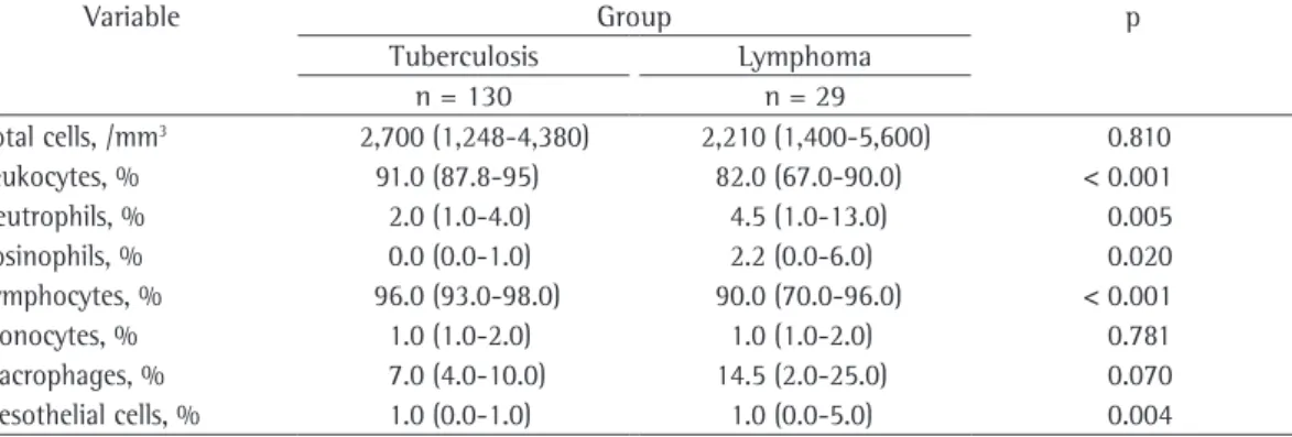
Regarding the cytological evaluation, the proportion of leukocytes and lymphocytes in pleural fluid was found to be higher in the tuberculosis group (Table 3). Screening for malignant cells was carried out in both groups and was found to be negative in most of the tuberculosis patients (96.9%); however, atypical lymphocytes have been found in 4 (3.1%). In those patients, culture for M. tuberculosis in pleural fluid or tissue was positive, and, during the follow-up period, there was a satisfactory response to specific treatment, with no evidence of malignancy.
Among the patients with lymphoma-related pleural effusions, cytology for malignant cells was positive, highly suspicious, and negative in 51.8%, 24.1%, and 24.1%, respectively. Closed pleural biopsy revealed lymphomatous infiltrate in 41.4%. In patients with suspicious cytology results and negative pleural biopsy, the diagnosis of lymphoma was confirmed by histology and immunophenotyping of lymphocytes in sites other than the pleura. Table 4 shows the histological/ phenotypic classification of the lymphoma cases. Effusions associated with Hodgkin’s lymphoma accounted for only 6.9% of the cases. In cases of effusions associated with non-Hodgkin’s lymphoma, B-cell subtypes predominated (66.7%). the variables. For categorical variables, we used
the chi-square test. A value of p < 0.05 was considered significant.
Results
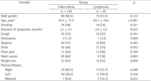
Table 1 shows the clinical and demographic data of the 159 patients (130 patients with tuberculosis and 29 patients with lymphoma) included in the present study. In both groups, there was a slight predominance of males. The mean age and the mean duration of symptoms were significantly lower in the tuberculosis group. Among the symptoms reported, only night sweats were significantly more common in the tuberculosis group than in the lymphoma group. Right pleural effusion predominated, regardless of the etiology.
The biochemical variables studied are shown in Table 2. The levels of protein, albumin, cholesterol, and amylase in pleural fluid were significantly higher in the tuberculosis group than in the lymphoma group. All of the patients in the tuberculosis group and 31% of those in the lymphoma group showed pleural fluid ADA levels higher than the upper normal limit (40 U/L). Serum levels of protein, albumin, and amylase were significantly higher in the tuberculosis group than in the lymphoma group, whereas
Table 1 - Demographic, clinical, and radiological data of 159 patients with tuberculosis-related or lymphoma-related pleural effusions.a
Variable Group p
Tuberculosis Lymphoma
n = 130 n = 29
Male gender 89 (68.5) 15 (51.7) 0.133
Age, yearsb 44.7 ± 17.4 64.1 ± 18.9 < 0.001
Smoking 39 (30) 10 (35) 0.761
Duration of symptoms, monthsb 2.1 ± 3.1 2.9 ± 3.2 0.037
Cough 93 (72) 16 (57) 0.193
Hemoptysis 2 (1.5) 1 (3.5) 0.969
Dyspnea 89 (77) 24 (85) 0.485
Fever 76 (66) 21 (75) 0.473
Chest pain 71 (62) 13 (46) 0.169
Night sweats 69 (60) 9 (28) 0.003
Weight loss 72 (63) 16 (57) 0.696
Pleural effusion
Right 79 (60.7) 15 (51.7) 0.489
Left 50 (38.5) 11 (38.0) 0.538
Bilateral 1 (0.8) 3 (10.3) 0.022
lymphoma group. Biochemical analysis showed that protein and ADA levels in pleural fluid were higher in the tuberculosis group, lactate dehydrogenase having been found to be unable to discriminate between the causes of effusion. Despite the lymphocytic nature of both diseases, the proportion of lymphocytes was found to be higher in the tuberculosis group. Pleural biopsy was found to play an unquestionable role in
Discussion
The present study evaluated clinical and laboratory characteristics of patients with lymphocytic pleural effusions secondary to tuberculosis or lymphoma. Although there were no differences between the groups in terms of the predominance of unilateral effusion, age and duration of symptoms were greater in the
Table 3 - Cytological variables in pleural fluid samples from 159 patients with tuberculosis-related or
lymphoma-related pleural effusions.a
Variable Group p
Tuberculosis Lymphoma
n = 130 n = 29
Total cells, /mm3 2,700 (1,248-4,380) 2,210 (1,400-5,600) 0.810
Leukocytes, % 91.0 (87.8-95) 82.0 (67.0-90.0) < 0.001
Neutrophils, % 2.0 (1.0-4.0) 4.5 (1.0-13.0) 0.005
Eosinophils, % 0.0 (0.0-1.0) 2.2 (0.0-6.0) 0.020
Lymphocytes, % 96.0 (93.0-98.0) 90.0 (70.0-96.0) < 0.001
Monocytes, % 1.0 (1.0-2.0) 1.0 (1.0-2.0) 0.781
Macrophages, % 7.0 (4.0-10.0) 14.5 (2.0-25.0) 0.070
Mesothelial cells, % 1.0 (0.0-1.0) 1.0 (0.0-5.0) 0.004
aValues expressed as median (interquartile range).
Table 2 - Biochemical variables analyzed in pleural fluid and blood samples from 159 patients with
tuberculosis-related or lymphoma-related pleural effusions.a
Variable Group p
Tuberculosis Lymphoma
n = 130 n = 29
Pleural fluid
Glucose, mg/dL 70.0 (55.0-82.0) 83.5 (53.0-103.0) 0.053
Protein, g/dL 5.3 (4.9-5.7) 4.1 (2.9-4.4) < 0.001
Albumin, g/dL 2.6 (2.3-3.0) 2.5 (2.0-2.8) 0.018
LDH, U/L 740 (553-952) 561 (355-1,567) 0.454
Cholesterol, mg/dL 85 (70-100) 68.0 (60.0-85.0) 0.008
Triglycerides, mg/dL 31.5 (25.0-40.0) 34.0 (22.0-75.0) 0.289
Amylase, U/L 54 (42-68) 39 (24-57) 0.012
ADA, IU/L 97 (77-128) 66 (41-99) < 0.001
Blood
Glucose, mg/dL 83.0 (73.0-92.0) 83.5 (71.0-98.0) 0.983
Protein, g/dL 7.7 (7.2-8.2) 6.8 (5.5-7.2) < 0.001
Albumin, g/dL 3.7 (3.3-4.1) 3.4 (2.9-3.7) 0.015
LDH, U/L 485 (424-569) 549 (413-1,009) 0.062
Cholesterol, mg/dL 157 (127-184) 190 (160-202) 0.029
Triglycerides, mg/dL 88.0 (68.8-121.0) 123.0 (95.0-173.0) < 0.001
Amylase, U/L 71 (55-91) 39 (24-57) < 0.001
Pleura/blood ratio
LDH 1.69 (1.20-2.20) 0.74 (0.56-1.38) < 0.001
Protein 0.69 (0.65-0.73) 0.61 (0.52-0.69) < 0.001
mesothelial cells (< 5%).(18) These characteristics were found in most of the patients investigated in the present study.
Pleural effusion secondary to lymphoproliferative diseases constitutes a major cause of malignant effusion. According to Sahn,(20) lymphoproliferative diseases represent the third leading cause of pleural effusion (10%), after breast carcinoma and lung carcinoma (25% and 36%, respectively). Depending on the mechanisms involved in pleural fluid formation, the fluid can be a transudate or an exudate. In the present study, all of the effusions were exudates, with a slight increase in the levels of protein and lactate dehydrogenase. Although increased triglyceride levels in pleural fluid have been reported in cases of lymphoma,(13,14,17) only 4 (≈14%) of the lymphoma patients investigated
in the present study showed triglyceride levels higher than 110 mg/dL, which explains the low mean values obtained in that group, as well as the absence of statistical significance when those values were compared with those obtained in the tuberculosis group. Although high levels of cholesterol in pleural fluid are associated with pseudochylous effusions, a condition that is generally associated with chronic fluid collections,(21) cholesterol in pleural fluid can be used in order to classify pleural exudates. In the present study, although cholesterol levels were found to be slightly increased in the two groups, they were higher in the tuberculosis group. Amylase, an enzyme that is increased in the pleural fluid of patients with pancreatic diseases or rupture of the esophagus, can also be moderately elevated in malignant effusions. Joseph et al.(22) and Villena et al.(23) reported increased amylase levels in patients with lung cancer, although a similar finding has also been reported in cases of lymphoma and tuberculosis. In the present study, 3 (10.3%) of the patients with lymphoma and 8 (6.0%) of those with tuberculosis had amylase levels in pleural fluid that were higher than those considered normal in serum.
An enzyme released by activated T lymphocytes and macrophages, ADA is considered an important biological marker for the diagnosis of pleural tuberculosis, especially in countries with a high prevalence of the disease. Cut-off points between 35 and 70 U/L result in sensitivity and specificity greater than 80%, with a negative predictive value establishing the diagnosis of pleural tuberculosis.
However, the contribution of pleural biopsy to the diagnosis of lymphoma was found to be less significant than was that of pleural fluid cytology.
The differential diagnosis between lymphomatous pleural effusions and benign diseases that progress with lymphocytic pleural effusion is generally difficult, given that the pleural fluid displays few monomorphic lymphoid cells with few morphological changes in both situations. (14,16-18) In addition, ADA levels are commonly
increased in the pleural fluid of patients with lymphoma, regardless of the cell subtype (T or B cells).(13)
From a clinical standpoint, both tuberculosis and lymphoma predominate in males in the second decade of life, although tuberculosis can also affect younger patients.(7,17-19) The clinical similarities between tuberculosis patients and lymphoma patients are noteworthy. In both types of patients, complaints are usually related to the magnitude of the effusion and to constitutional symptoms, such as weight loss.(7,18,19)
Although dry cough, pleuritic chest pain, and fever were more commonly reported by the patients with tuberculosis, those symptoms were also common in those with lymphoma. Night sweats, a common symptom in patients with pulmonary tuberculosis,(18) were found to be significantly more common in the patients with pleural tuberculosis (60%).
In patients with pleural tuberculosis, cytology typically reveals a predominance of mature lymphocytes (> 50%), together with scarce Table 4 - Classification and diagnostic frequency of
non-Hodgkin’s lymphoma cases.a
Classification Non-Hodgkin’s
lymphoma (n = 27)
B cells 18 (66.7)
Diffuse large B cells 6 (22.2)
Lymphocytic 4 (14.8)
Follicular 2 (7.4)
Burkitt 2 (7.4)
Lymphoplasmacytic 2 (7.4)
Primary effusion lymphoma 2 (7.4)
B- and T-cell precursors (lymphoblastic)
9 (33.3)
aOf the 29 cases evaluated, only 2 were diagnosed as
of mature B-cell lymphomas, which are easily identified by that method.
In conclusion, our results reinforce that patients with tuberculosis-related pleural effusion and those with lymphoma-related pleural effusion have similar clinical, radiological, and laboratory characteristics. Although protein and ADA levels in pleural fluid tended to be higher in the tuberculosis group than in the lymphoma group, even these variables showed an overlap. However, it should be highlighted that none of the tuberculosis patients investigated in the present study had pleural fluid ADA levels below the 40-U/L cut-off point.
References
1. Light RW. Approach to the patient. In: Light RW, editor. Pleural Diseases. Philadelphia: Lippincott Williams & Wilkins; 2007. p. 109-19.
2. Hooper C, Lee YC, Maskell N; BTS Pleural Guideline Group. Investigation of a unilateral pleural effusion in adults: British Thoracic Society Pleural Disease Guideline 2010. Thorax. 2010;65 Suppl 2:ii4-17. PMid:20696692. http://dx.doi.org/10.1136/thx.2010.136978
3. World Health Organization [homepage on the Internet]. Geneva: World Health Organization [cited 20 Sep 2011]. Global tuberculosis control: a short update to the 2009 report. [Adobe Acrobat document, 48p.]. Available from: http://whqlibdoc.who.int/ publications/2009/9789241598866_eng.pdf
4. Lee YC, Rogers JT, Rodriguez RM, Miller KD, Light RW. Adenosine deaminase levels in nontuberculous lymphocytic pleural effusions. Chest. 2001;120(2):356-61. PMid:11502629. http://dx.doi.org/10.1378/chest.120.2.356 5. Jiménez Castro D, Díaz Nuevo G, Pérez-Rodríguez E,
Light RW. Diagnostic value of adenosine deaminase in nontuberculous lymphocytic pleural effusions. Eur Respir J. 2003;21(2):220-4. PMid:12608433. http:// dx.doi.org/10.1183/09031936.03.00051603
6. Greco S, Girardi E, Masciangelo R, Capoccetta GB, Saltini C. Adenosine deaminase and interferon gamma measurements for the diagnosis of tuberculous pleurisy: a meta-analysis. Int J Tuberc Lung Dis. 2003;7(8):777-86. PMid:12921155.
7. Seiscento M, Conde MB, Dalcolmo MM. Tuberculous pleural effusions [Article in Portuguese]. J Bras Pneumol. 2006;32 Suppl 4:S174-81. PMid:17273621.
8. Jiang J, Shi HZ, Liang QL, Qin SM, Qin XJ. Diagnostic value of interferon-gamma in tuberculous pleurisy: a metaanalysis. Chest. 2007;131(4):1133-41. PMid:17426220. http://dx.doi.org/10.1378/chest.06-2273
9. Liang QL, Shi HZ, Wang K, Qin SM, Qin XJ. Diagnostic accuracy of adenosine deaminase in tuberculous pleurisy: a meta-analysis. Respir Med. 2008;102(5):744-54. PMid:18222681. http://dx.doi.org/10.1016/j. rmed.2007.12.007
10. Antonangelo L, Vargas FS, Seiscento M, Bombarda S, Teixera L, Sales RK. Clinical and laboratory parameters in the differential diagnosis of pleural effusion secondary to tuberculosis or cancer. Clinics (Sao Paulo).
2007;62(5):585-of nearly 100% for excluding pleural tuberculosis. (5,6-12) However, elevated values can be found in
pleural fluid samples from patients with rheumatoid arthritis, empyema, and neoplasms, especially lymphoproliferative malignancies.(7,9,10,12) In the present study, all of the patients with tuberculosis and 9 (31.0%) of those with lymphoma had ADA levels higher than 40 U/L.
All of the patients presented exudates with more than 50% of lymphocytes and a small number of mesothelial cells. Although increased eosinophil counts have been reported in lymphoma-related effusions,(24) only 2 of the patients investigated in the present study presented with more than 10% of eosinophils in pleural fluid. It is of note that, although eosinophilia does not predict the benign character of an effusion, none of the patients with tuberculosis presented with eosinophilia.
According to the literature, the sensitivity of pleural cytology in the diagnosis of lymphomatous pleural effusion ranges from 40% to 87%.(2) According to Billingham et al.,(25) exfoliative cytology represents an excellent method for diagnosing the condition, with an accuracy that is similar to that of histological examination. Santos et al.(26) evaluated 256 serous effusions (pleural or ascitic fluid) associated with lymphomas. Of the 197 pleural effusions tested, cytology results were positive in 52.7%, suspicious in 3.9%, and negative in 43.4%. Although Das et al.(27) found a high positivity in cases of non-Hodgkin’s lymphomas (16/17; 94%), Celikoglu et al.(28) reported positive results in only two cases of a series of 26 pleural effusion samples. In our study, we obtained positive results in 51.8%, a result that is similar to those reported by Santos et al. (26) However, if we add to that the cases that were
20. Sahn SA. Malignant pleural effusions. In: Fishman AP, Elias JA, Fishman JA, Grippê MA, Kaiser LR, Senior RM, editors. Fishman’s pulmonary diseases and disorders. New York: McGraw-Hill, Health Professions Division; 1998. p. 1429-38.
21. Huggins JT. Chylothorax and cholesterol pleural effusion. Semin Respir Crit Care Med. 2010;31(6):743-50. PMid:21213206. http://dx.doi. org/10.1055/s-0030-1269834
22. Joseph J, Viney S, Beck P, Strange C, Sahn SA, Basran GS. A prospective study of amylase-rich pleural effusions with special reference to amylase isoenzyme analysis. Chest. 1992;102(5):1455-9. PMid:1385051. http://dx.doi. org/10.1378/chest.102.5.1455
23. Villena V, Pérez V, Pozo F, López-Encuentra A, Echave-Sustaeta J, Arenas J, et al. Amylase levels in pleural effusions: a consecutive unselected series of 841 patients. Chest. 2002;121(2):470-4. PMid:11834659. http://dx.doi. org/10.1378/chest.121.2.470
24. Martínez-García MA, Cases-Viedma E, Cordero-Rodríguez PJ, Hidalgo-Ramírez M, Perpiñá-Tordera M, Sanchis-Moret F, et al. Diagnostic utility of eosinophils in the pleural fluid. Eur Respir J. 2000;15(1):166-9. http:// dx.doi.org/10.1183/09031936.00.15116600
25. Billingham ME, Rawlinson DG, Berry PF, Kempson RL. The cytodiagnosis of malignant lymphomas and Hodgkin’s disease in cerebrospinal, pleural and ascitic fluids. Acta Cytol. 1975;19(6):547-56. PMid:1061472.
26. Santos GC, Longatto-Filho A, de Carvalho LV, Neves JI, Alves AC. Immunocytochemical study of malignant lymphoma in serous effusions. Acta Cytol. 2000;44(4):539-42. PMid:10934945. http://dx.doi.org/10.1159/000328526 27. Das DK, Gupta SK, Ayyagari S, Bambery PK, Datta BN,
Datta U. Pleural effusions in non-Hodgkin’s lymphoma. A cytomorphologic, cytochemical and immunologic study. Acta Cytol. 1987;31(2):119-24. PMid:3548191. 28. Celikoglu F, Teirstein AS, Krellenstein DJ, Strauchen JA.
Pleural effusion in non-Hodgkin‘s lymphoma. Chest. 1992;101(5):1357-60. PMid:1582297. http://dx.doi. org/10.1378/chest.101.5.1357
90. PMid:17952319. http://dx.doi.org/10.1590/ S1807-59322007000500009
11. Morisson P, Neves DD. Evaluation of adenosine deaminase in the diagnosis of pleural tuberculosis: a Brazilian meta-analysis. J Bras Pneumol. 2008;34(4):217-24. PMid:18425258.
12. Sales RK, Vargas FS, Capelozzi VL, Seiscento M, Genofre EH, Teixeira LR, et al. Predictive models for diagnosis of pleural effusions secondary to tuberculosis or cancer. Respirology. 2009;14(8):1128-33. PMid:19909461. http:// dx.doi.org/10.1111/j.1440-1843.2009.01621.x 13. Das DK. Serous effusions in malignant lymphomas: a review.
Diagn Cytopathol. 2006;34(5):335-47. PMid:16604559. http://dx.doi.org/10.1002/dc.20432
14. Alexandrakis MG, Passam FH, Kyriakou DS, Bouros D. Pleural effusions in hematologic malignancies. Chest. 2004;125(4):1546-55. PMid:15078773. http://dx.doi. org/10.1378/chest.125.4.1546
15. Giusti G. Adenosine deaminase. In: Bergmeyer HU, editor. Methods of enzymatic analysis. New York: Academic Press; 1974. p. 1092-99.
16. Swerdlow SH; International Agency for Research on Cancer; World Health Organization. WHO classification of tumours of haematopoietic and lymphoid tissues. Lyon: International Agency for Research on Cancer; 2008. 17. Johnston WW. The malignant pleural effusion.
A review of cytopathologic diagnoses of 584 specimens from 472 consecutive patients. Cancer. 1985;56(4):905-9. http://dx.doi. org/10.1002/1097-0142(19850815)56:4%3C905::AID-CNCR2820560435%3E3.0.CO;2-U
18. Light RW. Update on tuberculous pleural effusion. Respirology. 2010;15(3):451-8. PMid:20345583. http:// dx.doi.org/10.1111/j.1440-1843.2010.01723.x 19. Elis A, Blickstein D, Mulchanov I, Manor Y, Radnay
J, Shapiro H, et al. Pleural effusion in patients with non-Hodgkin’s lymphoma: a case-controlled study. Cancer. 1998;83(8):1607-11. http://dx.doi.org/10.1002/ (SICI)1097-0142(19981015)83:8%3C1607::AID-CNCR16%3E3.3.CO;2-X
About the authors
Leila Antonangelo
Tenured Professor. University of São Paulo School of Medicine, São Paulo, Brazil. Francisco Suso Vargas
Full Professor. Department of Pulmonology, University of São Paulo School of Medicine, São Paulo, Brazil. Eduardo Henrique Genofre
Attending Physician. University of São Paulo School of Medicine Hospital das Clínicas, São Paulo, Brazil. Caroline Maris Neves de Oliveira
Medical Student. University of São Paulo School of Medicine, São Paulo, Brazil. Lisete Ribeiro Teixeira
Tenured Professor. University of São Paulo School of Medicine, São Paulo, Brazil. Roberta Karla Barbosa de Sales