THERAPEUTIC CHALLENGE
138 J Vasc Bras. 2016 Apr.-June; 15(2):138-141 http://dx.doi.org/10.1590/1677-5449.004215
Heparin induced thrombocytopenia in a
patient with acute arterial occlusion
Trombocitopenia induzida por heparina em paciente com oclusão arterial aguda
Rafael Elias Farres Pimenta1, Winston Bonetti Yoshida1, Hamilton Almeida Rollo1, Marcone Lima Sobreira1, Matheus Bertanha1, Jamil Victor de Oliveira Mariúba1, Rodrigo Gibin Jaldin1, Paula Angeleli Bueno de Camargo1
Abstract
Heparin induced thrombocytopenia (HIT) is a serious complication of heparin anticoagulation and is associated with formation of anti-platelet factor 4. It usually occurs from the ifth day of treatment onwards, with a fall in platelet count of at least 50%. Venous or arterial thrombosis may occur as a result of concomitant platelet activation, with serious clinical repercussions. We present the case of a patient with antiphospholipid antibody syndrome who presented with acute arterial occlusion and was treated surgically and given unfractionated heparin intraoperatively and postoperatively. On the ifth day of anticoagulant treatment he exhibited a platelet count decreased by more than 50% compared to the count prior to heparin administration. he suspicion of heparin-induced thrombocytopenia and its diagnostic and therapeutic features are addressed in this therapeutic challenge paper.
Keywords: thrombocytopenia; heparin; thrombophilia; diagnosis and therapy.
Resumo
A trombocitopenia induzida por heparina é uma complicação grave da terapêutica anticoagulante com heparina e está associada à formação de anticorpos antifator IV plaquetário. Costuma surgir a partir do quinto dia do tratamento, com queda de pelo menos 50% da contagem plaquetária. Em decorrência da ativação plaquetária concomitante, pode ocorrer quadro de trombose, venosa ou arterial, com repercussões clínicas graves. Apresentamos um caso de paciente portador de síndrome do anticorpo antifosfolípide, com quadro de oclusão arterial aguda, que foi tratado cirurgicamente e recebeu heparina não fracionada no intra e pós-operatório. No quinto dia de tratamento anticoagulante, apresentou queda maior de 50% da contagem de plaquetas em relação à contagem pré-heparina. A suspeita de trombocitopenia induzida por heparina e seus aspectos diagnósticos e terapêuticos serão abordados neste desaio terapêutico.
Palavras-chave: trombocitopenia; heparina; tromboilia; diagnóstico.
1 Universidade Estadual Paulista – UNESP, Faculdade de Medicina de Botucatu, Botucatu, SP, Brazil.
Financial support: None.
Conlicts of interest: No conlicts of interest declared concerning the publication of this article. Submitted: June 15, 2015. Accepted: March 31, 2016.
139 J Vasc Bras. 2016 Apr.-June; 15(2):138-141 Rafael Elias Farres Pimenta, Winston Bonetti Yoshida et al.
INTRODUCTION
Transitory thrombocytopenia after intravascular
injection of unfractionated heparin (UFH) was irst
described by Copley and Robb in experimental studies with dogs, in 1942.1 Development of thrombotic
complications in patients given UFH treatment was
irst described in 1958.2 The association between
developing thrombocytopenia and occurrence of a thromboembolic event in patients being treated with UFH was reported at the start of the 1970s.3
Later studies demonstrated that the incidence of heparin‑induced thrombocytopenia (HIT) was lower than observed in previous studies and that was when it was recognized that heparin can cause reduced platelet counts via two mechanisms.4
Heparin‑induced thrombocytopenia is an immunohematological syndrome that involves platelet activation in the presence of heparin, causing them to aggregate, and it can cause severe thrombotic complications. The frequency of HIT among patients
given heparin for more than 5 days is from 1 to 6%,5
with a higher probability of occurrence with UFH compared with low molecular weight heparin (LMWH), because it has a longer polysaccharide chain and higher level of sulfation from the bovine heparin.
Heparin-induced thrombocytopenia is classiied as
type I or type II.6
Type I HIT, a nonimmune heparin‑associated thrombocytopenia, is the more common form and
can occur in up to 30% of patients. It is characterized
by non‑immunological, benign, transitory and moderate suppression of platelet production and numbers. Clinical and laboratory diagnosis is made
within the irst 2 days after starting treatment with
heparin, when moderate thrombocytopenia sets in. Platelet counts rarely drop below 100,000 mm3.7
The mechanism of Type I HIT is probably related to the platelet pro‑aggregating effect, which results in increased platelet sequestration by the spleen and, as a result, in thrombocytopenia. The fall in platelet count
does not have clinical signiicance and the number
of platelets may normalize even if administration of heparin is maintained.8
Type II HIT, also known as heparin‑induced i m m u n o l o g i c a l t h r o m b o c y t o p e n i a , i s a n immunohematological syndrome mediated by an antibody that causes platelet activation in the presence of heparin and induces platelet aggregation. After
the irst exposure to heparin, between the ifth and
fourteenth days of treatment, the platelet count can
undergo a reduction greater than or equal to 50% in
relation to the pre‑heparin platelet count (generally
lower than 100,000/mm3) and this type can be
associated with severe thrombotic complications, with a risk of death.9
The suspicion of heparin‑induced thrombocytopenia and its diagnostic and therapeutic features are addressed in this therapeutic challenge.
PART I: CLINICAL SITUATION
A 40‑year‑old, white, male patient was admitted to the emergency room with a clinical status of pain, coldness, pallor and absent popliteal and distal pulses in the right lower limb (RLL), with onset 4 days previously. He was diagnosed with acute arterial
occlusion (AAO), with a IIa Rutherford classiication.
He denied any history of intermittent claudication, phlebitis, thromboses, diabetes mellitus, systemic arterial hypertension, ischemic cerebrovascular accident, acute myocardial infarction or cardiac arrhythmia. He was a smoker and had been alcoholic in the past. Vascular ultrasonography showed images compatible with
occlusion of the transition from the supericial branch
of the common femoral artery to the popliteal artery. He underwent emergency thromboembolectomy with a fogarty catheter technique, followed by angioplasty
of the supericial branch of the common femoral artery
and fasciotomy of the posterior compartment of the
right leg. He was given UFH intraoperatively (5,000 UI
intravenously in bolus) and postoperatively (initial dose of 30,000 UI/day intravenously, with later adjustment to maintain activated partial thromboplastin time
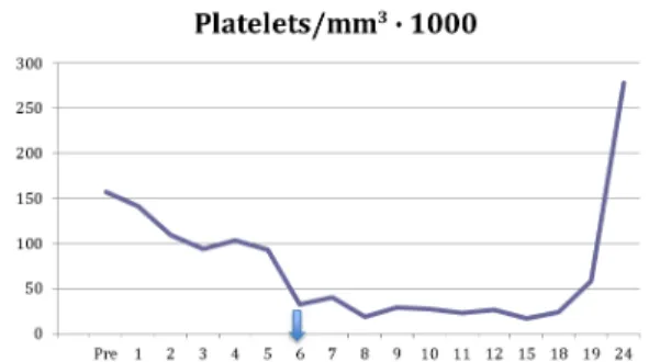
between 1.5 and 2.5). His preoperative platelet count was 157,000/mm3 (Figure 1). He was reoperated twice more for attempts at vascular repair, at 24 and 48h,
because of repeat arterial thrombosis at the same site. Starting on the third day of treatment with UFH, his platelet count began to fall (109,000/mm3),
continuing to drop until it reached 32,000/mm3 on
the sixth postoperative day. Administration of UFH
140 J Vasc Bras. 2016 Apr.-June; 15(2):138-141 Heparin-induced thrombocytopenia
was then suspended (Figure 1), due to a suspicion
of HIT (> 50% drop in platelet count after ive days
of UFH) (Figure 1).
PART II: WHAT WAS DONE
In response to complications of ischemia, on the seventh postoperative day, the patient underwent an RLL open guillotine amputation at the level of the ankle. In view of the patient’s age and absence of other risk factors for atherosclerosis, a clinical hypothesis of arteritis was suspected in response to the etiology of acute arterial ischemia, and so laboratory tests were requested for investigation of connective tissue diseases. Test results were normal for antinuclear antibodies (anti‑ENA = saliva extractable nuclear antigens; anti‑snRNP subtypes = small nuclear ribonucleoproteins; SM = anti‑Smith; dsDNA = anti‑double stranded DNA), LE cells and latex fixation test. Results were abnormal for erythrocyte sedimentation rate
(32 mm/h), titrated CRP (7.8 mg/dl), ibrinogen (579 mg/dl), alpha 1-acid glycoprotein (169 mg/dl) and antinuclear factor (1/5120). Pathology results
for the amputation specimen did not reveal anything other than thrombosis of the posterior tibial artery and necrosis of muscles. Sixteen days after admission, the patient underwent an RLL closed amputation at the proximal third of the leg. He recovered well and was discharged 30 days after admission. During post‑discharge follow‑up, tests for thrombophilia were conducted, including antithrombin, C and S proteins, lupus anticoagulant and IgM anticardiolipin assays, all of which were normal. Anticardiolipin IgG antibody levels were elevated (120 GPL/Uml,
reference value is ≤ 40 GPL/Uml). Anticardiolipin
IgG remained elevated when the assay was repeated
12 weeks later, at 60 GPL/Uml, which conirmed the
diagnosis of antiphospholipid antibody syndrome (AAS). One clinical criterion supporting this diagnosis was met (arterial thrombosis) and one laboratory criterion supporting this diagnosis was met (anticardiolipin IgG > 40 GPL/Uml in two assays, with an interval
of at least 12 weeks). It was not possible to conirm
HIT on the basis of test results because HIT antibodies had not been assayed at the hospital, but the clinical suspicion remained strong. Since the patient has AAS and had already had arterial thrombosis, we chose to put him on permanent anticoagulation with warfarin.
As shown in Figure 1, the platelet count rose when heparin was withdrawn, returning to normal 24 days after the operation.
DISCUSSION
Type II HIT is rare immunomediated disease that is generally severe, provoking thrombocytopenia
from 5 to 15 days after starting heparin treatment.
Paradoxically, it involves a high risk of thromboembolic complications.10 Type II HIT occurs in 1 to 6% of patients treated with UFH and in up to 0.9% of patients treated with LMWH. From 33 to 50% of these cases
progress to venous or arterial thromboses and there is a high risk of amputations.11
The etiologic basis of type II HIT is formation of IgG type antibodies against the heparin complex and
platelet factor 4, which are identiied as antigens.12
The imunocomplexes react with platelet FcyRIIA receptors13 activating them and provoking aggregation
and release of greater quantities of platelet factor. This culminates in activation of thrombin and the coagulation cascade, which stimulates formation of thrombi and destruction of platelets.10,11
It is recommended that all patients to be given heparin therapy should have a baseline platelet count at the start of treatment and the test should be repeated every 2 days. If it is observed that the platelet count
has fallen by more than 50% from the ifth day of
treatment on, HIT should be suspected and heparin suspended immediately.11,14 Diagnosis can be conirmed
by the functional method, which measured platelet activation caused by the heparin‑dependent antibody in vitro,11 but the test is not routinely available in
all hospitals.13 Risk scores designed to strengthen
diagnostic suspicion exist (4T or HEP score), but they are of little clinical utility because of their low
sensitivity and speciicity.13
In Brazil, fondaparinux is the drug used for treating patients with Type II HIT.15 It is possible that rivaroxaban may be an alternative possibility, but there are no studies of use of this drug for this purpose. In other countries, argatroban, hirudin, bivalirudin and danaparoid are recommended.13 Premature introduction of warfarin as
an alternative should be avoided, since there is a risk of increasing the pro‑thrombotic state, because of a rapid reduction in serum levels of C protein, which is a natural anticoagulant. However, as soon as platelet levels return to normal, it can be introduced at doses
below 5 mg/day, and then adjusted to maintain the
international normalized ratio between 2.0 and 3.0.13
Our patient had a comorbidity, AAS, which is an acquired thrombophilia that can provoke arterial
(30%) or venous (70%) thromboses, thrombocytopenia
141 J Vasc Bras. 2016 Apr.-June; 15(2):138-141 Rafael Elias Farres Pimenta, Winston Bonetti Yoshida et al.
antibody in patients with systemic lupus erythematosus
is approximately 40%, and clinical manifestations of AAS are probably present in 30 to 40% of the patients who have this antibody or in around 10 to 15% of
those with lupus.16 Just one criterion for systemic lupus
erythematosus was identiied in our case (positive
for antinuclear antibodies), but the patient did not meet the other minimum criteria for this diagnosis. As this is a thrombophilia with a high incidence of relapse, it is recommended that anticoagulation be
maintained for 12 months after a irst episode of venous thrombosis and indeinitely after a second
episode or arterial thrombosis.17
The presentation of HIT in this case was highly typical, since the platelet count reduction by more than
50% occurred on the ifth and sixth days of treatment
with heparin. The condition was also transitory, regressing after withdrawal of UFH. Unfortunately,
the tests needed for conirmation are not available at
our service, which prevents us from being absolutely certain of the diagnosis.
CONCLUSIONS
Heparin‑induced thrombocytopenia is an uncommon complication, but it should always be remembered, and it should be made routine to take a platelet count before and every 2 days after starting anticoagulant treatment with heparin.
REFERENCES
1. Copley AL, Robb TP. The effect of heparin in vivo on the platelet count in mice and dogs. Am J Clin Pathol. 1942;12(11):563-70. http://dx.doi.org/10.1093/ajcp/12.11.563.
2. Weismann RE, Tobin RW. Aterial embolism occurring during systemic heparin therapy. AMA Arch Surg. 1958;76(2):219-27. http://dx.doi. org/10.1001/archsurg.1958.01280200041005. PMid:13497418.
3. Rhodes GR, Dixon RH, Silver D. Heparin induced thrombocytopenia with thrombotic and hemorrhagic manifestation. Surg Gynecol Obstet. 1973;136(3):409-16. PMid:4688805.
4. Chong BH. Heparin induced thrombocytopenia. Blood Rev. 1988;2(2):108-14. http://dx.doi.org/10.1016/0268-960X(88)90032-X. PMid:3042056.
5. Oliveira SC. Trombocitopenia induzida por heparina: aspectos clínicos e laboratoriais [tese]. São Paulo: Universidade de São Paulo; 2008 [citado 2013 jul 21]. http://www.teses.usp.br/teses/ disponiveis/5/5146/tde-04112008-155406/
6. Ortel TL. Heparin induced thrombocytopenia. Semin Hematol. 1998;35(4, Supl 5):1-2. PMid:9855177.
7. Daneschvar HL, Daw H. Heparin induced thrombocytopenia (an overview). Int J Clin Pract. 2007;61(1):130-7. http://dx.doi. org/10.1111/j.1742-1241.2006.00874.x. PMid:16836645.
8. Chong BH. Annotation: heparin induced thrombocytopenia. Br J Haematol. 1995;89(3):431-9. http://dx.doi.org/10.1111/j.1365-2141.1995. tb08346.x. PMid:7734342.
9. Warkentin TE, Greinacher A. Heparin induced thrombocytopenia. 2. ed. New York: Marcel Dekker; 2001.
10. Baroletti SA, Goldhaber SZ. Heparin-induced thrombocytopenia. Circulation. 2006;114(8):e355-6. http://dx.doi.org/10.1161/ CIRCULATIONAHA.106.632653. PMid:16923760.
11. Maffei FH. Trombose venosa profunda de membros inferiores: tratamento anticoagulante. In: Maffei FH, editor. Doenças vasculares periféricas. 4. ed. Rio de Janeiro: GEN; 2008. vol. 2, p. 1603.
12. Junqueira DR, Carvalho M, Perini E. Heparin-induced thrombocytopenia: a review of concepts regarding a dangerous adverse drug reaction. Rev Assoc Med Bras. 2013;59(2):161-6. http://dx.doi.org/10.1016/j. ramb.2012.11.004. PMid:23582558.
13. Kelton JG, Arnold DM, Bates SM. Nonheparin anticoagulants for heparin-induced thrombocytopenia. N Engl J Med. 2013;368(8):737-44. http://dx.doi.org/10.1056/NEJMct1206642. PMid:23425166.
14. Warkentin TE. Heparin-induced thrombocytopenia: diagnosis and management. Circulation. 2004;110(18):e454-8. http://dx.doi. org/10.1161/01.CIR.0000147537.72829.1B. PMid:15520327. 15. Savi P, Chong BH, Greinacher A, et al. Effect of fondaparinux on
platelet activation in the presence of heparin-dependent antibodies: a blinded comparative multicenter study with unfractionated heparin. Blood. 2005;105(1):139-44. http://dx.doi.org/10.1182/ blood-2004-05-2010. PMid:15388575.
16. Harris EN, Khamashta MA, Hughes GR. Antiphospholipid antibody syndrome. In: Mccarty DJ, Koopman WJ, editors. Arthritis and allied conditions. 13. ed. Philadelphia: Lea & Febiger; 1997. p. 1201-13. 17. Franco H. Trombofilias adquiridas. In: Maffei FH, editor. Doenças
vasculares periféricas. 4. ed. Rio de Janeiro: GEN; 2008. vol. 2, p. 1593.
*
Correspondence
Rafael Elias Farres Pimenta Universidade Estadual Paulista – UNESP Faculdade de Medicina de Botucatu Departamento de Cirurgia e Ortopedia Avenida Prof. Montenegro, s/n - Distrito de Rubião Junior CEP 18618-970 - Botucatu (SP) - Brazil Tel.: +55 (14) 3811-6305 E-mail: [email protected]
Author information
REFP, JVOM, RGJ and PABC - Primary physicians, Vascular and Endovascular Surgery, Hospital das Clínicas de Botucatu, Universidade Estadual Paulista (UNESP). WBY - Full professor of Vascular and Endovascular Surgery, Faculdade de Medicina de Botucatu, Universidade Estadual Paulista (UNESP). HAR - Adjunct professor of Vascular and Endovascular Surgery, Faculdade de Medicina de Botucatu, Universidade Estadual Paulista (UNESP). MLS and MB - Assistant professors of Vascular and Endovascular Surgery, Faculdade de Medicina de Botucatu, Universidade Estadual Paulista (UNESP).
Author contributions
Conception and design: WBY Analysis and interpretation: REFP, WBY, MLS Data collection: REFP, WBY Writing the article: REFP, WBY, HAR Critical revision of the article: REFP, WBY, MLS, PABC, RGJ, JVOM, MB, HAR Final approval of the article*: REFP, WBY, MLS, PABC, RGJ, JVOM, MB, HAR Statistical analysis: N/A. Overall responsibility: REFP, WBY