Heart Institute (InCor), Hospital das Clínicas, Faculty of Medicine, University of São Paulo – São Paulo/SP, Brazil.
E-mail: [email protected]
Received for publication on November 09, 2004. Accepted for publication on Março 08, 2005.
ORIGINAL RESEARCH
CORONARY REVASCULARIZATION WITH THE LEFT
INTERNAL THORACIC ARTERY AND RADIAL
ARTERY. COMPARISON OF SHORT-TERM CLINICAL
EVOLUTION BETWEEN ELECTIVE AND
EMERGENCY SURGERY
Roberto Rocha-e-Silva, Antônio de Pádua Mansur, José Fabri Junior, Rogério Bicudo Ramos, Carlos Edson Campos Cunha Filho, Luis Alberto Oliveira Dallan, and Sérgio Almeida de Oliveira
Rocha-e-Silva R, Mansur A de P, Fabri Junior J, Ramos RB, Cunha Filho CEC, Dallan LAO et al. Coronary revascularization with the left internal thoracic artery and radial artery. Comparison of short-term clinical evolution between elective and emergency surgery. Clinics. 2005;60(3):227-32.
BACKGROUND: Left internal thoracic artery-to left anterior descending artery grafting has become a fundamental part of coronary artery bypass grafting. This grafting has led to increased use of other arterial conduits, of which the radial artery is most popular. Whether radial grafting can be used in the emergency patient is not known. This study compares the short-term clinical evolution between elective vs emergency coronary artery bypass grafting surgery with left internal thoracic artery and radial artery.
METHODS: A retrospective study of 47 patients who underwent elective or emergency coronary artery bypass grafting from 1996 to 2003. All patients had coronary stenosis ≥70% in all target vessels. Only the left internal thoracic artery and radial artery were used as grafts. Patients were divided into elective group (23 patients) and emergency group (24 patients). Emergency criteria were unstable angina and/or critical coronary stenosis with high risk for acute myocardial infarction. Groups were similar for age and number of diseased vessels.
RESULTS: The mean number of left internal thoracic artery grafts per patient in the elective and emergency groups were respectively 1.17 and 1.38 (P = .17). The mean number of radial artery grafts per patient in the elective and emergency
groups was respectively 2.26 and 2.08 (P = .48). The 30-day mortality was 0. There was no postoperative cardiogenic shock.
The elective group had 1 acute myocardial infarction (4.4%) postoperatively, and emergency group had 5 (20.8%). A nonsignificant trend towards acute myocardial infarction was noted in the emergency group (P = .18). Intensive care unit
and postoperative stay were similar in both groups.
CONCLUSION: Coronary artery bypass grafting using left internal thoracic artery and radial artery accomplishing complete revascularization can be performed in emergency patients with results similar to those for elective patients.
KEYWORDS: Coronary artery bypass surgery.Arterial conduits. Radial artery. Emergency. Coronary disease.
The left internal thoracic artery (LITA) is the conduit of choice in coronary artery bypass grafting (CABG) because of superior graft patency, reduced cardiac events, and
en-hanced short- and long-term survival.1 In the search for other
conduits for total arterial revascularization, Acar2
reintro-duced the radial artery (RA) graft after long-term observa-tion of patent RA conduits that were thought to have been occluded in the early postoperative period.
Other studies3-8 demonstrated RA patency rates of 82%
However, Khot9 reported a 51% patency rate for the RA
conduit in a study where all coronary angiography proce-dures were reviewed. Legare10 reported enhanced
morbid-ity with complete arterial revascularization compared to conventional use of saphenous vein conduits, but without a difference in mortality. Tatoulis11 reported that higher RA
patency rates were associated with more severe coronary stenosis; they also reported variable patency as a function of the grafted target coronary. Their 1- and 4-year patency rates were respectively 96% and 89%. Gaudino12
demon-strated that the main determinant of higher RA patency rate is the level of coronary stenosis. Zacharias13 demonstrated
that the use of the RA compared to saphenous veins as a second conduit associated to LITA-left anterior descending artery (LADA) decreases long-term mortality.
There are few reports on the use of arterial conduits in emergency CABG, but reported outcomes are good.14-15
Hayashi16 reported complete arterial revascularization in 9
emergency patients with good outcome. Nishida17 in a
simi-lar study with 37 patients, reported a 5-year survival rate of 97.1% and a patency rate of 100% for RA 3 to 4 weeks after surgery.
There are no comparative reports for complete arterial revascularization with the LITA and RA in elective vs emer-gency surgery.
OBJECTIVE
The main objective of this study was to compare the short-term clinical evolution between elective vs emergency surgery in patients undergoing complete arterial revascularization with the left internal thoracic artery (LITA) and radial artery (RA).
MATERIALS AND METHODS
A retrospective study in which all patients who were undergoing complete arterial revascularization with the ex-clusive use of LITA and RA performed by the same sur-geon, were included. The criterion for the use of this tech-nique was the presence of coronary stenosis ≥70% in all target vessels. Forty-seven patients were included; surgery was performed from 1996 to 2003.
Patients were classified into 2 groups, an elective group with 23 patients (17 males) and an emergency group with 24 patients (22 males). Emergency inclusion occurred in this series whenever unstable angina refractory to medical therapy and/or critical stenosis in coronary arteries impos-ing high risk for acute myocardial infarction (AMI) was present.
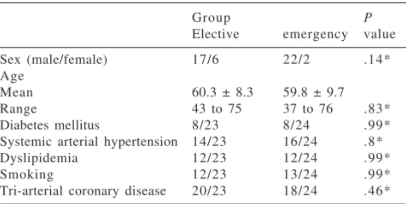
The main preoperative characteristics of both groups
were similar and are summarized in Table 1. Most of the patients were men (39/47), were between 50 and 60 years of age, and had triple-vessel coronary artery disease (38/ 47). Patients in the emergency group underwent surgery for unstable angina, and 1 had an associated hemodynamic in-stability (cardiac index of 1.9 L·min-1·m-2 with dopamine
infusion of 10 mg·kg-1·min-1). Eleven of these patients (46%)
underwent CABG up to 21 days after an acute myocardial infarct (AMI). There was no preoperative use of an intra-aortic balloon pump (IAB).
Two patients in the elective group and 1 in the emer-gency group had left ventricular aneurysms. The aneurisms were diagnosed during surgery.
The surgical approach was through sternotomy with car-diopulmonary bypass (CPB). From March 2003, off-pump surgery was performed when possible. The elective and emergency groups had off-pump surgery in 4/7 (57%) and 7/12 (58%) respectively.
The LITA was harvested in all but 1 case (reoperation with patent LITA-LADA graft). The RA was harvested in all cases. After harvesting, the grafts were left in situ with topical papaverine until the initialization of coronary graft-ing. The LITA was grafted to the LADA and (when needed and possible) to the diagonal branch (sequential anastomo-sis). The RA was grafted to all other target vessels except the LADA. When more than 1 target vessel was grafted with the RA, sequential anastomoses were performed. Each distal anastomosis was considered an independent graft. The RA was anastomosed proximally to the LITA as a Y graft in 21/ 23 and 20/24 in the elective and emergency groups respec-tively. In the other 6 cases, the RA was proximally anasto-mosed to the aorta because the LITA was subjectively evalu-ated as having a small caliber.
The 2 patients in the elective group and the 1 in the emergency group with ventricular aneurysms had left ven-tricular aneurysmectomies.
Table 1 - Preoperative characteristics.
Group P
Elective emergency value
Sex (male/female) 17/6 22/2 .14*
Age
Mean 60.3 ± 8.3 59.8 ± 9.7
Range 43 to 75 37 to 76 .83*
Diabetes mellitus 8/23 8/24 .99*
Systemic arterial hypertension 14/23 16/24 .8*
Dyslipidemia 12/23 12/24 .99*
Smoking 12/23 13/24 .99*
The two groups were compared for: (i) CPB time, dura-tion of ischemia, number of grafts (LITA and RA grafts per patient), bleeding volume, days under vasoactive drugs, ICU stay, and hospital stay through ANOVA; (ii) occurrence of AMI, reoperations, arrhythmia, pulmonary thromboem-bolism, pneumonia, neurologic complications, and medi-astinitis through the Fisher exact test. For both tests a P
value < .05 was considered statistically significant.
RESULTS
The 30-day mortality rate was 0 in both groups. There was 1 late death in each group both due to pulmonary com-plications In the elective group death occurred on the 30th
day due to acute pulmonary edema secondary to pleural drainage of severe left pleural effusion. In the emergency group death occurred on the 49th day as a consequence of
atypical pneumonia.
The mean number of LITA grafts per patient in the elec-tive and emergency groups were respecelec-tively 1.17 ± 0.49 and 1.38 ± 0.49 (P = .17). The mean number of RA grafts
per patient in the elective and emergency groups were re-spectively 2.26 ± 0.75 and 2.08 ± 0.93 (P = .48). There was
no difference between groups for LITA, RA, or total number of grafts.
Intensive care unit stay and postoperative stay were simi-lar in both groups (Table 2). There was no postoperative use of an IAB, nor did low cardiac output syndrome occur. The postoperative use of catecholamines in the elective and emergency groups ranged from 0 to 3 days (1.0 ± 0.9 days) and 0 to 4 days (1.2 ± 1.0 days) respectively (P = .58).
Two patients in the elective group had CPK-MB eleva-tion postoperatively, but with only 1 AMI confirmaeleva-tion (4.4%) by pyrophosphate scintigraphy. This last case pre-sented ventricular fibrillation during graft harvesting and urgent CPB initiation was necessary. Neither patient had postoperative electrocardiographic or echocardiographic alterations. In contrast, the emergency group had 5 patients with sustained AMI in the same period (20.8%); all of them had postoperative electrocardiographic and/or echocardiographic alterations, 1 went to surgery in cardiogenic shock, 1 presented ventricular fibrillation dur-ing graft harvestdur-ing and required urgent CPB initiation, and 3 underwent surgery 1 to 5 days after an AMI associated with unstable angina. A nonsignificant trend toward AMI was noted in the emergency group (P =. 18).
The CPB and ischemic time for both groups was simi-lar (Table 2).
One patient in the emergency group presented a bleed-ing volume of 2730 mL up to the end of the day after sur-gery. Since he had an associated coagulopathy,
re-interven-tion was postponed to the second postoperative day, and no active bleeding was found; neither was any blood clot found near the grafts, but only directly behind the sternum. All other patients had postoperative bleeding compatible with the procedure (Table 2).
There were 3 postoperative neurological complications: (i) transient mental confusion that lasted 1 day (elective group); (ii) seizure in a patient with previous history of sei-zures (emergency group); (iii) low level of conscience due to cardiogenic shock entering surgery with a full week be-fore recovering normal mental status. This last patient was having an AMI and required rescue CABG surgery. This pa-tient had a previous history of insulin-dependent diabetes mellitus, required prolonged mechanical ventilation, and was the only one who developed mediastinitis, which was successfully treated with antibiotic therapy. Later, an ab-dominal muscle rotation was used to cover the open wound. There was 1 case of pulmonary thromboembolism dur-ing the second postoperative day (emergency group). The patient had previous significant ventricular dysfunction, moderate tricuspid valve insufficiency, and a definitive im-planted pacemaker for 8 years. He underwent off-pump sur-gery and had a postoperative period without CPK-MB or electrocardiographic alteration. He recovered well and was discharged from the hospital on the 11th day. Table 2 lists
all observed complications. No statistical differences be-tween groups occurred for any of these.
DISCUSSION
No immediate mortality and no low cardiac output
syn-Table 2 - Postoperative characteristics.
Group P
elective emergency value
CPB time (min) 112 ± 27 111 ± 30 .93**
Duration of ischemia (min) 54 ± 14 61 ± 27 .30** Grafts/patient 3.43 ± 0.79 3.46 ± 1.02 .93** LITA grafts/patient 1.17 ± 0.49 1.38 ± 0.49 .17** RA grafts/patient 2.26 ± 0.75 2.08 ± 0.93 .48** Days under vasoactive drugs 1.0 ± 0.9 1.2 ± 1.0 .58**
Days in ICU 2.4 ± 0.9 2.8 ± 2.0 .45**
Days in hospital 8.1 ± 2.5 12.8 ± 12.2 .10**
Postoperative AMI 1/23 5/24 .18*
Bleeding (24 hours) 699 ± 332 755 ± 576 .73**
Reoperations 0/23 1/24 1.0*
Atrial arrhythmia 3/23 2/24 .67*
Pulmonary thromboembolism 0/23 1/24 .99*
Pneumonia 0/23 2/24 .49*
Neurological complications 1/23 2/24 .99* (no sequelae)
Mediastinitis 0/23 1/24 .99*
drome occurred in either the elective or the emergency group, suggesting that complete arterial revascularization can be used in patients with multiple and critical stenosis in emergencies as well as in elective scenarios. The 2 late deaths were due to pulmonary complications.
No differences were noted between groups in the type or number of grafts, suggesting that the emergency situa-tion does not limit the use of arterial conduits for complete revascularization.
Considering that: (i) all patients had critical coronary stenosis in all target vessels; (ii) RA conduits represented more than 60% of all grafts in both groups; (iii) the post-operative period evolved with no cardiogenic shock, no use of IAB, and a low need for catecholamines, it may be con-cluded that the patency and flow in the RA grafts must have been adequate for these critical patients.
The transient postoperative mental confusion is attrib-utable to the use of CPB.18 The other neurological
compli-cations were attributable to the preoperative conditions of the patients. All of these complications were transient and reversed with no sequelae.
The only case of mediastinitis was in a patient who had rescue emergency surgery in cardiogenic shock. He also had prolonged mechanical ventilation because of a low level of consciousness, with a history of insulin-dependent dia-betes mellitus, all of which represent strong risk factors for infection.19
The only case of pulmonary thromboembolism occurred in a patient with previous ventricular dysfunction, tricus-pid valve disease, and a definitive implanted pacemaker. As his surgery was uneventful, this complication may be attributed to his previous heart condition.
Transoperative AMI occurred in 1 patient of the elective group (4.4%), which is in accordance to the 0.8% to 7.7% found in other published series.20-21 The emergency group
had a higher rate of AMI (5 patients—20.8%), but these pa-tients presented signs of significant ischemia (cardiogenic shock, ventricular fibrillation, and refractory angina) before coronary grafting. As none evolved with postoperative cardiogenic shock, the CABG brought beneficial effects to the ischemic muscles, probably reducing cardiac tissue loss. This morbidity difference, which was not statistically signifi-cant, was not unexpected because of the intrinsically higher severity of the conditions of the emergency group.22
Even though bleeding control of the RA conduit is more time consuming because of its numerous branches, there were no bleeding complications attributable to the use of RA grafting. Total arterial myocardial revascularization may be achieved safely and effectively with the use of 1 RA in conjunction with the internal thoracic artery.23
Conse-quently, RA harvesting is a perfectly sound indication even in emergency surgery.
The superiority of LITA as a graft for CABG is beyond dispute.1 However, there is controversy over the kinds of
grafts that should be associated with LITA, especially in the emergency scenario. Patients included in this report had excellent immediate clinical evolution with CABG using exclusively LITA and RA for complete revascularization in elective as well as emergency surgeries. These cases still require angiographic study for demonstration of graft pat-ency. If postoperative angiography patency is demonstrated, the long-term evolution should be similar to that of elec-tive surgery, which is excellent in most reports.3-8 11-13
CONCLUSION
CABG using LITA and RA grafts to accomplish com-plete revascularization can be performed in emergency tients with results similar to those obtained in elective pa-tients.
RESUMO
Rocha-e-Silva R, Mansur A de P, Fabri Junior J, Ramos RB, Cunha Filho CEC, Dallan LAO et al. Revascularização coronariana com uso de artéria torácica interna esquerda e artéria radial esquerda. Estudo comparativo da evolução clínica imediata entre cirurgias eletivas e emergências. Clinics. 2005;60(3):227-32.
O uso de artéria torácica interna esquerda para descendente anterior se tornou fundamental na revascularização do miocárdio. Este enxerto levou ao aumento da utilização de enxertos arteriais, dos quais a artéria radial é muito
popu-lar. O uso de artéria radial em pacientes de emergência foi pouco estudado. Este estudo compara a evolução clínica imediata entre revascularização do miocárdio eletiva vs. emergência com artéria torácica interna esquerda e artéria radial.
grupos: eletivo (23 casos) e emergência (24 casos). Critérios de emergência foram angina instável e/ou estenose coronariana crítica com alto risco de infarto agudo do miocárdio. Os grupos eram homogêneos para idade e artérias acometidas.
RESULTADOS: A média de enxertos de artéria torácica interna esquerda por paciente eletivo e de emergência foi respectivamente 1,17 e 1,38 (P=.17). A média de enxertos
de artéria radial por paciente eletivo e de emergência foi respectivamente 2,26 e 2,08 (P=.48). A mortalidade até 30
dias foi zero. No pós-operatòrio não ocorreram casos de choque cardiogênico. Um paciente eletivo (4,4%) e 5pacientes de emergência (20,8%) apresentaram infarto
agudo do miocárdio no pós-operatório; tendência não significativa para ocorrência de infarto agudo do miocárdio no grupo de emergência (P=.18). Tempo de internação na
unidade de terapia intensiva e hospitalar foi semelhante nos dois grupos.
CONCLUSÃO: A revascularização do miocárdio com utilização de artéria torácica interna esquerda e artéria ra-dial pode ser realizada em pacientes de emergência com resultados equivalentes aos pacientes eletivos.
UNITERMOS: Revascularização do miocárdio. Condutos arteriais. Artéria radial. Emergência. Doença coronária.
REFERENCES
1 . Zeff RH, Kongtahworn C, Iannone LA, Gordon DF, Brown TM, Phillips SJ, et al. Internal mammary artery versus saphenous vein graft to the left anterior descending coronary artery prospective randomized study with 10-year follow-up. Ann Thorac Surg. 1988;45:451-4.
2 . Acar C, Jebara VA, Portoghese M, Beyssen B, Pagny JY, Grare P, et al. Revival of the radial artery for coronary artery bypass grafting. Ann Thorac Surg. 1992;54:652-60.
3 . Sundt TM 3rd, Barner HB, Camillo CJ, Gay WA Jr. Total arterial revascularization with an internal thoracic artery and radial artery T graft. Ann Thorac Surg. 1999;68:399-405.
4 . Royse AG, Royse CF, Tatoulis J, Grigg LE, Shah P, Hunt D, et al. Postoperative radial artery angiography for coronary artery bypass surgery. Eur J Cardiothorac Surg. 2000;17:294-304.
5 . Possati G, Gaudino M, Alessandrini F, Luciani N, Glieca F, Trani C, et al. Midterm clinical and angiographic results of radial artery grafts used for myocardial revascularization. J Thorac Cardiovasc Surg. 1998;116:1015-21.
6 . Bhan A, Gupta V, Choudhary SK, Sharma R, Singh B, Aggarwal R, et al. Radial artery in CABG: could the early results be comparable to internal mammary artery graft? Ann Thorac Surg. 1999;67:1631-6. 7 . Fremes SE, Christakis GT, Del Rizzo DF, Musiani A, Mallidi H, Goldman BS. The technique of radial artery bypass grafting and early clinical results. J Card Surg. 1995;10:537-44.
8 . Calafiore AM, Teodori G, Di Giammarco G, D’Annunzio E, Angelini R, Vitolla G, et al. Coronary revascularization with the radial artery: new interest for an old conduit. J Card Surg. 1995;10:140-6.
9 . Khot UN, Friedman DT, Pettersson G, Smedira NG, Li J, Ellis SG. Radial artery bypass grafts have an increased occurrence of angiographically severe stenosis and occlusion compared with left internal mammary arteries and saphenous vein grafts. Circulation. 2004;109:2086-91.
10. Legare JF, Buth KJ, Sullivan JA, Hirsch GM. Composite arterial grafts versus conventional grafting for coronary artery bypass grafting. J Thorac Cardiovasc Surg. 2004;127(1):160-6. 11. Tatoulis J, Buxton BF, Fuller JA. Patencies of 2127 arterial to
coronary conduits over 15 years. Ann Thorac Surg. 2004;77(1):93-101.
12. Gaudino M, Alessandrini F, Pragliola C, Cellini C, Glieca F, Luciani N, et al. Effect of target artery location and severity of stenosis on mid-term patency of aorta-anastomosed vs. internal thoracic artery-anastomosed radial artery grafts. Eur J Cardiothorac Surg. 2004;25(3):424-8.
13. Zacharias A, Habib RH, Schwann TA, Riordan CJ, Durham SJ, Shah A. Improved survival with radial artery versus vein conduits in coronary bypass surgery with left internal thoracic artery to left anterior descending artery grafting. Circulation 2004;109(12):1489-96.
14. Kamata S. Emergency and subemergency coronary artery bypass graft: active use of arterial grafts. Kyobu Geka. 1999;52(8 Suppl):693-6.
15. Hirotani T, Kameda T, Kumamoto T, Shirota S, Yamano M. Should arterial grafts be used for urgent coronary artery bypass surgery? Kyobu Geka. 2000;53(1):69-73.
17. Nishida H, Tomizawa Y, Endo M, Koyanagi H. Complete arterial revascularization in emergency CABG. Kyobu Geka. 1999;52(8 Suppl):688-92.
18. Harrison MJ, Schneidau A, Ho R, Smith PL, Newman S, Treasure T. Cerebrovascular disease and functional outcome after coronary artery bypass surgery. Stroke. 1989;20(2):235-7.
19. The Parisian Mediastinitis Study Group. Risk factors for deep sternal wound infection after sternotomy: a prospective multicenter study. J Thorac Cardiovasc Surg. 1996;11:1200-7.
20. Berdat PA, Muller K, Schmidli J, Kipfer B, Eckstein F, Carrel T. Total arterial off-pump versus on-pump coronary revascularization: comparison of early outcome. J Card Surg. 2004;19(6):489-94.
21. Tatoulis J, Royse AG, Buxton BF, Fuller JA, Skillington PD, Goldblatt JC, et al. The radial artery in coronary surgery: a 5-year experience—clinical and angiographic results. Ann Thorac Surg. 2002;73(1):143-7.
22. Hirose H, Amano A, Yoshida S, Nagao T, Sunami H, Takahashi A, et al. Surgical management of unstable patients in the evolving phase of acute myocardial infarction. Ann Thorac Surg. 2000;69(2):425-8.