232
Rev Dor. São Paulo, 2016 jul-sep;17(3):232-5
ABSTRACT
BACKGROUND AND OBJECTIVES:Multiple sclerosis is an autoimmune, inlammatory, demyelinating and chronic disease of the central nervous system. As from the understanding of its pathophysiology and of thermoregulating dysfunctions caused by the disease, it is clear that, whenever possible, infrared ther-mography should be done. herther-mography helps understanding how the disease afects diferent body areas, by investigating asymmetries, contractures and neurogenic patterns. his study aimed at documenting by infrared thermography a case of mul-tiple sclerosis in crisis of pain.
CASE REPORT: Female patient, 63 years old, diagnosed with multiple sclerosis in 2007 after magnetic resonance and lumbar puncture. Six month ago she started complaining of progressive decrease in lower limbs muscle strength in addition to increased spinal pain, especially in lumbar spine and right hemibody. Pa-tient was submitted to new exams (head and cervical spine reso-nance), which have shown the same pattern found in previous exams, resulting from old injuries by demyelinating substract. hermometry has shown asymmetry of the whole right hemi-boby with central neurogenic patterns and temperature difer-ence (∆T 0.8ºC), thus conirming initial diagnosis. With regard to major complaint, there was asymmetry between paralumbar regions and presence of lumbar paravertebral hyperradiation, suggesting local muscles contracture.
CONCLUSION:Multiple sclerosis has a wide range of symptoms, especially the installation of chronic pain and inadequate thermo-regulation, which directly interfere with quality of life of patients.
Keywords: Lumbar pain, Multiple sclerosis, hermography.
Infrared thermography to evaluate pain in a multiple sclerosis patient.
Case report
Termografia por infravermelho na avaliação da dor em paciente com esclerose múltipla Relato
de caso
Rosa Maria Papaléo1, Manoel Jacobsen Teixeira2,Marcos Leal Brioschi3
1. Universidade de São Paulo, Faculdade de Medicina, São Paulo, SP, Brasil.
2. Universidade de São Paulo, Faculdade de Medicina, Hospital das Clínicas, Departamento de Neurologia, São Paulo, SP, Brasil.
3. Universidade de São Paulo, Faculdade de Medicina, Hospital das Clínicas, Coordenador da Especialização em Termologia e Termograia, São Paulo, SP, Brasil.
Submitted in October 27, 2015. Accepted for publication in June 23, 2016. Conlict of interests: none – Sponsoring sources: none.
Correspondence to:
Av. Dr. Arnaldo, 455 - Cerqueira César 01246-903 São Paulo, SP, Brasil. E-mail: [email protected]
© Sociedade Brasileira para o Estudo da Dor
RESUMO
JUSTIFICATIVA E OBJETIVOS: A esclerose múltipla é uma doença autoimune, inlamatória, desmielinizante e crônica do sistema nervoso central. A partir do entendimento da sua i-siopatologia e das disfunções termorreguladoras decorrentes da doença, ica claro que quando possível, a termograia por in-fravermelho deve ser feita. A termograia facilita o entendimento de como a doença atinge as diversas áreas do corpo, investigando assimetrias, contraturas e padrões neurogênicos. O objetivo deste estudo foi documentar por termograia infravermelha um caso de esclerose múltipla em crise álgica.
RELATO DO CASO: Paciente do gênero feminino, 63 anos, diagnosticada com esclerose múltipla em 2007, após realiza-ção de ressonância magnética e punrealiza-ção liquórica. Ha seis me-ses começou a queixar-se de diminuição de força muscular nos membros inferiores de caráter progressivo, além de aumento nas dores da região da coluna vertebral, principalmente na coluna lombar e no dimídio direito. Realizou novos exames (ressonância de crânio e coluna cervical), que mostraram o mesmo padrão encontrado em exames anteriores, resultantes de lesões antigas por substrato desmielinizante. A termometria demonstrou as-simetria de todo hemicorpo direito, com padrão neurogênico central, e diferença de temperatura (∆T 0,8ºC), conirmando assim o diagnostico inicial. Em relação à queixa principal, foram encontradas assimetria entre regiões paralombares e presença de hiper-radiação paravertebral lombar, sugerindo contratura da musculatura local.
CONCLUSÃO: A esclerose múltipla possui vastos sintomas, destacando-se aqui a instalação de quadros álgicos crônicos e ter-morregulação inadequada que interferem diretamente na quali-dade de vida de seus portadores.
Descritores: Dor lombar, Esclerose múltipla, Termograia.
INTRODUCTION
Multiple sclerosis (MS) is an autoimmune disease afecting the central nervous system (CNS), more speciically the white matter, causing demyelination and inlammation. It usually afects more females than males, aged 20 to 40 years, but there have been also cases outside these limits. Females are more likely to develop MS as compared to males in a ratio of three females for each male. In Brazil, its prevalence is ap-proximately 15 cases for every 100 thousand inhabitants1,2.
CASE REPORT
233
Infrared thermography to evaluate pain in a multiple sclerosis patient. Case report
Rev Dor. São Paulo, 2016 jul-sep;17(3):232-5
here are four types of clinical evolution: remittent-recurrent (RR-MS), primarily progressive (PP-MS), primarily progres-sive with outbreak (PP-MS with outbreak) and secondarily progressive (SP-MS). Most common type is RR-MS, repre-senting 85% of all cases at onset3,4.
here are many MS symptoms which afect each individual diferently. hey vary according to the magnitude of nervous injury and to where in the CNS it has occurred. MS individu-als may experience virtually any neurological sign or symptom, including alterations: 1) sensory or motor, such as loss of tac-tile sensitivity or tingling, paresthesia, muscle fatigue, muscle spasms; 2) in coordination and balance (ataxia); 3) in speech (dysarthria) or swallowing (dysfagia); 4) visual such as phos-phene, diplopia, nistagmus, in the sequence of optic neuritis. Fatigue, acute or chronic pain and urinary and bowel move-ment diiculties, the latter with secondary constipation5,6.
Although not exclusive to MS, there are also Uhthof phe-nomenon, worsening of symptoms due to exposure to tem-perature higher than normal, and Lhermitte signal, sensa-tion of electric current irradiating through the spine when binding the neck6. Diagnosis is primarily based on reviewed
McDonald criteria7, being these adopted by the world
scien-tiic community to diagnose MS. Brain magnetic resonance (MRI) shows injuries typical of deymelination. For being a very broad diferential diagnosis and having a variety of symp-toms, new complementary evaluation methods may be used, such as digital infrared thermography.
As decribed by Brioschi8 & Brioschi, Lin & Teixeira9, this is a
noninvasive method to evaluate skin neurovegetative system by mapping thermal distribution, which does not require contrast or physical contact with patients, the results of which allow determining the functioning of vascular, nervous and muscu-loskeletal systems, of inlammatory, dermatologic, endocrine and oncologic processes. hermal image is changed in cases of disease and functional and structural alterations, relecting abnormal thermal patterns, such as areas of hypo or hyper-ra-diation, thermal asymmetry between contralateral body regions or hemibodies with temperature diferentials above 0.3º C10,11.
Taking this aspect into consideration, thermoregulatory dys-function in MS patients should not be neglected, being primar-ily induced by immaturity of CNS cooling system. his system works as from a complex vascular network in face and skull, closely linked to inner brain. Repercussions of such system immaturity are complex, from mere diiculty in taking warm bath to major risk factor of sudden death in this population when individuals are exposed to high temperatures12. Knowing
this, it is possible to notice the usefulness of the study of ther-moregulation in this population since the onset of the disease13.
Ueno et al.14 have performed thermography in a child
diag-nosed with MS and complaining of unilateral sweating. An-other study from the 1960s has shown asymmetric temperature pattern in MS patients15, however this was only evaluated in
the face and diagnostic impressions are not clearly described, making diicult the comparison with current studies. In addi-tion to these, a recent literature review recommends the use of thermography in the clinical practice in patients with
neuro-logical disorders, where it may be used as auxiliary diagnostic tool or as method for clinical follow up of the disease13. Other
thermographic studies in this population have not been found. So, as from results, the investigation of thermoregulation changes and of the relationship with neurovegetative system may open a new study ield around MS pathophysiology. So, this study aimed at documenting with infrared thermography a case of MS with pain crisis.
CASE REPORT
Female patient, 63 years old, since 2007 started to have se-vere pain in cervical and lumbosacral spine, with episodes of numbness in upper (UULL) and lower (LLll) limbs, which have evolved to strength deicit during the practice of exer-cises and gait changes (shift to the left and widening of the base), suggesting balance disorders.
In this year, after consultation with a neurologist, she was sub-mitted to cervical magnetic resonance (MRI) which has evi-denced mild signs of degenerative disc disease, evidences sug-gesting demyelinating disease, involving cervical spinal cord. Presence of vertebral hemangioma in vertebral body T1; skull MRI showing multiple images compatible with zones of mag-netic signal changes in supratentorial white matter, predomi-nantly located close to corpus callosum and frontal regions, the largest to the right, with hyposignal in T1 and hypersignal in long TR sequences, which is compatible with demyelinating injuries. his MRI has shown good alignment of vertebral ele-ments without bony eleele-ments conformation changes, focuses of fatty tissue deposition inL3 and L4. Decreased height and sig-nal of intervertebral discs L4/L5 and L5/S1 in T2 images with mild signal modiication in the anteroinferior rim of L4 and anterosuperior of L5 due to degenerative subchondral change. Spinal canal and conjugation foramina with good amplitude. Conus with normal signal shape, situation, dimensions and intensity. Joint processes and pre and paravertebral elements without apparent anatomic changes. Lack of abnormal post-contrast enhancements. No signs of posterior disc protrusion indicative of disc hernia. CSF puncture was also performed which has shown absence of neoplastic cells and negative bio-chemistry for viral infections. Non-reagent VDRL.
After ruling out other possible diseases and relating to symp-toms referred by patient and to recurrence of episodes in pre-vious months, MS was diagnosed and treatment was started. Patient started using interferon 44 associated to baclofen (an-tispasmodic) and luoxetine, being referred to physiotherapy, occupational therapy and psychology. Currently patient is us-ing natalizumab to replace interferon.
234
Papaléo RM, Teixeira MJ and Brioschi ML Rev Dor. São Paulo, 2016 jul-sep;17(3):232-5
Skull MRI in 2010 has shown T2 images with areas of hyper-signal in white matter of brain hemispheres in paracallosum and periventricular situation, being of interest corpus callo-sum and callocallo-sum-septal interface some of them with areas of hyposignal in T1 and, which for their disposition, show disease with demyelinating substrate. here is moderate wid-ening of cortical sulci in the convexity of brain hemispheres and Sylvian issures, with brain ventricles with normal dimen-sions. Lack of extra-axial luid collections.
Skull MRI in 2011 has shown areas of hypersignal in callosum and paracallosum situation, well suggestive of MS, without relevant changes in posterior fossa, presence of a very subtle area of hypersignal in the bulb-medullary transition, in mid-line, however slightly more to the right, which should corre-spond to a demyelinating injury. Cortical sulci, basal cisterns and brain ventricles with normal amplitude and dimensions. Six months ago patient started to complain of progressive de-crease of muscle strength in LLll, in addition to inde-creased spinal pain, especially in lumbar spine and right hemibody. Under antidepressants, ansiolytic and anticonvulsants. Patient was submitted to new exams (skull and cervical spine MRI), which have shown the same pattern found in previous exams, resulting from older injuries by demyelinating sub-strate. So, we decided to perform total body skin thermogra-phy to clarify diagnosis and lumbar pain screening.
Description of skin thermometry technique
Exam was started after patient’s thermal stabilization, and for such she remained naked for 15 minutes in thermally con-trolled environment (23oC), with minimum air convection
(0.2m/s) and relative humidity below 60%. Equipment was a FLIR brand infrared camera, modelT420, focus of 25o 60Hz
with long infrared Wi-i, 7.5-13m, spatial resolution of 1.36 mrad, thermal sensitivity of 0.045oC and resolution 320x240.
With the patient in the standing position, 45 neurovascular territories were bilaterally analyzed, in each hemibody, total-ing 90 territories.
Diagnostic impression
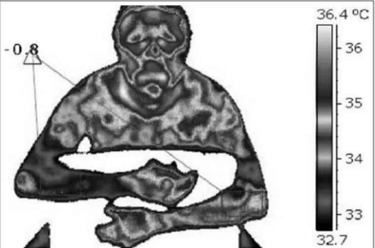
hermometry has shown asymmetry of the whole right hemi-body, with central neurogenic pattern (Figure 1) and temper-ature diference (∆T 0,8º C), symmetry in medial eye corners, internal carotid extracranial terminal branches territory, well delimited and homogeneous symmetric periocular hyper-radiation, thermal asymmetry of the whole hemibody, head, trunk and extremities, asymmetry between posterosuperior cervical faces. Presence of extensive linear hyper-radiation, in descending and oblique band, with well-delimited isothermal distribution, on projection of posterior cervical muscles, thus conirming MS-induced thermoregulatory dysfunction. With regard to lumbar spine pain (Figure 2), images analysis has shown paralumbar regions asymmetry. Presence of lumbar paravertebral linear vertical hyper-radiation, with ill-deined borders, irregular thermal distribution in paralumbar region, suggesting contracture of posterior cervical muscles and of lumbar paravertebral region (osteoarthropathy).
DISCUSSION
Irregular thermal distribution suggestive of posterior muscles contracture could explain patient’s severe pain. Pain evalua-tion is still a challenge because it is unique to each individual, and most available evaluation methods are very subjective.
Figure 1. Infrared image showing central neurogenic pattern with temperature difference of 0.8oC
Figure 2. A. Infrared image showing lumbar paravertebral hyper--radiation and irregular thermal distribution. B. Image showing asym-metry between hemibodies and cervical spine hyper-radiation area, suggesting posterior muscles contracture.
A
235
Infrared thermography to evaluate pain in a multiple sclerosis patient. Case report
Rev Dor. São Paulo, 2016 jul-sep;17(3):232-5
However, thermography may be applied with this objective16,
since studies using thermal distribution evaluation by means of thermography in the investigation of pain episodes, includ-ing of neurological origin, are many and increasinclud-ing, and their results have been proven, even comparing them to other al-ready consolidated evaluation methods16-18.
A study with 65 chronic low back pain patients which related thermography to other imaging exams, has shown that infra-red thermography showed abnormal thermal pattern in 92% of cases, while MRI has shown structural changes in 89%, com-puterized tomography (CT) in 87% and myelography in 80% of patients. From 22 patients with positive disc alteration in MRI, 21 have shown thermographic changes and all cases with root involvement in CT and myelography have shown signii-cant changes in thermography, especially in LLll19.
However, it has to be stressed that changes in local tempera-ture are not always relex of installed painful processes, and in-adequate motor activity resulting from sympathetic inluences might be present11,20. hermography has shown signiicant
ther-mal asymmetry in the patient, with central neurogenic pattern, by involving right hemibody (face, trunk and extremities). Simi-lar case was reported in a study14 with a child with MS, who
presented excessive sweating on right face and shoulder. Unilat-eral hyperhydrosis of the whole hemibody is a central neuro-vegetative phenomenon which, comparatively, is similar to skin vasomotor change found in this patient. However, temperature changes were not evidenced by thermography, which makes this study the irst to describe this phenomenon in case of MS. Hyperhydrosis may be associated to hypothermia in some MS cases, as described by other studies21-23. Authors suggest
that these symptoms, in addition to peripheral vasoconstric-tion, are associated to hypothalamic injuries, because this is the region controlling normal physiologic body responses to temperature changes. However, MRI images are not always able to detect changes in this brain region, making diicult the identiication of the origin of thermal changes in MS pa-tients, as well as their repercussions.
CONCLUSION
MS is still one medical mystery; it is not avoidable or cur-able and has broad symptoms, especially chronic pain and inadequate thermo-regulation, which directly interfere with quality of life of patients. To adequately treat the disease, it is necessary a correlation between symptoms and imaging ex-ams results and in this case thermography would act as com-plementary diagnostic method, since many diseases result in similar pain presentations, however with temperature pattern
diferent from MS, such as CNS infections, radiculopathies, ibromyalgia, among others24. In addition, another
objec-tive would be to screen painful acute and chronic symptoms, helping the understanding of the symptoms of the disease.
REFERENCES
1. Associação Brasileira de Esclerose Múltipla – ABEM [Homepage na internet]. O que é esclerose múltipla? [acesso em 27 de dez de 2015]. Disponível em: www.abem.org.br 2. Callegaro D, Goldbaum M, Morais L. he prevalence of multiple sclerosis in the city
of São Paulo, Brazil. Acta Neurol Scand. 2001;104(4):208-13
3. Fragoso YD, Peres M. Prevalence of multiple sclerosis in the city of Santos, SP, Brazil. Rev Bras. Epidemiol. 2007;10(4):479-82.
4. Ferreira ML, Machado MI, Vilela ML, Guedes MJ, Ataíde L Jr, Santos S, et al. [Epi-demiology of 118 cases of multiple sclerosis after 15 years of follow-up on the refer-ence center of Hospital da Restauração de Pernambuco, Brazil.] Arq Neuropsiquiatr. 2004;62(4):1027-32. Portuguese.
5. Milo R, Kahana E. Multiple sclerosis: geoepidemiology, genetics and the environ-ment. Autoimmun Rev. 2010;9(5):A387-94.
6. Rudick RA. Esclerose múltipla e distúrbios relacionados. In: Goldman L, Bennett JC. (editores) Cecil: tratado de medicina interna. 21a ed. Rio de Janeiro: Guanabara
Koogan; 2001. 2387-96p.
7. Polman CH, Reingold SC, Edan G, Filippi M, Hartung HP, Kappos L, et.al. Diag-nostic criteria for multiple sclerosis: 2005 revisions to the “McDonald Criteria”. Ann Neurol. 2005;58(6):840-6.
8. Brioschi ML. Metodologia de normalização de análise do campo de temperaturas em imagem infravermelha humana. Curitiba. [Engenharia Mecânica]. Universidade Fed-eral do Paraná; 2011.
9. Brioschi ML, Lin TY, Teixeira MJ. Estudo da dor por imagem infravermelha. Rev Dor. 2005;6(3):589-99.
10. Brioschi ML, Yeng LT, Pastor EM, Colman D, Silva FM, Teixeira MJ. Documen-tação da síndrome dolorosa miofascial por imagem infravermelha. Acta Fisiátrica. 2007;14(1):41-8.
11. Uematsu S, Jankel WR, Edwin DH, Kim W, Kozikowski J, Rosenbaum A, et al. Quantiication of thermal asymmetry Part 2: Application in low-back pain and sci-atica. J Neurosurg. 1988;69(4):556-61.
12. Baker DG. Multiple sclerosis and thermoregulatory dysfunction. J Appl Physiol. 2002;92(5):1779-80.
13. Neves EB, Alves JV, Rosa C, Reis VM. hermography in neurologic practice. Open Neurol J. 2015;9:24-7.
14. Ueno M, Tokunaga Y, Terachi S, Gondo K, Hara T. Asymmetric sweating in a child with multiple sclerosis. Pediatr Neurol. 2000;23(1):74-6.
15. Goldberg HI, Heinz ER, Taveras JM. hermography in neurological patients. Prelimi-nary experiences. Acta Radiol Diagn.1966;5:786-95.
16. Marcos ML, Abramavicus S, Corrêa CF. Valor da imagem infravermelha na avaliação da dor. Rev Dor. 2005;6(1):514-24.
17. Brioschi ML, Yeng LT, Teixeira MJ. Diagnóstico avançado em dor por imagem in-fravermelha e outras aplicações. Prática Hospitalar. 2007;50(IX):93-8.
18. Lima RP, Brioschi ML, Teixeira MJ, Neves EB. Análise termográica de corpo inteiro: indicações para investigação de dores crônicas e diagnóstico complementar de disfun-ções secundárias. Pan Am J Med hermol. 2015;2(2):70-7.
19. homas D, Cullum D, Siahamis G, Langlois S. Infrared thermographic imaging, magnetic resonance imaging, CT scan and myelography in low back pain. Br J Rheu-matol. 1990;29(4):268-73.
20. Kurz A, Sessler DI, Tayefeh F, Goldberger R. Poikilothermia syndrome. J Intern Med. 1998;244(5):431-6.
21. Edwards S, Lennox G, Robson K, Whiteley A. Hypothermia due to hypothalamic involvement in multiple sclerosis. J Neurol Neurosurg Psychiatry. 1996;61(4):419-20. 22. Darlix A, Mathey G, Monin ML, Sauvée M, Braun M, Schaf JL, et al. Hypothalamic
involvement in multiple sclerosis. Rev Neurol. 2012;168(5):434-43.
23. Martinez-Rodriguez JE, Munteis E, Roquer J. Periodic hyperthermia and abnor-mal circadian temperature rhythm in a patient with multiple sclerosis. Mult Scler. 2006;12(4):515-7.