DENTOFACIAL DEFORMITIES:
OROFACIAL MYOFUNCTIONAL CHARACTERISTICS
Deformidades Dentofaciais: características miofuncionais orofaciais
Janayna de Aguiar Trench (1), Roberto Paulo Correia de Araújo (1)
One or both jawbones could be altered in the vertical, horizontal and transverse planes, either in
isolation or combination, leading to diferent types of deformities5.
Serious problems of dental and skeletal maloc -clusion require combined treatment with orthodontics
and orthognathic surgery. This treatment aims to achieve facial, dental and functional harmony6.
The intimate relationship between the hard and soft tissues and the need to perform stomatognathic functions for survival lead to the occurrence of functional adaptations that enable the performance of these functions regardless of existing changes7.
To quantify the function of the masticatory system, many parameters are being studied, including chewing eiciency, maximum bite force, electromyographic activity of masticatory muscles and range of mandibular movement8.
Knowing how stomatognathic functions occur on the normality and how performance is modiied according to the placement of bone and tooth bases is essential for the speech therapist to be able to plan the myofunctional treatment, before and after
INTRODUCTION
Human mandibular growth is characterized
by great variations that determine the vertical and sagittal dimensions of the craniofacial complex. Cephalometric analyses have shown that variations in craniofacial growth are related to the direction of mandibular condyle growth1.
Dentofacial deformities (DFD) can be deined as conditions in which the facial skeleton diverges from normality; additionally, there is malocclusion, and the facial appearance is afected. These deformities may be minimal, such as a slight projection of the chin, or extreme, such as a severe vertical maxillary excess or hemifacial microsomia2. This condition
may be evident at birth or appear during growth and development, creating functional, degenerative, aesthetic and psychosocial problems. The time of
surgical intervention can be critical and should occur
during or after complete growth3,4.
ABSTRACT
Purpose: to analyze and describe the performance of the stomatognathic functions according to
the diferent types of dentofacial deformities and compare the characteristics of these functions in subjects with dentofacial deformities and subjects without changing of the facial skeleton. Methods:
this descriptive, analytical and transversal study comprised 50 patients with dentofacial deformities. The control group consisted of 46 healthy individuals. Data collection occurred between the months of July and September 2013, and the orofacial myofunctional analysis was performed by the application of the Marchesan, Berrentin-Felix, Genaro, Rehder protocol. The statistical protocol was based on descriptive data analysis. Results: all dentofacial deformities studied had some change in the
implementation of the stomatognathic functions and these changes varied according to the type of dentofacial deformities. Conclusion: diferent types of dentofacial deformities are related to changes
detected in the performance of the stomatognathic functions.
chewing speed, atypical muscle contractions and pain or noise in the temporomandibular joint (TMJ). Three tests were applied to evaluate swallowing, as follows: swallowing of solids, habitual swallowing of liquid and directed swallowing of liquid. The irst test (swallowing solids) was analyzed by ilming
chewing. For the second test (habitual swallowing
of liquid), the participant was instructed to drink 200 mL of water as usual. For the third test (directed swallowing), the subject was instructed to take and keep a sip of water in the mouth and swallow only after requested by the evaluator. The presence of tongue interposition, hyperfunction of the perioral muscles and remains were evaluated after
swallowing.
The speech evaluation was performed through ive tests. In the irst test, the subject was instructed to count from zero to twenty (0-20) and say the days of the week followed by the months of the year. In the second test, a board was used with phonetically balanced igures, and the volunteer was asked to name those igures. The third test was aimed as assessing the motor coordination of speech. In this
test, the subject was instructed to articulate the
syllables /pa/ /ta/ /ka/, irst separately and, then, in sequence. The fourth test evaluated sponta
-neous speech, with the participant being asked to articulate their full name, age and talk about their job/profession or describe a trip or outing that they experienced. The ifth test was only conducted with patients who had some type of phonetic alteration.
In this case, the subject was instructed to repeat the altered phoneme and add the vowel e, for example,
if the altered phoneme was /s/, the subject was then asked to repeat it. The following criteria were evaluated: the presence of saliva, lip movement, presence of articulatory imprecision, speech rate and pneumo-phono-articulatory coordination.
The respiratory mode was evaluated using an
Altmann graph mirror, which is a metal plate that is mirrored on both sides with a graph on the upper side, through which was observed the air route
(oral, nasal and oronasal). Mouth breathing was deined as when the route was only through the oral cavity. Nasal breathing occurred when respiration was performed only through the nose, and oronasal took place when the route was mixed, i.e. nasal and
oral cavities.
As this is a sample plan to the entire target
population and that was performed through a stomatognathic functions according to the diferent
types of dentofacial deformities and confront the characteristics of these functions in subjects with DFD and subjects without changes of the facial skeleton.
METHODS
This study was approved by the Ethics Committee of the School of Dentistry at the Federal University of Bahia under Protocol # 301.251. Patients were informed about the aims of the present study, and permission was voluntarily granted by signing an informed consent form.
Individuals of both sexes participated in this study, including patients with DFD who were 16 to 55 years of age and with indication for orthognathic surgery from the Division of Oral and Maxillofacial Surgery and Treatment in the School of Dentistry at the Federal University of Bahia; the Division of Speech Therapy of the Professor Edgar Santos University Hospital Complex; and the Orthodontics staf at the Center for Dental Studies (CENO) between July and September 2013.
Exclusion criteria people who have dental laws
or wearing dentures and or dental implants. Was provided to all participants the right to withdraw at
any predetermined stage of the research, despite having signed the informed consent form. These criteria also apply to the control group.
The stomatognathic functions of breathing,
swallowing, speech and chewing were evaluated in
all volunteers. Therefore, the MBGR (Marchesan, Berrentin-Felix, Genaro and Rehder) protocol9
was used as an investigative tool along with the
following materials: French bread, water, disposable cup, examination gloves, Altmann graph mirror and
disposable tongue depressor.
The characteristics of masticatory function were assessed by examining the ability to chew French bread by each individual, who was instructed to proceed as usual with daily life. The performance of this function was ilmed with a SONY DSC – W 620
digital camera; the obtained results were evaluated
by counting the number of chewing cycles, and
the vertical or lateral mandibular movements
RESULTS
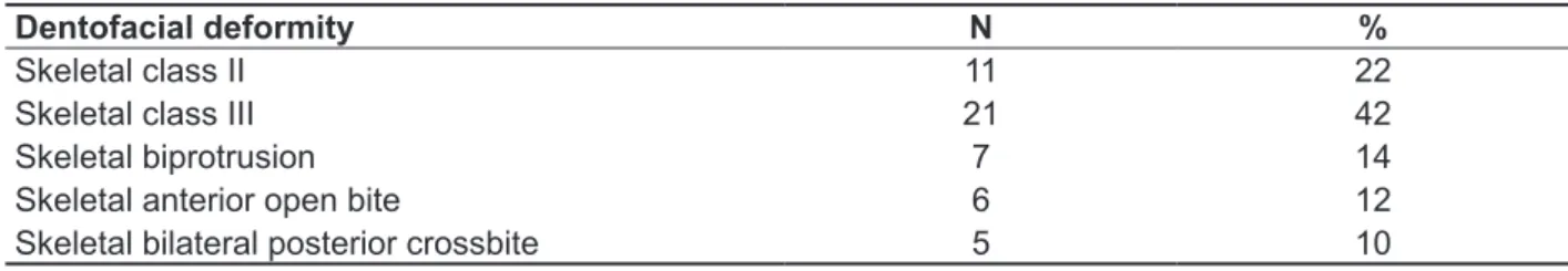
Fifty individuals, including 26 women and 24 men, with a mean age of 26.7 years comprised the group with Dentofacial Deformities (DFD). The control group (CG) consisted of 46 subjects, including 25 men and 21 women, with a mean age of 25.3 years.
The DFD group included individuals with diferent types of deformities, as follows: 11 patients with a skeletal Class II deformity; 21 patients with a skeletal Class III deformity; 7 with skeletal bipro
-trusion; 6 with skeletal anterior open bite; and 5 with skeletal posterior crossbite (Table 1).
an adequate measure of the standard error and therefore the performance of statistical inference, no inferential statistics (statistical test of hypothesis or conidence interval) was used, as they are completely inadequate to the context of inferential statistics theory and theories of probability that support them, which will not be seen in this study12-16. Therefore the
statistical analysis was performed using descriptive
statistics. For the variables measured in qualitative
scale (DFD, functional characeristics), respective measurements of proportion were obtained.
Analyses were performed in the R statistical package version17.
Table 1 – Characterization of dentofacial deformity group
Dentofacial deformity N %
Skeletal class II 11 22
Skeletal class III 21 42
Skeletal biprotrusion 7 14
Skeletal anterior open bite 6 12
Skeletal bilateral posterior crossbite 5 10
Myofunctinoal orofacial features
The data will be described according to the type of DFD because each type features speciic myofunctional behaviors.
Control group
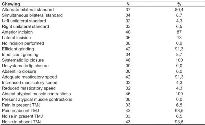
The alternating bilateral chewing pattern was present in 80.4% of subjects in this group, followed by simultaneous bilateral (8.7%), unilateral right
simultaneous bilateral (9.1%). Cutting into food was performed with the front teeth in 63.2% of cases, and chewing was ineicient in 72.7%. Lip closure was exhibited by all patients; however, it occurred unsystematically in 63.2% of subjects evaluated. Atypical muscle contractions were not common and observed in only 36.4% of subjects. The chewing speed was faster in 72.7% of patients with this type of deformity. While performing masticatory function, pain and noise in the TMJ were present in only 9.1% and 18.2% of participants in this group, respectively.
During the evaluation of speech articulation, an accumulation of saliva at the labial commissure was observed in 54.5% of the subjects, and lip movement was adequate in most participants. The speech articulation was unsystematically inaccurate in 45.5% of cases. The speech rate was adequate in 90.9% of subjects. The pneumo-phono-articulatory coordination was altered in 63.6% of patients with skeletal Class II DFD. Most of these individuals had some type of phonetic distortion (Table 3).
Speech articulation occurred with saliva being swallowed, lip motion and adequate speech rate in
93.5% of the CG. The pneumo-phono-articulatory coordination was adequate in 97.8% of cases evaluated. Precise speech articulation without phonetic distortions was observed in 100% of
subjects in this group.
Swallowing occurred without tongue interposition
or remains after the food was swallowed. Only 4.3% of subjects without DFD exhibited hyperfunction of
the perioral muscles during swallowing.
The nasal respiratory mode was observed in 93.5% of the control group subjects, which was followed by oronasal (6.5%). There were mouth
breathers in this group.
Group with dentofacial deformities
1. SKELETAL CLASS II
The left unilateral chewing pattern was present in 54.5% of subjects with skeletal Class II DFD, which was followed by unilateral right (36.4%) and
Table 2 – Distribuition based on occurence of chewing characteristics of control group
Chewing N %
Alternate bilateral standard 37 80,4 Simultaneous bilateral standard 04 8,7
Left unilateral standard 02 4,3
Right unilateral standard 03 6,5
Anterior incision 40 87
Lateral incision 06 13
No incision performed 00 0,0
Eicient grinding 42 91,3
Inneicient grinding 04 8,7
Systematic lip closure 46 100
Unsystematic lip closure 00 0,0
Absent lip closure 00 0,0
Adequate masticatory speed 42 91,3
Increased masticatory speed 02 4,3
Reduced masticatory speed 02 4,3
Absent atypical muscle contractions 46 100
Present atypical muscle contractions 00 0,0
Pain in present TMJ 03 6,5
Pain in absent TMJ 43 93,5
Noise in present TMJ 03 6,5
faster rate (23.8%) and slower rate (38.1%). Pain and noise in the TMJ were reported by 23.8% and 33.3% of subjects, respectively (Table 4).
Accumulation of saliva in the labial commissure
during speech articulation was observed in 33.3%
of subjects with this type of DFD, while 66.7% of these exhibited swallowing of saliva during speech production, thereby preventing it from accumulating in the oral cavity. The lip movement, speech rate and pneumo-phono-articulatory coordination were adequate in 66.7%, 85.7% and 76.2%, respectively, of the cases evaluated. Articulatory imprecision was absent in 71.4% of participants. Some type of phonetic distortion was present in 90.5% of subjects.
The interposition of the tongue during swallowing occurred in 81% of cases, and hyperfunction of
the perioral muscles was seen in 72.2%. Remains
after swallowing was observed in 19% of subjects
evaluated.
The oronasal respiratory mode was present in 47.4% of participants, followed by oral (28.6%) and
nasal (23.8%).
Swallowing function with tongue interposition occurred in 45.5% of subjects; hyperfunction of
the perioral muscles was seen in 81.8%, and the
presence of food remains after swallowing was observed in 27.3% of cases.
The oral respiratory pattern was the most common among patients with this type of DFD, observed in 81.8% of cases.
2. SKELETAL CLASS III
In individuals with skeletal Class III deformities,
the bilateral simultaneous chewing pattern was
observed in 90.5% of subjects, versus the unilateral right and left that was observed in 4.8% of cases. Cutting was performed using the front teeth by 54.5% of participants, while the remaining partici
-pants did not cut into the food (47.6%). The chewing of the food was inefective in 71.4% of subjects. Lip closure occurred, but in an unsystematic way, in 71.4% of cases. Atypical muscle contractions were present in 52.4% of those evaluated. The chewing rate results in patients with this type of DFD were
balanced between an adequate rate (38.1%),
Table 3 – Distribuition of ocurrence of phono-articulatory characteristics on the bearers of skeletal class II dentofacial deformity
Phono-articulation N %
Swallowed saliva 04 36,4
Accumulation of saliva in labial commissure 06 54,5
Accumulation of saliva in lower lip 01 9,1 Adequate lip movement 08 72,7
Exaggerated lip movement 03 27,3
Reduced lip movement 00 0,0
Absent articulatory imprecision 03 27,3
Unsystematic articulatory imprecision 05 45,5
Systematic articulatory imprecision 03 27,3 Adequate speech speed 10 90,9 Increased speech speed 01 9,1 Reduced speech speed 00 0,0
Adequate pneumo-phono-articulatory coordination 04 36,4
Altered pneumo-phono-articulatory coordination 07 63,6
Present phonetic distortion 08 72,7
Regarding the evaluation of speech articu
-lation, 42.9% of the subjects had accumulation of saliva in the labial commissure. The movement of
the lips during speech production was adequate
in 85.7% of cases evaluated. Articulatory impre -cision was not observed, and the speech rate was
adequate in 85.7% of participants evaluated. The pneumo-phono-articulatory coordination was also adequate in 71.4% of cases. Phonetic distortion was observed in 85.7% of subjects.
The interposition of the tongue during swallowing was present in 42.9% of subjects evaluated, while hyperfunction of the perioral muscles was observed in 71.4% of cases (Table 5).
Among patients with skeletal biprotrusion, the oronasal respiratory mode was present in 42.9% of subjects, which was followed by nasal (28.6%) and oral (28.6%) (Table 5).
3. SKELETAL BIPROTRUSION
In the skeletal biprotrusion DFD, the unilateral left chewing pattern occurred in 71.4% of cases, followed by unilateral right (14.3%) and simulta
-neous bilateral (14.3%). The cutting into food was performed using the front teeth by 100% of partici
-pants in this group, and the chewing of the food was ineicient, with a faster chewing speed in 57.1% of subjects evaluated. Of patients with the skeletal biprotrusion type of DFD, 71.4% exhibited unsys
-tematic lip closure, and there was an absence of atypical muscle contractions. The presence of noise in the TMJ was not reported by any of the partici
-pants during masticatory function; however, 14.3% of this population reported pain in the TMJ during
chewing.
Table 4 – Distribuition based on occurence of masticatory characteristics on the bearers of skeletal class III dentofacial deformity
Chewing N %
Alternate bilateral standard 00 0,0 Simultaneous bilateral standard 19 90,5
Left unilateral standard 01 4,8
Right unilateral standard 01 4,8
Anterior incision 11 54,5
Lateral incision 00 0,0
No incision performed 10 47,6
Eicient grinding 06 28,6
Inneicient grinding 15 71,4
Systematic lip closure 04 19
Unsystematic lip closure 15 71,4
Absent lip closure 02 9,5
Adequate masticatory speed 08 38,1
Increased masticatory speed 05 23,8
Reduced masticatory speed 08 38,1
Absent atypical muscle contractions 10 47,6
Present atypical muscle contractions 11 52,4
Pain in present TMJ 05 23,8
Pain in absent TMJ 16 76,2
Noise in present TMJ 07 33,3
Table 5 – Distribuition based on occurrence of swallowing and breathing characteristics of bearers of skeletal biprotrusion dentofacial deformity
Swallowing N %
Tongue interposition presence 03 42,9
Perioral muscle hyperfunction 05 71,4
Presence of residues after swallowing 00 0,0
Breathing
Oral 02 28,6
Nasal 02 28,6
Oronasal 03 42,9
Table 6 – Distribuition based on occurrence of phono-articulatory characteristics of bearers of skeletal anterior open bite dentofacial deformity
Phono-articulation N %
Swallowed saliva 02 33,3
Accumulation of saliva in labial commissure 02 33,3
Accumulation of saliva in lower lip 02 33,3 Adequate lip movement 02 33,3
Exaggerated lip movement 04 66,7
Reduced lip movement 00 0,0
Absent articulatory imprecision 04 66,7
Unsystematic articulatory imprecision 01 16,7
Systematic articulatory imprecision 01 16,7 Adequate speech speed 04 66,7 Increased speech speed 01 16,7 Reduced speech speed 01 16,7
Adequate pneumo-phono-articulatory coordination 02 33,3
Altered pneumo-phono-articulatory coordination 04 66,7
Present phonetic distortion 06 100
Absent phonetic distortion 00 0,0
4. SKELETAL ANTERIOR OPEN BITE
Anterior open bite led to the unilateral right
chewing pattern in 50% of subjects, followed by simultaneous bilateral (33.3%) and unilateral left (14.3%). None of these subjects performed anterior incision, and 50% cut into food using the lateral teeth, with the other half not cutting. All patients with this DFD exhibited ineicient chewing. Additionally, 66.7% performed masticatory function without lip closure, while 33.3% performed masticatory function with unsystematic lip closure. The chewing rate was faster in 83.3% of subjects. Atypical muscle contrac
-tions were observed in 50% of the cases evaluated. Noises in the TMJ were reported by only 33.3% of
participants. None of the patients with anterior open bite DFD reported TMJ pain during chewing.
During the speech of these individuals, saliva was
observed to accumulate in the labial commissure and the lower lip and to be swallowed at the same
proportion, i.e., in 33.3% of cases. The speech artic
-ulation featured exaggerated lip movement in 66.7% of participants. The speech rate was adequate in 66.7%; however, the pneumo-phono-articulatory
coordination was altered at the same proportion as
patients evaluated (66.7%). Articulatory imprecision
was absent in most cases. All patients with anterior
of cases evaluated. The oral respiratory mode was observed in 100% of subjects with anterior open bite (Table 7).
Swallowing with the tongue interposition was
present in all participants with skeletal anterior open bite. Hyperfunction of the perioral muscles did not occur at a great frequency but was present in 50%
Table 7 – Distribution based on occurrence of characteristics of swallowing and breathing of bearers of skeletal anterior open bite dentofacial deformity
Swallowing N %
Tongue interposition presence 06 100
Perioral muscle hyperfunction 03 50
Presence of residues after swallowing 00 00
Breathing
Oral 06 100
Nasal 00 0,0
Oronasal 00 0,0
5. SKELETAL BILATERAL POSTERIOR CROSS BITE
In patients with skeletal bilateral posterior cross bite DFD, the unilateral left chewing pattern was observed in 80% of subjects, which was followed by unilateral right (20%). The cutting into food was performed using the front teeth in 100% of cases evaluated. During mastication, the rate was adequate, with systematic lip closure, and without atypical muscle contractions in 60% of partici
-pants; however, there was ineicient chewing in 80% of subjects evaluated. Noise in the TMJ was not reported by any of the participants, but painful symptoms were reported by 40% of patients with bilateral posterior cross bite DFD (Table 8).
During the evaluation of speech articulation, 80% of individuals swallowed saliva, thus preventing it from accumulating in the oral cavity, while accumulation
in the labial commissure was observed in the
remaining 20% of participants. The lip movement was adequate in 80% of cases evaluated. The speech rate and pneumo-phono-articulatory coordi
-nation were adequate in all subjects. Only 20% of patients with bilateral posterior crossbite exhibited some type of phonetic distortion, and imprecise speech was evident, albeit in an unsystematic way, in 40% of subjects.
The only swallowing disorders exhibited by
patients with bilateral posterior crossbite remained
after the act of swallowing, which was observed in 60% of these individuals.
functional adaptations such as unilateral chewing to facilitate the chewing process21,22.
The lip closure during chewing occurred unsys
-tematically in most cases evaluated. Reports addressing that topic during chewing were not found; however, studies were found that show the change in the lip closure at rest. These indings reveal the occurrence of parted lips at rest and closing with diiculty, which involved the chin muscles.
Phonetic distortion was the phono-articulatory change that occurred most in skeletal Class II DFD, which is consistent with the scientiic literature20-23.
Articulatory imprecision also occurred, although unsystematically, that can be explained by the accumulation of saliva in the labial commissure. As an attempt to contain the saliva in the oral cavity,
the individual results in impaired speech articulation
as a whole. Altered pneumo-phono-articulatory coordination, which was present in more than half of individuals, may also have contributed to the unsystematic articulatory imprecision; however, this did not afect the speech rate.
DISCUSSION
For the most part, participants in the control group
showed no signiicant myofunctional changes, and those present were limited to isolated and temporary situations. The chewing pattern most commonly
observed in this group was bilateral alternating, which is the ideal mastication pattern because it
allows the load to be distributed evenly, alternating the efort and rest of the muscles and joints18,19.
Patients with DFD have myofunctional features that vary according to the type of deformity. The
musculature adapts so that the stomatognathic
functions can be performed. These adaptations occur according to the pattern of the maxillofacial skeleton jawbones.
Regarding patients with skeletal Class II DFD, the present study found masticatory changes such as unilateral chewing, faster chewing rate and ineicient chewing. The literature20 shows that the
states that chewing occurs in these individuals with
rapid and shorter cycles that directly inluence the
Table 8 – Distribuition based on occurrence of masticatory characteristics of bearers of skeletal bilateral posterior crossbite dentofacial deformity
Chewing N %
Alternate bilateral standard 00 0,0 Simultaneous bilateral standard 00 0,0
Left unilateral standard 04 80
Right unilateral standard 01 20
Anterior incision 05 100
Lateral incision 00 0,0
No incision performed 00 0,0
Eicient grinding 01 20
Inneicient grinding 04 80
Systematic lip closure 03 60
Unsystematic lip closure 02 40
Absent lip closure 00 0,0
Adequate masticatory speed 03 60
Increased masticatory speed 02 40
Reduced masticatory speed 00 0,0
Absent atypical muscle contractions 03 60
Present atypical muscle contractions 02 40
Pain in present TMJ 02 40
Pain in absent TMJ 03 60
Noise in present TMJ 00 0,0
phenomenon may be due to the ability to seal the lip becoming diicult due to an increase in the vertical dimension. Phonetic distortion in speech articulation occurred in most individuals with this type of DFD, which can be explained by the advancement of the maxilla and mandible, allowing forward movement of the tongue and a change in lip tone4. The interpo
-sition of the tongue was found during swallowing, as well as hyperfunction of the perioral muscles, which may have contributed to the emergence of phonetic
changes in speech.
The most common respiratory mode for individuals with skeletal biprotrusion was oronasal. No published data were found to conirm this inding, but it can be explained by the diiculty of
maintaining the lip seal because there is an advance
of two jawbones, the maxilla and mandible, which is a condition that makes it possible to breathe through
both routes, oral and nasal.
For the anterior open bite DFD, the most common chewing pattern was unilateral with a faster chewing rate. These data are similar to the results of a recent study29 in which the authors claim that individuals
with an anterior open bite chew with shorter cycles, which increases the rate of the masticatory process and reduces the function eiciency. Most participants of the present study did not exhibit lip closure during mastication, and it was unsystematic in those who did. This result can occur due to the loss of tone and strength of the orbicularis oris muscle. This element also explains the fact that half of the individuals exhibited atypical muscle contractions during this function because it is not only the orbicularis oris
that has less tone but the buccinator and elevator
muscles of the mandible4. Therefore, it is necessary
for the individual to use other muscle groups or for the contraction of these muscles to be stronger to provide greater control of food during chewing.
All subjects with an open bite had some type of phonetic distortion. Moreover, an exaggerated lip movement was observed during speech. The liter -ature states that the tongue can interpose during the
production of some phonemes, such as sibilants4.
Furthermore, with the reduced lip tone, the individual can increase the lip movements in an attempt to better articulate the speech sounds, but these
changes do not afect the accuracy of articulation in
the individuals. However, the
pneumo-phono-articu-latory coordination was altered in most individuals, which can be explained by exclusive oral breathing. the perioral muscles is the hallmark of swallowing in
patients with skeletal Class II DFD20,24.
Regarding this respiratory mode in patients with skeletal Class II DFD, there was a higher prevalence of oral breathing, which is consistent with reports in
the literature20,24.
In patients with skeletal Class III DFD, the masti
-catory alterations found most commonly were the simultaneous bilateral chewing pattern, ineicient chewing of food, unsystematic lip closure and atypical muscle contractions. The literature showed
that chewing in these individuals occurs with the
presence of vertical mandibular movements, without lateralization of the jaw and through inei
-cient grinding due to the loss of tone in the elevator muscles of the mandible, buccinator and lips20,25.
The main change in speech articulation was
phonetic distortion, which is similar to the data
reported in the literature on this topic. Phonetic distortion occurs mainly by hyperfunction of the upper lip so that the articulation points of some phonemes, such as bilabials and fricatives, are produced using
inverse lip movements20,25. To compensate for the
structural and functional changes, patients with this DFD exhibit changes in articulation points of phonemes; however, this condition did not afect the accuracy of speech articulation in the patients in question because only a few participants showed articulatory imprecision, while the majority showed this phenomenon unsystematically as reported in
the literature25,26.
The function of swallowing led to the key features of tongue interposition and hyperfunction of the perioral muscles, as reported in the indings of other
research20,25. Remains after swallowing were found
in a small number of patients evaluated. This type of swallowing is a consequence of the loss of tone and consequent function of the buccinator muscle during mastication and is responsible for returning the foods that fall into the vestibule to the occlusal surfaces of teeth27. When this does not happen,
the remains are observed to be lodged in the oral
cavity after swallowing, especially in the region of the vestibule. The literature shows that the chewing by patients with skeletal Class III occurs with little or no activity of the buccinator muscle20.
Regarding respiration, the results obtained
Speech articulation in the presence of a bilateral posterior cross bite was not signiicantly diferent,
appearing normal in almost all aspects evaluated.
However, it was found that articulatory imprecision was present unsystematically in almost half of the cases evaluated. This inding is explained by a reduction of the internal horizontal space that could make the articulation relatively imprecise.
The function of swallowing was performed ineiciently by over half of these individuals, with remains observed after swallowing. This inding runs
opposite to the results presented in the literature35
that reported that 87.5% of the subjects showed no remains after swallowing. Those authors claim that
the buccinator muscle is stronger on the chewing
side; however, the study cited was conducted on
individuals with unilateral posterior crossbite, while
in the present study we evaluated the bilateral DFD.
CONCLUSION
The diferent types of facial skeleton interfere in a particular way in the performance of stomato
-gnathic functions, being these changes related to the position of bone and tooth base and the insertion of orofacial and chewing muscles on those bases. Chewing functions, speech articulation, swallowing
and breathing, are altered according to the variation
in the positioning of bone and tooth bases. Therefore, a detailed evaluation of the functional aspects must be performed so that the treatment can be planned more individually as possible.
The oral respiratory mode was found in all of the subjects with anterior open bite. The literature reports that open bite is one of the main character
-istics of mouth breathers30,31.
The bilateral posterior cross bite DFD featured a unilateral chewing pattern and ineicient food chewing by most individuals. The relationship
between the unilateral chewing and the presence
of a posterior cross bite can be explained by the decrease of vertical space and the impossibility of performing the rocking motion, thus causing the
individual to chew on the opposing side32. In the
present study, the posterior cross bite evaluated was bilateral; however, the only masticatory pattern found was unilateral, and most individuals chewed on the left side. Taking into account the reasoning
above and even with a crossbite on both sides,
there may be one side with smaller vertical dimen -sions, which would be the chewing side.
Additionally, regarding the chewing in patients with bilateral posterior crossbite, it was found
that the chewing rate was adequate, and the lip
closure during this chewing was systematic in more than half of the cases evaluated. The symptom “pain” was reported by approximately half of the individuals. However, no results that could explain these indings were obtained, although the scientiic literature relates the occurrence of skeletal posterior
crossbite with temporomandibular disorders33,34.
It would be a mistake to consider only reports of painful symptoms during chewing as reliable data to ensure the diagnosis of this disorder.
RESUMO
Objetivo: analisar e descrever as funções estomatognáticas de acordo com os diferentes tipos de
deformidades dentofaciais e confrontar as características dessas funções em sujeitos com defor
-midade dentofacial e sujeitos sem alterações do esqueleto facial. Métodos: trata-se de um estudo
descritivo, analítico e de caráter transversal, envolvendo uma amostra de 50 indivíduos portadores de deformidades dentofaciais frente ao grupo controle constituído por 46 indivíduos saudáveis. A coleta de dados aconteceu entre os meses de julho a setembro de 2013, foi realizada a avaliação miofun
-cional orofacial, mediante a aplicação do protocolo Marchesan, Berrentin-Felix, Genaro, Rehder. O protocolo estatístico fundamentou-se na análise descritiva dos dados. Resultados: todos os sujeitos
portadores de deformidades dentofaciais avaliados apresentaram alterações na execução das fun
-ções estomatognáticas sendo que tais altera-ções variaram de acordo com o tipo de deformidades dentofaciais apresentada. Conclusão: os diferentes tipos de deformidades dentofaciais estão rela
-cionados às alterações detectadas no desempenho das funções estomatognáticas.
DESCRITORES: Anormalidades Craniofaciais; Músculo Masseter; Músculo Temporal; Oclusão
16. Vacha-Haase T. Statistical signiicance should not be considered one of life’s guarantees: efect sizes are needed. Educ. psychol. measur.
2001;61(2):219-24.
17. R Development Core Team. R: A language and environment for statistical computing. Vienna, Austria: R Foundation for Statistical Computing, 2012. Disponível em: <http://www.R-project.org>. 18. Ferrario VF, Sforza C, Colombo A, Ciusa VA. A electromyographic investigation of masticatory muscles symmetry in normo-occlusion subjects. J
Oral Rehabil. 2000;27(1):33-40.
19. Felicio CM, Melchior MO, Silva MAMR, Celeghini RMS. Desempenho mastigatório em adultos
relacionado com desordem temporomandibular
e com a oclusão. Pró-Fono R Atual Cient.
2007;19(2):151-8.
20. Coutinho TA, Abath MB, Campos GJL, Antunes AA, Carvalho RWF. Adaptações do sistema estomatognático em indivíduos com desproporções maxilo-mandibulares: revisão da literatura. Rev Soc
Bras Fonoaudiol. 2001;14(2):275-9.
21. Bianchini EMG. Avaliação fonoaudiológica da motricidade orofacial: distúrbios miofuncionais orofaciais ou situações adaptativas. Rev Dent Press
Ortodon Ortoped Facial. 2001;6(3):73-82.
22. Pereira JBA, Bianchini EMG. Caracterização das funções estomatognáticas e disfunções temporomandibulares pré e pós cirurgia ortognática e reabilitação fonoaudiológica da deformidade dentofacial classe II esquelética. Rev CEFAC.
2011;13(6):1086- 94.
23. Taucci RA, Bianchini EMG. Veriicação da interferência das disfunções temporomandibulares na articulação da fala: queixas e caracterização
dos movimentos mandibulares. Rev Soc Bras Fonoaudiol. 2007;12(4):274-80.
24. Kasai RCB, Portela MQ. Intervenção fonoaudiológica em pacientes submetidos ao tratamento ortodôntico cirúrgico. Rev Dent Press Ortodon Ortoped Maxilar. 2001;6(2):79-84.
25. Aléssio CV, Mezzomo CL, Körbes D. Intervenção fonoaudiológica nos casos de pacientes classe III com indicação à cirurgia ortognática. Arq Odontol.
2007;43(3):102-10.
26. Santos IF, Pereira SAA. A prevalência de alterações de fala em indivíduos portadores de
classe III. Fono atual. 2001;15(4):16-21.
27. Mory MR, Tessitore A, Pfeilsticker L, Junior
REFERENCES
1. Bjork A. Variations in the growth pattern of the human mandible: longitudinal radiographic study by the implant method. J Dent Res. 1963;(42):400-11. 2. Fish LC, Epker BN, Sullivan CR. Orthognathic surgery: the correction of dentofacial deformities. J. Oral Maxillofac Surg. 1993;(51):28-41.
3. American Association of Oral And Maxillofacial Surgeons. Parameters of care: clinical practice guidelines for oral and maxillofacial surgery (AAOMS ParCare2012). J Oral Maxillofac Surg.
2012;70(11):107-36.
4. Benevides SD. Fonoterapia no pré e pós operatório de cirurgia ortognática. In: Associação
Brasileira de Odontologia.Pró-odonto cirurgia. Ciclo
6, vol 4. Porto Alegre: Artmed Panamericana; 2013. P. 117-44.
5. Sproncen V. Long-face craniofacial morphology: cause or efect of weak masticatory musculature?
Seminars Orthond. 2010;16(2):99-117.
6. Sinko K, Jagsch B, Benes G, Millesi F,
Fischmeister R. Facial aesthetics and the
assignment of personality traits before and after orthognathic surgery. Int. J. Oral Maxillofac Surg.
2012;41:469-76.
7. Mezzono CL, Machado, PG, Pacheco AB, Gonçalves BFT, Hofmann CF. As implicações da classe II de Angle e da desproporção esquelética tipo classe II no aspecto miofuncional. Rev CEFAC.
2011;13(4):728-34.
8. Van Den Braber W, et al. Masticatory function in retrognathic patients before and after mandibular advancement surgery. J. Oral Maxillofac Surg.
2004;62(5):549-54.
9. Genaro KF, Berretin-Felix G, Rehder MIBC, Marchesan IQ. Avaliação miofuncional orofacial:
Protocolo MBGR. Rev CEFAC. 2009;11(2):237-55. 10. Felício CM, Ferreira CLP. Protocol of orofacial myofunctional evaluation with scores. Int J Pediatr Otorhinolaryngol. 2008;73(3):367-75.
11. Amaral DB. Mastigação unilateral x oclusão normal: um estudo sobre sua ocorrência em crianças
de 4 a 5 anos. Rev CEFAC. 2000;2(2):23-30.
12. Cowger CD. Statistical signiicance tests: scientiic ritualism or scientiic method? Soc. serv.
rev. 1984;58(3):358-72.
13. Gentry DL, Hoftyzer J. The misuse of statistical
techniques in evaluating sampling data. J. acad.
32. Marchesan IQ. The speech pathology treatment with alterations of the stomatognathic system. Int J Orofacial Myology. 2000;26:5-12.
33. Egermark I, Magnusson T, Carlsson GE. A 20-year follow-up of signs and symptoms of
temporomandibular disorders and malocclusions in subjects with and without orthodontic treatment in childhood. Angle Orthod. 2003;73(2):109-15.
34. Mcnamara JRJA, Seligman DA, Okeson JP. Occlusion, orthodontic treatment and temporomandibular disorders: a review. J Orofac Pain. 1995;9:73-90.
35. Pastana SG, Costa SM, Chiappetta ALML. Análise da mastigação em indivíduos que apresentam mordida cruzada unilateral na faixa-etária de 07 a 12 anos. Rev CEFAC. 2005;9(3):351-7. tipos faciais. R Dental Press Ortodon Ortop Facial.
2005;10(6):111-9.
29. Piancino MG, Isola G, Merlo A, Dalessandri D, Debernardi C, Bracco P. Chewing pattern
and muscular activation in open bite patients. J
Electromyogr Kinesiol. 2012;22(2):273-9.
30. Andrade FV, Andrade DV, Araújo AS, Ribeiro ACC. Alterações estruturais de órgãos fonoarticulatórios, más oclusões dentárias em
respiradores orais de 6 a 10 anos. Rev CEFAC. 2005;7:318-25.
31. Rodrigues HOSN, Faria RS, Paula FSG, Motta AR. Ocorrência de respiração oral e alterações miofuncionais orofaciais em sujeitos em tratamento
ortodôntico. Rev CEFAC. 2005;7(3):356-62.
Received on: July 22, 2014 Accepted on: November 13, 2014
Mailing address:
Janayna de Aguiar Trench Institute of Health Sciences, Federal University of Bahia