Karine Laskos SAKODA(a) Paula Karine JORGE(a)
Cleide Felício Carvalho CARRARA(b) Maria Aparecida de Andrade Moreira MACHADO(a) Fabrício Pinelli VALARELLI(c) Arnaldo PINZAN(a)
Thais Marchini OLIVEIRA(a)
(a) Universidade de São Paulo – USP, Bauru Dental School, Department of Pediatric Dentistry, Bauru, São Paulo, Brazil.
(b) Universidade de São Paulo – USP, Hospital for Rehabilitation of Craniofacial Anomalies, Department of Pediatric Dentistry, Bauru, São Paulo, Brazil.
(c) Universidade Ingá, Department of Orthodontics, Maringá, Paraná, Brazil.
3D analysis of effects of primary
surgeries in cleft lip/palate children
during the first two years of life
Abstract:
This study aimed at monitoring the maxillary growth of
children with cleft lip/palate in the irst two years of life, and to evaluate
the effects of primary surgeries on dental arch dimensions. The sample
consisted of the three-dimensional digital models of 25 subjects with
unilateral complete cleft lip and palate (UCLP) and 29 subjects with
isolated cleft palate (CP). Maxillary arch dimensions were measured
at 3 months (before lip repair), 1 year (before palate repair), and at
2 years of age. Student’s
t
test was used for comparison between the
groups. Repeated measures ANOVA followed by Tukey’s test was used
to compare different treatment phases in the UCLP group. Paired
t
test
was used to compare different treatment phases in the CP group. P<0.05
was considered statistically signiicant. Decreased intercanine distance
and anterior arch length were observed after lip repair in UCLP. After
palate repair, maxillary dimensions increased signiicantly, except
for the intercanine distance in UCLP and the intertuberosity distance
in both groups. At the time of palate repair and at two years of age,
the maxillary dimensions were very similar in both groups. It can be
concluded that the maxillary arches of children with UCLP and CP
changed as a result of primary surgery.
Keywords:
Cleft Lip; Cleft Palate; Growth and Development; Dental
Models.
Introduction
Children with orofacial clefts undergo surgical and non-surgical
multidisciplinary procedures that frequently cause adverse psychological
consequences to the individuals and their families.
1The treatment of
individuals with cleft lip/palate is complex, and its outcome is judged by
obtaining a balance among factors of esthetics, speech, and facial growth.
Problems of complex craniofacial growth are frequently observed in
individuals with cleft lip and palate, and are generally relected in transverse,
anterior-posterior and vertical
2dental relationships.
Some studies suggest that repair surgeries play an important role in
altering craniofacial growth and development.t
3,4,5At the same time, other
factors are also related to modiications in maxillo-mandibular growth:
cleft width, amount of tissue present at birth, individual growth potential,
6surgical technique employed in the primary repair surgeries,
7surgical
Declaration of Interests: The authorscertify that they have no commercial or associative interest that represents a conflict of interest in connection with the manuscript.
Corresponding Author: Thais Marchini Oliveira E-mail: [email protected]
https://doi.org/10.1590/1807-3107BOR-2017.vol31.0046
Submitted: Mar 21, 2016
outcome,
8and the surgeon’s ability.
4,9Some of the
frequently reported adverse consequences of primary
surgeries have been midface reduction,
4,10collapse of
maxillary arches
11and presence of cross bite.
12Currently, the literature lacks information on the
individual effects of lip and palate repair surgeries on
maxillo-mandibular growth in the irst years of life.
A good understanding of the effects of primary surgeries
is essential for the rehabilitation of the individuals with
cleft lip/palate. The search for techniques to decrease
the iatrogenic effects of the rehabilitative process may
uncover more favorable outcomes that may consequently
improve the quality of life of affected individuals.
This study aimed at monitoring the maxillary growth
of children with unilateral complete cleft lip and palate
and isolated cleft palate in the irst two years of life,
and to evaluate the effects of primary surgeries on the
dental arch dimensions.
Methodology
The Ethical Research Committee of the Hospital
for the Rehabilitation of Craniofacial Anomalies of
the University of São Paulo approved the protocol
of this study (#517.324). The inclusion criteria were
children with cleft lip and palate and children with
isolated cleft palate, born between 2010 and 2012,
of both genders. Children presenting syndromes
or associated malformations, and Simonart’s band,
and those having incomplete documentation were
excluded from the study.
The sample size was calculated so that the number
of selected children met the representative rating
to conduct the study. Considering a prior study by
Prahl et al.,
13with a signiicance level of 5%, test power of
80% and difference to be detected of 1.15, the minimum
sample size was calculated to be 24 individuals per
group. Thus, the sample comprised 25 children with
unilateral complete cleft lip and palate (group UCLP)
and 29 children with isolated cleft palate (group CP).
Surgical procedures of lip repair followed Millard’s
technique. In regard to palate repair, von Langenbeck’s
technique was used for both groups. One surgeon
performed all the surgical procedures on the same
patient. Dental study casts of each patient were
obtained at the following stages: T1 – Lip repair (UCLP),
T2 – Palate repair (UCLP and CP) and T3 - 2 years of
age (UCLP and CP). The impressions to obtain the casts
of stages T1 and T2 were made before the surgeries.
The study casts were digitized (Scanner 3Shape
R700
TMScanner, Copenhagen, Denmark) and 3D
OrthoAnalyzer
TMsoftware (Copenhagen, Denmark)
was used to evaluate the measurements and to deine
the landmarks
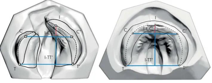
8(Figure 1).
Figure 1. Landmarks and distances used for assessment: CC’ (intercanine distance: where the lateral sulcus crosses the crest of the ridge); TT’ (intertuberosity distance: at the junction of the crest of the ridge with the outline of the tuberosity); I-CC’ (anterior arch length. perpendicular from point I to line CC’); I-TT’ (total arch length: perpendicular from point I to line TT’)
I
C’
C
I-CC’
I-TT’
T’
T
I
C’
C
I-CC’
I-TT’
Statistical analyses were performed with
Statistica
software (
Statistica for Windows - Version 7.0 - StatSoft
),
by adopting a 5% level of signiicance. Intraexaminer
error was analyzed by repeating the measurements
15 days after the irst assessment, in 20 randomly
selected study casts. Paired
t
test was used to calculate
the systematic error. The casual error was determined
by Dahlberg’s formula. The
t
test was applied for
intergroup comparisons. Repeated measures ANOVA
was used for intragroup comparisons regarding
different treatment stages in group UCLP, followed
by Tukey’s test. Paired
t
test was applied to carry out
the intragroup comparison for different treatment
stages in group CP.
Results
The intraexaminer test showed no statistically
signiicant differences in the repeated measurements
(Table 1). The mean ages of the children were
compared at the different treatment stages, and
are presented in Table 2. The lack of statistically
signiicant differences among the evaluated mean
ages enabled the comparison between the groups.
Maxillary dimensions of group UCLP at the
different treatment stages are described in Table 3.
The intercanine distance (CC’) decreased after lip
repair, but remained stable from palate repair to 2 years
of age. The intertuberosity distance (TT’) showed a
signiicant increase after lip repair. However, after palate
repair, this distance did not exhibit signiicant changes.
The anterior arch length (I-CC’) decreased after lip repair
and increased after palate repair. At 2 years of age, this
variable showed smaller values than those obtained
before the surgical procedures. The total arch length
(I-TT’) increased signiicantly in all periods evaluated.
The changes in the maxillary dimensions of group
CP from palate repair to 2 years of age can be seen in
Table 4. No signiicant changes were observed in TT’
through the evaluated stages. The CC’, I-CC’, and I-TT’
distances increased throughout the period studied.
The comparison of the maxillary dimensions
between groups UCLP and CP at stages T2 and T3 is
displayed in Table 5. None of the evaluated dimensions
showed statistically signiicant differences between
the groups.
Table 6 exhibits the changes in the maxillary
dimensions occurring between stages T2 and T3. From
palate repair to 2 years of age, only CC’ exhibited a
signiicantly greater increase in group CP.
Table 2. Mean age (years) for UCLP and CP groups at different treatment stages.
Stage UCLP CP P
Mean (SD) Mean (SD)
T1 0.39 (0.12) -
-T2 1.13 (0.10) 1.16 (0.17) 0.339
T3 2.21 (0.19) 2.18 (0.36) 0.697
T3-T2 1.09 (0.13) 1.02 (0.27) 0.249
T1: lip repair; T2: palate repair; T3: 2 years of age
Table 1. Intraexaminer test – Paired ttest and Dahlberg’s formula.
Dimension 1
st measurement 2nd measurement
Dahlberg p Mean (SD) Mean (SD)
CC’ 29.78 (2.85) 29.66 (2.84) 0.432 0.410
TT’ 34.80 (3.92) 34.68 (3.78) 0.475 0.434
I-CC’ 9.11 (1.94) 9.10 (1.98) 0.300 0.867
I-TT’ 27.75 (2.90) 27.81 (2.72) 0.367 0.639
Table 3. Maxillary dimensions (mm) of group UCLP – ANOVA, followed by Tukey’s test.
Dimension T1 T2 T3 P
Mean (SD) Mean (SD) Mean (SD)
CC’ 31.24 (2.83)a 29.60 (3.01)b 30.20 (2.80)b < 0.001*
TT’ 34.71 (2.52)a 35.57 (2.55)b 35.85 (3.08)b 0.006*
I-CC’ 9.29 (1.26)a 7.55 (1.27)b 8.14 (1.56)c < 0.001*
I-TT’ 26.96 (2.08)a 28.78 (2.69)b 30.46 (2.34)c < 0.001*
T1: lip repair; T2: palate repair; T3: 2 years of age. Groups with the same letter are not statistically different from each other (horizontal line); *Statistically significant difference.
Table 4. Maxillary dimensions (mm) of group CP – Paired ttest.
Dimension T2 T3 P
Mean (SD) Mean (SD)
CC’ 29.12 (2.19) 31.00 (1.99) < 0.001*
TT’ 34.67 (2.48) 34.84 (2.75) 0.671
I-CC’ 7.15 (1.50) 8.19 (1.47) < 0.001*
Discussion
The present study results corroborate previous
research on the analysis of dental arch dimensions
before and after lip repair.
13,14,15In group UCLP, the
maxillary dimensions for the anterior arch region
(CC’ and I-CC’) diminished after lip repair, but I-TT’
and TT’ showed a sizable increase. These results suggest
that the modeling action and pressure exerted by the
surgery after lip repair modiied the maxillary segments
in the anterior arch region
4, rotating the segments
towards the midline and decreasing the transverse
diameter of the cleft.
14The reduced anterior maxillary
dimensions indicated that the distorting effect of the
surgery starts early
16and the immediate postoperative
period is the most critical for maxillary retrusion.
17,18Growth restrictions caused by lip repair depend on cleft
extension. Complete clefts impair maxillary growth
to a greater extent, because they exhibit less resistance
to the pressure exerted by the repaired lip, due to the
lack of continuity of the alveolar ridge and palate.
18From when the palate was repaired up to two
years of age, the anterior-posterior dimensions
(I-CC’ and I-TT’) increased significantly in both
groups. The CC’ remained stable in group UCLP,
but increased significantly in group CP. The TT’
did not undergo any change at this time (2 years),
in the groups evaluated. This fact may indicate a
greater interference of palate repair in group UCLP,
by inhibiting transverse growth in the anterior and
posterior regions. In group CP, growth inhibition
was more pronounced in the posterior region. This
difference may be related to the presence of the
alveolar cleft and the lack of arch continuity. Similar
results for the indings of group CP were observed
by Mazaheri et al.
19The authors related the similarity
of the intertuberosity distance to the closure of the
posterior palate. Honda et al.
15observed a decrease
in CC’ and total arch length two years after palate
closure in groups UCLP and CP.
When palate repair was performed, the maxillary
dimensions in group UCLP were very similar to
those of group CP. Other studies found similar
results.
15,19,20The modeling action promoted by lip
repair led to medial repositioning of the lateral-shifted
maxillary segments in children with UCLP, resulting
in good alignment of the dental arch and maxillary
dimensions similar to those of children with CP,
without alveolar cleft.
At two years of age, the maxillary dimensions
between the groups continued to be similar, as also
observed by Mazaheri et al.
19Nevertheless, a greater
increase in CC’ occurred in group CP. At 4 years of
age, Honda et al.
15found no statistically signiicant
differences in the intercanine and intertuberosity
distances between children with UCLP and CP.
However, the anterior and total arch length exhibited
smaller values in children with UCLP in their study.
Conlicting data were found by Mazaheri et al.,
19Table 5. Comparison of maxillary dimensions (mm) between groups UCLP and CP at stages T2 (palate repair) and T3 (2 years of age) – Paired ttest.
Dimension
T2 T3
UCLP CP
P UCLP CP P
Mean (SD) Mean (SD) Mean (SD) Mean (SD)
CC’ 29.60 (3.01) 29.12 (2.19) 0.505 30.20 (2.80) 31.00 (1.99) 0.225
TT’ 35.57 (2.55) 34.67 (2.48) 0.195 35.85 (3.08) 34.84 (2.75) 0.205
I-CC’ 7.55 (1.27) 7.15 (1.50) 0.304 8.14 (1.56) 8.19 (1.47) 0.909
I-TT’ 28.78 (2.69) 28.11 (1.92) 0.296 30.46 (2.34) 30.80 (2.08) 0.583
T2: palate repair; T3 : 2 years of age;
Table 6. Changes in maxillary dimensions (mm) between stages T2 (palate repair) and T3 (2 years of age) – Paired ttest.
Dimension UCLP CP P
Mean (SD) Mean (SD)
CC’ 0.60 (1.06) 1.88 (1.53) < 0.001*
TT’ 0.29 (1.79) 0.17 (2.13) 0.828
I-CC’ 0.59 (1.09) 1.04 (1.40) 0.205
I-TT’ 1.69 (2.07) 2.69 (2.21) 0.095
who observed similar dimensions of the dental
arches between the groups at four and ive years
of age. Generally, it is difficult to compare the
results of different studies. The definition of the
investigated parameters, therapeutic approaches
and the observation period should be considered.
21No consensus has been reached as to which primary
surgeries cause the greatest changes in maxillary
growth. Additionally, few studies have evaluated
isolated surgical effects.
22,23Studies comparing adults
with UCLP, who had only their lip repaired, with
those who had both their lip and palate repaired,
reported the presence of maxillary retrusion in both
groups, indicating that lip repair has an important
restrictive effect on maxillary growth.
22,23On the other
hand, many authors agree that palate repair causes
adverse effects of variable severity on transverse
and anterior-posterior maxillary growth.
16,18,19,24Kramer et al.
25veriied that sagittal maxillary growth
slows down immediately after hard palate closure.
Kremenak et al.
26and Wijdeveld et al.
27conirmed
this relationship with animal studies. Their studies
showed that the healing tissue from palate repair led
to the restriction of sagittal palatal growth. Thus, it
can be assumed that both surgeries can inluence
maxillary growth, with greater interference of lip
repair on the anterior arch region, and of palate repair
on the transverse and sagittal direction of the maxilla.
The children in this study were treated at
the same time periods, according to a uniform
treatment protocol. Nevertheless, the study has some
limitations. Although some studies indicate that
sexual dimorphism may play a role in growth
6,28,
the maxillary arch dimensions of boys and girls did
not exhibit statistically signiicant differences among
the observation periods. For this reason, the sample
evaluated comprised both genders.
This study enabled the analysis of the early effects
of primary surgeries on the dental arches of children
with cleft lip/palate. Further studies following up both
mixed and permanent dentition could provide better
perspectives about the effect of primary surgeries on
craniofacial growth and development.
Conclusion
From the results of the present study, it can be
concluded that the maxillary arches of children with
unilateral complete cleft lip and palate (UCLP) and
isolated cleft palate (CP) changed due to primary
surgeries. Lip repair showed greater inluence on
the anterior arch region in group UCLP. Palate repair
inhibited growth transversally in both groups, but
this inhibition seemed to be greater in group UCLP.
Acknowledgements
The authors would like to acknowledge the inancial
support of the São Paulo Research Foundation (FAPESP
grants # 2010/13724-9 at TMO) and all the patients and
families who helped us carry out this study.
1. World Health Organization. Global strategies to reduce the healthcare burden of craniofacial anomalies. Geneva: WHO; 2002. 2. Lilja J, Mars M, Elander A, Enocson L, Hagberg C,
Worrell E et al. Analysis of dental arch relationships in Swedish unilateral cleft lip and palate subjects: 20-year longitudinal consecutive series treated with delayed hard palate closure. Cleft Palate Craniofac J. 2006;43(5):606-11. https://doi.org/10.1597/05-069
3. Athanasiou AE, Mazaheri M, Zarrinnia K. Longitudinal study of the dental arch dimensions in hard and soft palate clefts. J Pedod. 1987;12(1):35-47.
4. Ross RB. Treatment variables affecting facial growth in complete unilateral cleft lip and palate. Cleft Palate J. 1987;24(1):5-77.
5. Nakamura N, Suzuki A, Takahashi H, Honda Y, Sasaguri M, Ohishi M. A longitudinal study on influence of primary facial deformities on maxillofacial growth in patients with cleft lip and palate. Cleft Palate Craniofac J. 2005;42(6):633-40. https://doi.org/10.1597/03-151.1
6. Reiser E, Skoog V, Andlin-Sobocki A. Early dimensional changes in maxillary cleft size and arch dimensions of children with cleft lip and palate and cleft palate. Cleft Palate Craniofac J. 2013;50(4):481-90. https://doi.org/10.1597/11-003 7. Dahl E, Hanusardóttir B, Bergland O. A comparison of
occlusions in two groups of children whose clefts were repaired by three different surgical procedures. Cleft Palate J. 1981;18(2):122-7.
8. Witzel MA, Salyer KE, Ross RB. Delayed hard palate closure: the philosophy revisited. Cleft Palate J. 1984;21(4):263-9. 9. Andersson EM, Sandvik L, Semb G, Abyholm F. Palatal
fistulas after primary repair of clefts of the secondary palate. Scand J Plast Reconstr Surg Hand Surg. 2008;42(6):296-9. https://doi.org/10.1080/02844310802299676
10. Doğan S, Onçağ G, Akin Y. Craniofacial development
in children with unilateral cleft lip and palate. Br J Oral Maxillofac Surg. 2006;44(1):28-33. https://doi.org/10.1016/j.bjoms.2005.07.023 11. Heidbuchel KL, Kuijpers-Jagtman AM, Kramer GJ,
Prahl-Andersen B. Maxillary arch dimensions in bilateral cleft lip and palate from birth until four years of age in boys. Cleft Palate Craniofac J. 1998;35(3):233-9.
https://doi.org/10.1597/1545-1569(1998)035<0233:MADIBC>2.3.CO;2 12. Reiser E, Skoog V, Gerdin B, Andlin-Sobocki A. Association
between cleft size and crossbite in children with cleft palate and unilateral cleft lip and palate. Cleft Palate Craniofac J. 2010;47(2):175-81. https://doi.org/10.1597/08-219.1 13. Prahl C, Kuijpers-Jagtman AM, van’t Hof MA,
Prahl-Andersen B. A randomised prospective clinical trial into the effect of infant orthopaedics on maxillary arch dimensions in unilateral cleft lip and palate (Dutchcleft). Eur J Oral Sci. 2001;109(5):297-305. https://doi.org/10.1034/j.1600-0722.2001.00056.x 14. Wada T, Miyazaki T. Growth and changes in maxillary arch
form in complete unilateral cleft lip and cleft palate children. Cleft Palate J. 1975;12(00):115-30.
15. Honda Y, Suzuki A, Ohishi M, Tashiro H. Longitudinal study on the changes of maxillary arch dimensions in Japanese children with cleft lip and/or palate: infancy to 4 years of age. Cleft Palate Craniofac J. 1995;32(2):149-55. https://doi.org/10.1597/1545-1569(1995)032<0149:LSOTCO>2.3.CO;2 16. Garrahy A, Millett DT, Ayoub AF. Early assessment of dental
arch development in repaired unilateral cleft lip and unilateral cleft lip and palate versus controls. Cleft Palate Craniofac J. 2005;42(4):385-91. https://doi.org/10.1597/03-159.1 17. Bardach J, Bakowska J, McDermott-Murray J,
Mooney MP, Dusdieker LB. Lip pressure changes following lip repair in infants with unilateral clefts of the lip and palate. Plast Reconstr Surg. 1984;74(4):476-81. https://doi.org/10.1097/00006534-198410000-00003 18. Kramer GJ, Hoeksma JB, Prahl-Andersen B. Palatal
changes after lip surgery in different types of cleft lip and
palate. Cleft Palate Craniofac J. 1994;31(5):376-84. https://doi.org/10.1597/1545-1569(1994)031<0376:PCALSI>2.3.CO;2 19. Mazaheri M, Harding RL, Cooper JA, Meier JA,
Jones TS. Changes in arch form and dimensions of cleft patients. Am J Orthod. 1971;60(1):19-32. https://doi.org/10.1016/0002-9416(71)90179-5
20. Wada T, Mizokawa N, Miyazaki T, Ergen G. Maxillary dental arch growth in different types of cleft. Cleft Palate J. 1984;21(3):180-92. 21. Braumann B, Keilig L, Stellzig-Eisenhauer A, Bourauel C,
Bergé S, Jäger A. Patterns of maxillary alveolar arch growth changes of infants with unilateral cleft lip and palate: preliminary findings. Cleft Palate Craniofac J. 2003;40(4):363-72. https://doi.org/10.1597/1545-1569(2003)040<0363:POMAAG>2.0.CO;2 22. Capelozza Filho L, Normando AD, da Silva Filho OG.
Isolated influences of lip and palate surgery on facial growth: comparison of operated and unoperated male adults with UCLP. Cleft Palate Craniofac J. 1996;33(1):51-6. https://doi.org/10.1597/1545-1569(1996)033<0051:IIOLAP>2.3.CO;2 23. Li Y, Shi B, Song QG, Zuo H, Zheng Q. Effects of lip repair
on maxillary growth and facial soft tissue development in patients with a complete unilateral cleft of lip, alveolus and palate. J Craniomaxillofac Surg. 2006;34(6):355-61. https://doi.org/10.1016/j.jcms.2006.03.005
24. Saperstein EL, Kennedy DL, Mulliken JB, Padwa BL. Facial growth in children with complete cleft of the primary palate and intact secondary palate. J Oral Maxillofac Surg. 2012;70(1):e66-71. https://doi.org/10.1016/j.joms.2011.08.022
25. Kramer GJ, Hoeksma JB, Prahl-Andersen B. Early palatal changes after initial palatal surgery in children with cleft lip and palate. Cleft Palate Craniofac J. 1996;33(2):104-11. https://doi.org/10.1597/1545-1569(1996)033<0104:EPCAIP>2.3.CO;2 26. Kremenak CR, Jr., Huffman WC, Olin WH. Maxillary growth
inhibition by mucoperiosteal denudation of palatal shelf bone in non-cleft beagles. Cleft Palate J. 1970;7:817-25.
27. Wijdeveld MG, Maltha JC, Grupping EM, De Jonge J, Kuijpers-Jagtman AM. A histological study of tissue response to simulated cleft palate surgery at different ages in beagle dogs. Arch Oral Biol. 1991;36(11):837-43. https://doi.org/10.1016/0003-9969(91)90033-Q 28. Stellzig A, Basdra EK, Hauser C, Hassfeld S, Komposch G.
Factors influencing changes in maxillary arch dimensions in unilateral cleft lip and palate patients until six months of age. Cleft Palate Craniofac J. 1999;36(4):304-9.