Abstract
Submitted: March 21, 2017
Modification: June 2, 2017 Accepted: July 16, 2017
The maxillary lateral incisor in the
rehabilitation of cleft lip and palate
Objective: This study analyzed the maintenance of lateral incisors in the dental rehabilitation of individuals with cleft lip and palate. Material and Methods: The study was conducted on a tertiary craniofacial center and comprised retrospective analysis of panoramic and periapical radiographs of Caucasoid individuals with non-syndromic complete unilateral cleft lip and palate, analyzing all radiographs available on the records of each
individual, from the first to the last up to 12 years of age. Overall, 2,826 records were reviewed to achieve a sample of 1,000 individuals. Among these, 487 individuals presented the permanent lateral incisors on both
cleft and non-cleft sides, which were included in this study. Results: The results were evaluated in percentages and by descriptive statistics. The association between maintenance of the lateral incisor and timing of alveolar
bone graft were analyzed by the t test. Among the 487 individuals, 265 had not completed treatment, 62 presented insufficient information, and 44 concluded the treatment elsewhere. Among the remaining 116 individuals, the lateral incisor was extracted from 88 (75.86%) of them on the cleft side (CS) and from 23 (19.83%) people on the non-cleft side (NCS). The age at accomplishment of alveolar bone graft was significantly associated with maintenance of the lateral incisor on the cleft side (p<0.01). Most extractions were indicated because of the inadequate positioning on the CS and for midline correction on the NCS. Rehabilitation was primarily completed by orthodontic movement (53 individuals on the CS and 13 individuals on the NCS). Conclusion: In conclusion, the lateral incisor on the cleft side
was not maintained in most individuals. Positive relationship was observed between extraction of the lateral incisor and age at accomplishment of the alveolar bone graft, suggesting the need to anticipate the initial radiographic evaluation to enhance its maintenance and reduce the procedures required for rehabilitation.
Keywords: Incisor. Cleft lip. Cleft palate. Oral rehabilitation. Guida Paola Genovez TEREZA1
Marcos Antônio Corrêa dos SANTOS1 Vivian Patricia Saldias Vargas
WINCKLER1 Ana Lúcia Pompeia Fraga de
ALMEIDA2 Gisele da Silva DALBEN1
1Universidade de São Paulo, Hospital de Reabilitação de Anomalias Craniofaciais, Bauru, São Paulo, Brasil.
2Universidade de São Paulo, Faculdade de Odontologia de Bauru, Departamento de Prótese; Hospital de Reabilitação de Anomalias Craniofaciais, Bauru, São Paulo, Brasil.
Introduction
Individuals with cleft lip and palate present complex
skeletal deformities and are subjected to a treatment load that requires several procedures, which begin in childhood and continue up to adulthood, aiming to restore the normal morphology and function.
The treatment of alveolar defects usually requires alveolar bone graft21. Even though the alveolar bone graft is widely accepted by professionals for cleft treatment, there is still no consensus concerning the technique, timing and donor site13. This procedure
was used in the 1960s in an early and primary
manner, aiming to stabilize the premaxilla, allow tooth eruption in the cleft area and increase the alar base18. Unfortunately, the long-term follow-up revealed severe interferences in maxillary growth and frequent need for procedures of secondary alveolar bone graft10,15. Thus, the secondary bone graft was introduced into alveolar defects of individuals in the mixed dentition stage before eruption of the permanent canine, aiming to minimize late complications1. However, recently, it was demonstrated that earlier accomplishment of bone graft, in the deciduous or early mixed dentition, might support the eruption of the lateral incisor8,14,19. The results of studies on early bone graft demonstrated favorable graft healing without interference in maxillary growth8,11,19; additionally, when the graft is performed to facilitate the eruption of the lateral incisor, the cleft space may be orthodontically repaired
in 100% of individuals8.
Despite the high success rates reported in the literature for the secondary alveolar bone graft, there are controversies concerning the age of accomplishment, suggesting the need to establish a
specific treatment protocol2,9. However, we believe no study has demonstrated the utilization of lateral incisors in the dental rehabilitation of individuals with cleft lip or palate. The lateral incisor is directly related to the rehabilitation of these individuals, and the knowledge of its impact is fundamental to develop effective treatment protocols while minimizing the
burden of care. This study analyzed (1) the prevalence of extraction of maxillary lateral incisors, (2) the reasons for extraction indication, (3) the association
between maintenance of the lateral incisor on the cleft side and age at accomplishment of the alveolar
bone graft; and (4) the types of treatment delivered
for dental rehabilitation of individuals with cleft lip
and palate.
Material and methods
This study was approved by the Institutional Review Board of HRAC/USP (protocol no. 241/2011).
The inclusion criteria were: 1) Caucasoid individuals
with non-syndromic complete unilateral cleft lip and
palate, 2) presence of panoramic and periapical radiographs in the individual’s files, from the first radiographs obtained up to the last up to 12 years
of age, in addition to thorough dental history in the records to analyze the presence or absence of the permanent lateral incisors on the cleft and non-cleft
sides, 3) individuals originally presenting permanent
lateral incisors on both cleft and non-cleft sides.
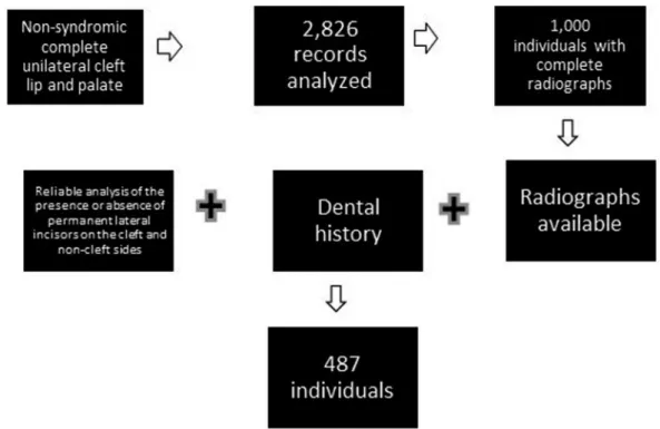
A single examiner reviewed 2,826 records of
individuals with non-syndromic complete unilateral cleft lip and palate, regularly registered in the institution. Among these individuals, we selected those whose records contained panoramic and periapical
radiographs from the first to the last up to 12 years of age, which led to a sample of 1,000 individuals.
Among these, an additional selection was performed to include only individuals whose radiographs available and dental history allowed reliable analysis of the presence or absence of permanent lateral incisors on both cleft and non-cleft sides. This led to a sample of
487 individuals who presented the permanent lateral
incisors on the cleft and non-cleft sides, who were
included in this study (Figure 1).
The authors did not find any study analyzing the
extraction of lateral incisors for orthodontic/dental
rehabilitation. For this reason, sample size calculation
could not be performed. Thus, after study completion, the post-hoc power of the study was calculated using
the following parameters: sample size 487 individuals/
percentage of extraction of lateral incisors in this
sample 76%/putative percentage of extraction of lateral incisors in the overall population 1%/alpha error of 0.05, which revealed a post-hoc power of 100%.
This study comprised retrospective analysis of such records, searching for information about maintenance of the permanent lateral incisor on the cleft and non-cleft sides for completion of dental rehabilitation.
In cases with indication of lateral incisor extraction,
achieved, in addition to the treatment indicated
for space closure in the cleft region. Secondarily,
the study also evaluated the association between maintenance of the lateral incisor on the cleft side and age at accomplishment of alveolar bone graft,
using the t test, at a significance level of p<0.05. The
other results were evaluated in percentages and by descriptive statistics.
Results
Among the 487 individuals previously selected who
presented the permanent lateral incisor on both cleft
and non-cleft sides, 265 individuals had not completed
the dental rehabilitation until the onset of this study,
62 individuals exhibited insufficient information on the
records, and 44 completed the treatment elsewhere. Thus, the other analyses were conducted on a sample
of 116 individuals with complete unilateral cleft lip
and palate, with presence of one permanent lateral incisor on both cleft and non-cleft sides, and dental rehabilitation concluded before the onset of this study.
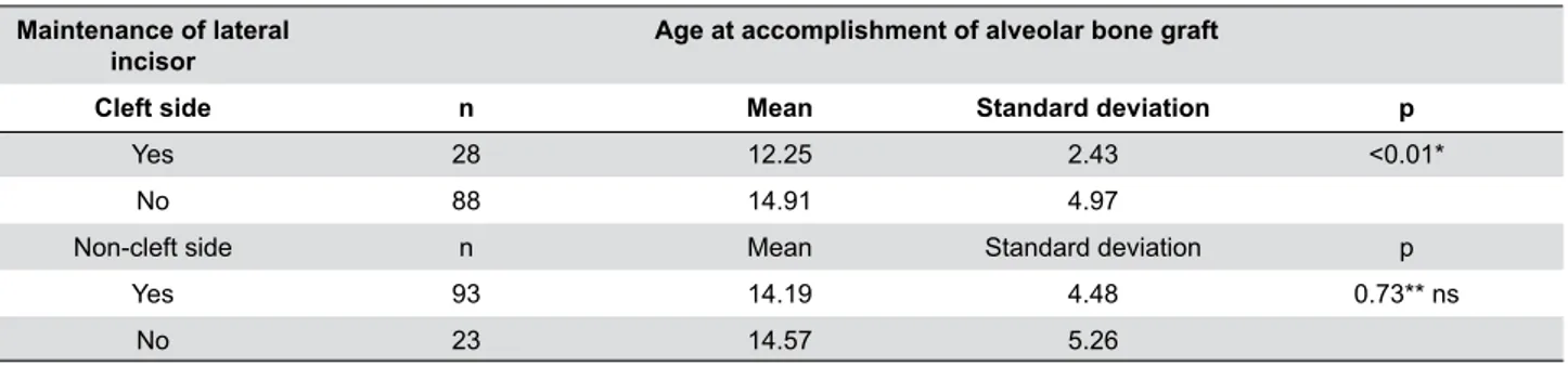
The lateral incisor on the cleft side was maintained
in 28 individuals (24.14%), while the lateral incisor on the non-cleft side was maintained in 93 individuals (80.17%).
The mean age at accomplishment of alveolar bone
graft was 14.30 years (SD=4.63), ranging from 8 to 30 years. Statistically significant association was found
between age at bone graft and maintenance of the
lateral incisor only for the cleft side (Table 1).
In individuals submitted to extractions, the
specialist indicating them was informed only on
the records of 26 individuals, with predominance of orthodontists (21), followed by maxillofacial surgeons (4) and prosthodontist (1). The reason for extraction indication was described in 16 cases (Table 2), including inadequate positioning (13), facilitation of orthodontic mechanics (1), lack of space in the dental arch (1) and lack of periodontal support (1). In cases
with extraction of the lateral incisor, the corresponding space was rehabilitated by orthodontic movement with
mesial movement of the canine (53), fixed prosthesis (17) and dental implant (10), without information
available for the other eight individuals.
The specialist who indicated the extraction of the lateral incisor on the non-cleft side was described
in the records of 13 individuals, all of which were
indicated by the orthodontist. The reason for extraction indication was described in nine cases, including
midline correction (4), inadequate positioning (3)
and achievement of space at the posterior region
(2). In cases of extraction of the lateral incisor, the
corresponding space was rehabilitated by orthodontic
movement with mesial movement of the canine (13) and fixed prosthesis (1), without information available
for the other nine individuals.
Discussion
The rehabilitation of individuals with complete unilateral cleft lip and palate is complex and involves several stages, including alveolar bone graft, which is fundamental to join the alveolar segments1. The main objectives of alveolar reconstruction are closure
of nasal fistula, unification of maxillary segments,
providing bone support for eruption of anterior teeth and for the nasal base, and allow prosthetic reconstruction including dental implants1,3,5.
Bone graft should be performed whenever possible to facilitate the eruption of teeth close to the cleft or orthodontic movement, either of the canine or lateral incisor8. Most studies consider the roots of canines to establish the timing for accomplishment of bone graft, considering one fourth and/or two thirds of its length4,12,16,21. However, recent studies demonstrated the possibility of alveolar bone graft in the deciduous
dentition, providing sufficient bone support for eruption
of central and lateral incisors with more favorable positioning of maxillary teeth20. Also, orthodontic
space closure may be possible in 100% of cases when
the bone graft is performed to facilitate the eruption of lateral incisor7,8. Additionally, individuals older
than 12 years are four times more likely to present
postoperative complications after alveolar bone graft13. Recently, the accomplishment of alveolar graft before eruption of the lateral incisor reduced the frequency of permanent canine impaction. This intervention does not change the risk, even though it was smaller in the group submitted to early bone graft6.
The eruption and adequate positioning of the permanent lateral incisor in the cleft area maintains the bone graft by mechanical stimulation, but also achieves stable functional occlusion, allowing normal maxillary growth and more harmonious facial and dental esthetics22.
In this study, the mean age at accomplishment of alveolar bone graft was 14.30 years (SD=4.63), ranging from 8 to 30 years. This may have been
influenced by the continental dimensions of the country and predominantly low socioeconomic status of the population assisted at the institution; and this may have contributed to the low percentage of maintenance of the lateral incisor on the cleft
side, as demonstrated by the statistically significant
association between maintenance of the lateral incisor and age at accomplishment of alveolar bone
graft (Table 1). However, this is the first study on
this subject conducted at the institution and the
Maintenance of lateral incisor
Age at accomplishment of alveolar bone graft
Cleft side n Mean Standard deviation p
Yes 28 12.25 2.43 <0.01*
No 88 14.91 4.97
Non-cleft side n Mean Standard deviation p
Yes 93 14.19 4.48 0.73** ns
No 23 14.57 5.26
* t test with Welch correction; ** t test
Table 1- Maintenance of lateral incisor according to the age at accomplishment of alveolar bone graft
Reasons for indication of extraction Cleft side Non-cleft side
Inadequate positioning 13 3
Facilitation of orthodontic movement 1
Lack of space in the dental arch 1 2
Lack of periodontal support 1
Midline correction 4
No information 72 14
Table 2- Reasons for the indication of lateral incisor extraction
Treatment Cleft side Non-cleft side
Mesial movement of canine 53 13 Dental prosthesis 17 1
Dental implant 10
-No information 8 9
findings should be carefully interpreted. Additionally,
the presence, position and root morphology of the lateral incisor should be carefully analyzed to assure the possibility of maintenance of this tooth18. Thus, ideally, the maxillofacial surgeon should evaluate both the individual and the radiographs before the mixed dentition stage to properly determine the better timing for bone graft, thus allowing more favorable positioning of the lateral incisor11.
Our results revealed indication of 17 prostheses and 10 implants, and the space corresponding to the
maxillary lateral incisor on the cleft side was closed
by orthodontic mesial movement in 53 individuals (Table 3). When necessary and possible, the mesial
movement of canine is favorable to reduce the utilization of prostheses and implants. Most lateral incisors on the non-cleft side were maintained. The accomplishment of bone graft before eruption of the incisors provides bone support for the eruption of these teeth, restoring the maxillary arch shape and enhancing their retention and gingival health. The burden of care may be reduced by minimizing the treatment stages and allowing earlier treatment completion, eliminating the need for rehabilitation with prostheses in adulthood11,17 and providing more favorable esthetic results by closing the space with a natural tooth. This aspect highlights the importance of bone graft and orthodontics in individuals with the lateral incisor.
Unfortunately, several records were incompletely filled by the different professionals treating the individuals in the institution. This is an inherent limitation of retrospective studies, especially those conducted in large institutions involving different specialists, such as in this case.
Based on these results, the authors concluded that
1) most of the lateral incisors (76%) adjacent to the cleft were extracted; 2) the main reason for extracting the lateral incisor was its inadequate position; 3) the
earlier the bone grafting procedure is accomplished, the greater are the chances of maintaining the
lateral incisor; and 4) the rehabilitation of the cleft
area in most of the cases was achieved by means of orthodontic space closure.
Thus, analyzing the maintenance of lateral incisor for dental rehabilitation of individuals with complete unilateral cleft lip and palate, we observed that evaluation for alveolar bone graft is mostly performed considering the canine, i.e. when the lateral incisor
is already erupted, thus missing the ideal timing of bone graft for this tooth, despite present and in good conditions. Considering the reports of success of secondary alveolar graft performed before eruption of the lateral incisor and the low rate of use of this tooth when bone graft is performed after eruption of the lateral incisor, as demonstrated in this study, the possibility of a slight anticipation in the timing of alveolar bone graft might be considered to increase the possibility of utilization of the lateral incisor. These
findings suggest the need to customize the timing
of orthodontic evaluation and alveolar bone graft in individuals with lateral incisor in the cleft area, considering the possibility of its eruption through the bone graft to increase the maintenance of the lateral incisor, when present, and reduce the burden of care for dental rehabilitation of individuals with cleft lip and palate.
Conclusion
The lateral incisor on the cleft side was not maintained in most individuals. There was positive relationship between extraction of the lateral incisor and age at accomplishment of the alveolar bone graft, suggesting the need to anticipate the initial radiographic evaluation to enhance its maintenance and reduce the steps required for rehabilitation.
Acknowledgement
The authors thank FAPESP for funding this study (research grant 2010/14481-2).
References
1- Boyne PJ, Sands NR.Combined orthodontic-surgical management of residual palato-alveolar cleft defects. Am J Orthod. 1976;70(1):20-37. 2- Freihofer HP, Borstlap WA, Kuijpers-Jagtman AM, Voorsmit RA, van Damme PA, Heidbüchel KL, et al. Timing and transplant materials for closure of alveolar clefts: a clinical comparison of 296 cases. J Cranio Maxillofac Surg. 1993;21(4):143-8.
3- Freitas JA, Almeida AL, Soares S, Neves LT, Garib DG, Trindade-Suedam IK, et al. Rehabilitative treatment of cleft lip and palate: experience of the Hospital for Rehabilitation of Craniofacial Anomalies/ USP (HRAC/USP) – Part 4: Oral Rehabilitation. J Appl Oral Sci. 2013;21(3):284-92.
5- Kearns G, Perrott DH, Sharma A, Kaban LB, Vargervik K. Placement of endosseous implants in grafted alveolar clefts. Cleft Palate Craniofac J. 1997;34(6):520-5.
6- Kleinpoort F, Ferchichi H, Belkhou A, Tramini P, Bigorre M, Captier G. Early secondary bone grafting in children with alveolar cleft does not
modify the risk of maxillary permanent canine impaction at the age of 10 years. J Craniomaxillofac Surg. 2017;45(4):515-9.
7- Lilja J. Alveolar bone grafting. Indian J Plast Surg. 2009;42 Suppl:S110-5.
8- Lilja J, Kalaaji A, Friede H, Elander A. Combined bone grafting and delayed closure of the hard palate in patients with unilateral cleft
lip and palate: facilitation of lateral incisor eruption and evaluation of indicators for timing of the procedure. Cleft Palate Craniofac J. 2000;37(1):98-105.
9- Luque-Martín E, Tobella-Camps ML, Rivera-Baró A. Alveolar graft in the cleft lip and palate patient: review of 104 cases. Med Oral Patol Oral Cir Bucal. 2014;19(5): e531-7.
10- Matic DB, Power SM. Evaluating the success of gingivoperiosteoplasty versus secondary bone grafting in patients with unilateral clefts. Plast Reconstr Surg. 2008;121(4):1343-53.
11- Miller LL, Kauffmann D, St John D, Wang D, Grant JH, Waite PD. Retrospective review of 99 patients with secondary alveolar cleft repair. J Oral Maxillofac Surg. 2010;68(6):1283-9.
12- Ochs MW. Alveolar cleft bone grafting (Part II): Secondary bone grafting. J Oral Maxillofac Surg. 1996;54(1):83-8.
13- Pessoa EA, Braune A, Casado PL, Tannure PN. Alveolar bone graft: clinical profile and risk factors for complications in oral cleft patients. Cleft Palate Craniofac J. 2017;54(5):530-4.
14- Precious DS. Alveolar bone grafting at 6 years of age. J Oral Maxillofac Surg. 2009;67(10):2045-53.
15- Rehrmann AH, Koberg WR, Koch H. Long-term postoperative results of primary and secondary bone grafting in complete clefts of the lip and palate. Cleft Palate J. 1970;7:206-21.
16- Seifeldin SA. Is alveolar cleft reconstruction still controversial? (Review of literature). Saudi Dent J. 2016;28(1):3-11.
17- Shashua D, Omnell ML. Radiographic determination of the position of the maxillary lateral incisor in the cleft alveolus and parameters for assessing its habilitation prospects. Cleft Palate Craniofac J. 2000;37(1):21-5.
18- Skoog T. The use of periosteal flaps in the repair of clefts of the primary palate. Cleft Palate J. 1965;2:332-9.
19- Talmant JC, Lumineau JP, Rousteau G. Cleft lip, maxilla and palate treatment by Dr. Talmant's team in Nantes. Ann Chir Plast Esthet. 2002;47(2):116-25.
20- Touzet-Roumazeille S, Vi-Fane B, Kadlub N, Genin M, Dissaux C, Raoul G, et al. Osseous and dental outcomes of primary gingivoperiosteoplasty with iliac bone graft: a radiological evaluation. J Craniomaxillofac Surg. 2015;43(6):950-5.
21- Trindade IK, Mazzottini R, Silva Filho OG, Trindade IE, Deboni MC. Long-term radiographic assessment of secondary alveolar bone grafting outcomes in patients with alveolar clefts. Oral Surg Oral Med Oral Pathol Oral Radiol Endod. 2005;100(3):271-7.