1N e u rology Clinical Division, Hospital das Clínicas, São Paulo University, Brazil (USP);2Beth Israel/Deaconess Medical Center, Harv a rd
Medical School, USA; 3Radiology Department, Hospital das Clínicas, USP.
Received 13 September 2005, received in final form 29 December 2005. Accepted 15 February 2006.
Dra. Adriana Bastos Conforto - Neurology Division, Hospital das Clínicas / USP - Av. Dr. Enéas de Carvalho Aguiar 255 / 5084 05403-000 São Paulo SP - Brasil. E-mail: abconf@usp.br
COMPARISON BETWEEN DIGITAL SUBTRACTION
ANGIOGRAPHY AND MAGNETIC RESONANCE
ANGIOGRAPHY IN INVESTIGATION OF NONLACUNAR
ISCHEMIC STROKE IN YOUNG PATIENTS
Preliminary results
Adriana Bastos Conforto
1, Felipe Fregni
2, Paulo Puglia Jr.
3, Claudia da Costa Leite
3,
Fabio Iuji Yamamoto
1, Karen F. Coracini
1, Milberto Scaff
1ABSTRACT - Purpose:We preliminarily investigated the relevance of perf o rming digital subtraction angiog-raphy (DSA) in addition to magnetic resonance angiogangiog-raphy (MRA) in definition of ischemic stroke etiol-ogy in young patients. Method:DSAs and MRAs from 17 young patients with nonlacunar ischemic stro k e were blindly analyzed and their impact on stroke management was evaluated. Results:Etiologies were the same considering results of either DSA or MRA in 12/17 cases. In 15/17 patients no changes would have been made in treatment, regardless of the modality of angiography considered. Conclusion:These pre-l i m i n a ry resupre-lts suggest that DSA may be redundant in two thirds of ischemic strokes in young patients. Further larger prospective studies are necessary to determine indications of DSA in this age group. KEY WORDS: TOAST, stroke etiology, embolism, arterial lesions, cryptogenic stroke.
Comparação entre arteriografia digital e angioressonância na investigação de acidente vascu-lar cerebral isquêmico não-lacunar em pacientes jovens: resultados preliminares
RESUMO -Propósito do estudo:Investigar de forma preliminar a relevância da realização de angiografia digital (AD) adicionalmente a angioressonância (AR) na definição de etiologias de acidente vascular cere-bral isquêmico (AVCI) em pacientes jovens. Método:ADs e ARs de 17 pacientes jovens com AVCIs não-l a c u n a res foram ananão-lisadas. Avanão-liamos o impacto destes exames no manejo cnão-línico dos casos. R e s u l t a d o s :
Em 12/17 casos, as etiologias dos AVCIs de acordo com os resultados de AD ou de AR foram idênticas. Em 15/17 pacientes, nenhuma mudança de conduta terapêutica seria realizada, independentemente da modali-dade de exame considerada. Conclusão:Estes resultados preliminares sugerem que os resultados da AD podem ser redundantes em relação à AR em até dois terços dos pacientes jovens com AVCI. Estudos pro s p e c-tivos maiores são necessários para otimizar o estabelecimento de indicações de AD nesta faixa etária. PA L AV R A S - C H AVE: T O A S T, etiologia de acidente vascular cerebral, embolia, lesões arteriais, criptogênico.
High incidences of stroke in young individuals ha-ve been re p o rted in deha-veloping countries1 - 3. In Brazil, it has been estimated that 10.6% of ischemic stro k e s a ffect patients aged 15-40 years2. In young patients, c ryptogenic strokes account for 8.3-55% of ischemic s t rokes worldwide, reflecting the challenge of deter-mining stroke mechanisms in this age gro u p1 - 1 1. While ethnical and regional lifestyle diff e rences are likely to influence incidence and etiological mechanisms o f
s t roke in the young in diff e rent populations, other factors such as use of strict criteria and extent of neu-rovascular investigation are equally important for d i a g n o s i s4 , 7 - 9. Given that therapeutic decisions depend on stroke etiology, accurate diagnosis is crucial for proper management of these patients.
re-ve focused predominantly on patients older than 45 years16-20or in lacunar strokes in younger patients21. T h e re f o re, it remains to be determined whether lim-itations in MRA accuracy and underperf o rmance of DSA have implications in definition of etiology of nonlacunar stroke in the young and in therapeutic decisions.
We have preliminarily investigated the clinical re l-evance of perf o rming DSA in addition to MRA in def-inition of stroke etiology and treatment in a Brazilian sample of patients with ischemic cere b rovascular dis-ease aged less than 45 years.
METHOD
We reviewed cases of ischemic stroke or transient ische-mic attack (TIA) consecutively re f e rred to our neuro l o g y ward. The study was part of a larger stroke database pro-tocol approved by the Ethics Committee of Hospital das Clínicas/São Paulo University. The study was re t ro s p e c t i v e and informed consent was considered unnecessary as long as patients were not identified. Inclusion criteria were: age less than 45 years; perf o rmance of intracranial MRA (iMRA), c e rvical MRA (cMRA) and DSA. Images that did not fulfil quality criteria were excluded. MRAs and DSAs were re v i e w-ed independently by two senior neuroradiologists (PP and CCL). Both were blind to clinical data and lesional topogra-phies in cranial magnetic resonance imaging (MRI).
S t a n d a rd imaging protocols for our institution were used. iMRAs were perf o rmed with a 1.5 T system with unen-hanced 3D time-of-flight (TOF) multiple overlapping thin-slab acquisition (MOTSA) sequences, three-dimensional ma-ximal intensity projection (MIP) images (TR/TE 33.3/3 ms, flip angle 20º, thickness=0.8 mm, matrix 256x192, field of view from 13x13 to 22x22 cm, 0 mm interval). cMRAs were p e rf o rmed with contrast-enhanced 3D TOF MOTSA sequen-ces with MIP images (TR/TE 7-23/4.4 ms, flip angle 45-50º, thickness=2.6 mm, matrix 256x160, field of view 26x26 cm, 0 mm interval) and a sm art preparation technique. Intra-a rteriIntra-al 4-vessel selective DSA wIntra-as perf o rmed viIntra-a the femo-ral art e ry, starting with imaging of the aortic arch followed by selective injections of contrast material into both caro t i d and vertebral arteries. DSA (GE Medical Systems) was per-f o rmed in the antero p o s t e r i o r, lateral and oblique pro j e c-tions. Non-ionic contrast of low osmolarity (Iopamiro n®, Schering) was administered with a power injector at a rate of 6 mL/s in the common carotid and subclavian art e r i e s (volume: 8 mL) and a rate of 4 mL/s in the internal carotid and vertebral arteries (6 mL). Hydrophilic coated guide
wi-fied as: normal/stenosis50%; stenosis >50%; occlusion. Final diagnos es were defined based on characteri stics of lesions and overall findings. Signs considered suggestive of embolism were: carotid “T” or basilar tip occlusion; abru p t and isolated occlusion of an intracranial arterial branch in the absence of signs of progressive disease in other arter-ies. Sources of emboli in proximal vessels were evaluated. All of the patients underwent clinical and neuro l o g i c a l evaluation, electro c a rdiogram, hemogram, blood biochem-i s t ry, lbiochem-ipbiochem-id profbiochem-ile, ery t h rocyte sedbiochem-imentatbiochem-ion rate, biochem- immuno-logic and coagulation testing, cranial computed tomogra-phy and MRI. Other investigations included transesophageal e c h o c a rdiogram (TEE) (16), duplex ultrasonography of extra-cranial vessels (15), transthoracic echocardiogram (TTE) (7), s e rology for syphilis and Chagas' disease (15), cere b ro s p i n a l fluid analysis (16), transcranial Doppler (8), Holter monito-ring (7). The only patient who underwent TTE but not TEE had a positive serology for Chagas' disease and evidence of cardiomyopathy on TTE. Chagas' disease is caused by Trypanosoma cruziinfection, endemic in some South Ame-rican re g i o n s2 2. Two experienced neurologists (MS and FY) defined by consensus the most likely etiological diagnosis a c c o rding to TOAST criteria2 3: larg e - a rt e ryathero s c l e ro s i s , c a rdioembolism, small-vessel occlusion, stroke of other d e t e rmined etiology and st roke of undetermined etiolo-g y. In addition, when seroloetiolo-gy for Chaetiolo-gas' disease was pos-itive and there was evidence of card i o m y o p a t h y, the diag-nosis was cardioembolism. Results of DS A and MRA were given for each case; neurologists were blind to the modal-ity of angiography and the order of DSA and MRA results was randomized. Treatment of each patient (antiplatelet d rugs/ anticoagulants/ other) was defined, considering clin-ical and laboratorial investigation as well as results of MRA or DSA. We evaluated agreement between etiology of stro-ke and agreement between treatment, considering re s u l t s of either exam.
Given the small sample size of this study (17 patients), this study was underpowered for perf o rmance of statisti-cal tests. There f o re, the two methods of neuro i m a g i n g were compared in a descriptive way.
RESULTS
not evaluated. There f o re, iMRAs, cMRAs and DSAs f rom 17 patients aged 17-44 years (mean±s t a n d a rd deviation, 33±9 years), 8 male, were analyzed.
All DSAs and 12/17 MRAs revealed abnorm a l i t i e s . Clinical features, affected arterial territories, DSA and MRA results as well as diagnosis according to TOAST criteria considering either DSA or MRA results are shown in the Table. None of the infarcts was lacunar. The most frequently affected artery was the middle c e rebral art e ry (MCA) (8/17 in both MRA and DSA) ( Table). MRA and DSA final diagnoses were
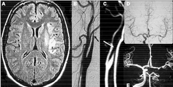
complete-ly concordant in oncomplete-ly 4 patients (Table, patients 1, 2, 4, 5). Fig 1 (A-E) shows images from patient 1. Inter-vals between examinations were (mean±s t a n d a rd deviation): iMRA and DSA, 3.5±5.6 days, cMRA and DSA, 1.4±7.1 days.
MRA had lower resolution than DSA for detec-tion of arterial branch lesions (Patients 6-8, 10-13, 1 5 ) . Fig 2 shows images from case 15. Also, MRA did not reveal a posterior cerebellar inferior art e ry (PICA) le-sion (patient 3), a lele-sion of fibromuscular dysplasia (patient 14) and 2 arterial dissections (patients 16 Fig 1. Patient 1, a 26yearold male, had sudden onset of headache, nausea, right hemiparesis and dysarthria after sexual inter -course. (A) Fluid attenuated inversion-re c o v e ry(FLAIR) sequence in cranial MRI showed an infarction involving the terr i t o ry of left lenticulostriate and insular MCA branches. (B) DSA demonstrated left cervical ICA segmental stenosis (arrow) suggestive of arte rial dissection and left intracavernous ICA occlusion. (D) left ACA and MCA were filled by the Willis circle. (C, E) MRA disclosed sim -ilar findings.
and 17) (Table). In case 9, DSA disclosed left VA dis-section and MRA, left MCA lesions but the patient had a right striatocapsular infarction. There f o re, DSA probably showed a false positive result in this case. In patients 3, 6-8 and 10-13, DSA revealed focal lesions suggestive of embolism in arteries supplying the infarcted are a3 , 6 , 7 , 1 2. Yet, emboligenic sources were only detected in patients 11 and 12.
Etiologies according to TOAST criteria were the same considering results of either DSA or MRA in 12/ 17 cases (Table, patients 1 to 12) and were different in 5/17 cases (Table, patients 13-17). In patient 14, diagnosis was larg e - a rt e ry athero s c l e rosis based on MRA findings but DSA revealed fibromuscular dys-plasia. In patient 13, the diagnosis was larg e - a rt e ry a t h e ro s c l e rosis considering the MRA finding of MCA stenosis but was undetermined considering the ab-sence of large-artery stenosis and the preab-sence of a lesion suggestive of embolism in DSA. In 9/17 cases (patients 5-10, 15-17) etiological diagnosis was unde-termined considering MRA results. In 3 out of these 9 cases, DSA contributed significantly to definition of stroke etiology: vasculitis (patient 15) and arteri-al dissection (patients 16 and 17). In the re m a i n i n g cases, etiological diagnosis remained undeterm i n e d in spite of DSA results.
N e u rologists chose diff e rent treatments in patients 16 and 17 only (anticoagulation based on DSA re s u l t s , antiplatelet drugs based on MRA results). In all oth-er 15 cases, no changes would have been made in t reatment, re g a rdless of the modality of angiogra-phy evaluated.
DISCUSSION
To our knowledge, this is the first study to dire c t-ly compare MRA and DSA findings in nonlacunar ischemic stroke in young adults. Concordance bet-ween final angiographic diagnoses obtained by dif-f e rent imaging modalities was low. On the other hand, concordance between stroke etiologies accord-ing to clinical and imagaccord-ing criteria was high, as well as concordance between therapeutic decisions based on results of either MRA or DSA.
DSA identified abnormalities in at least one art e ry in all patients and MRA, in more than two thirds of the patients. It has been re p o rted that lacunar stro k e s are highly predictive of normal DSAs in young pati-ents with ischemic stro k e2 1. None of the strokes in our series was lacunar (Table) and this is a likely re a-son to explain the high rate of abnormal DSAs in the present series.
Occlusion suggestive of embolism has been des-cribed as the most frequent DSA abnormality in young stroke patients1 2 , 1 4. In this series, embolism to intracranial arteries may have been an important eti-ology of stroke. First, the most commonly aff e c t e d artery was the MCA, an usual target for emboli. Se-cond, DSA revealed lesions suggestive of embo-l i s m3 , 6 , 7 , 1 2in arterial branches in 8 cases (Patients 3, 6-8 and 10-13). In 4 of these cases the lesions were not detected in MRA. In addition, in patient 15 lesions in c o rtical areas were demonstrated by DSA only. These findings may be explained by lower MRA re s o l u t i o n for visualization of terminal carotid branches and intracranial vessels24.
The detection of intracranial branch arterial lesi-ons in DSA in the absence of identifiable sources of embolism in the present series supports the view that underdiagnosed embolism may be partially respon-sible for a higher rate of cryptogenic strokes in youn-ger individuals. Other authors have described angio-graphic findings suggestive of embolism in the absen-ce of identifiable sourabsen-ces in stroke of undeterm i n e d c a u s e6 , 2 5. Some thrombi may lyse from their sourc e s or may be too small to be detected by current tools of investigation26,27. Therefore, quoting L.R. Caplan, it is possible that DSA may have revealed the “bird s ” while their “nests” remained unknown in some of the presented cases28.
H o w e v e r, in order to consider embolism as the etiology of an infarction, it is crucial to identify an emboligenic source. In patients 11 and 12, emboli-genic sources were determined and stroke etiology would be cardiac embolism re g a rdless of the modal-ity of angiography considered. In 5 of 8 patients ( Table, cases 6-8, 10 and 13), DSA revealed lesions suggestive of embolism but no emboligenic sources w e re found and causes of stroke remained undeter-mined. These results indicate that diff e rences in MRA and DSA sensitivity for detection of lesions sugges-tive of embolism did not substantially influence the final etiological diagnosis in these patients.
e-the risk of neurologic complications reported in e-the l i t e r a t u re3 1 , 3 2: Wilinsky and colleagues re p o rted 1.3% n e u rologic complications in 2,899 cerebral angiogra-p h i e s1 5. Complications were transient in 0.2%, re v e r-sible in 0.7% and permanent in 0.5% of the cases. Age greater than 55 years, presence of cardiovascu-lar disease and fluoroscopic times of 10 minutes sig-nificantly increased risk associated with DSA. It is still a matter of controversy whether ischemic stroke in-c reases DSA risk, in-compared to other in-conditions suin-ch as subarachnoid hemorrhage15.
DSA may be safer in younger patients without dif-fuse vasculopathy than in older individuals1 2 , 1 5. Howe-v e r, risk-benefit ratios should be considered and inHowe-va- inva-sive pro c e d u res should be avoided whenever addi-tional information does not substantially contribute to definition of diagnosis and treatment. There f o re , DSA in stroke in the young must be more clearly defi-ned. These pre l i m i n a ryfindings emphasize the need to enhance identification of sources of embolism and the importance of an integrated clinical/radiological a p p roach. Due to small sample size of this study, we performed a descriptive analysis of the data. There-f o re, we do not know iThere-f the results oThere-f this study may be externally generalized. However, the findings of this study encourage future prospective studies invol-ving larger number of patients to determine in which cases DSA is more likely to have a relevant impact on definition of ischemic stroke etiology and tre a t m e n t in young patients.
REFERENCES
1. Radhakrishnan K, Ashok PP, Sridharan R, Mousa ME. Stroke in the young: incidence and pattern in Benghazi, Lybia. Acta Neurol Scand 1986;73:434-438.
2. Siqueira JI Neto, Santos AC, Fabio SR, Sakamoto AC. Cerebral infarc-tion in patients aged 15 to 40 years. Stroke 1996;27:2016-2019. 3. Awada A. Stroke in Saudi Arabian young adults: a study of 120 cases.
Acta Neurol Scand 1994;89:323-328.
4. Adams HP J r., Kappelle LJ, Biller J, et al. Ischemic stroke in young adults: experience in 329 patients enrolled in the Iowa Registry of stro k e in young adults. Arch Neurol 1995;52:491-495.
5. Bogousslavsky J, Pierre P. Ischemic stroke in patients under age 45. Neurol Clin 1992;10:113-124.
6. Kristensen B, Malm J, Carlberg B, et al. Epidemiology and etiology of ischemic stroke in young adults aged 18 to 44 years in northern Sweden. Stroke 1997;28:1702-1709.
7. Jacobs BS, Boden-Albala B, Lin IF, Sacco RL. Stroke in the young in the northern Manhattan stroke study. Stroke 2002;33:2789-2793.
12. Smoker WR, Biller J, Hingtgen WL, Adams HP J r., To ffol GJ. A n g i o-graphy of nonhemorrhagic cerebral infarction in young adults. Stro k e 1987;18:708-711.
13. Stillman MJ, Ronthal M, Kleefield J, et al. Cerebral infarction: short-comings of angiography in the evaluation of intracranial cere b ro v a s-cular disease in 25 cases. Medicine (Baltimore) 1987;66:297-308. 14. Lisovoski F, Rousseaux P. Cerebral infarction in young people: a study
of 148 patients with early cerebral angiography. J Neurol Neuro s u rg Psychiatry 1991;54:576-579.
15. Willinsky RA, Taylor SM, Te r B rugge K, Farb RI, Tomlinson G, Montanera W. Neurologic complications of cerebral angiography: p rospective analysis of 2,899 pro c e d u res and review of the literature . Radiology 2003;227:522-528.
16. Cosottini M, Pingitore A, Puglioli M, et al. Contrastenhanced thre e -dimensional magnetic resonance angiography of athero s c l e rotic inter-nal carotid stenosis as the noninvasive imaging modality in re v a s c u-larization decision making. Stroke 2003;34:660-664.
17. Phan T, Huston J, 3rd, Bernstein MA, Riederer SJ, Brown RD Jr. Con-trast-enhanced magnetic resonance angiography of the cervical ves-sels: experience with 422 patients. Stroke 2001;32:2282-2286. 18. Nederkoorn PJ, van der Graaf Y, Hunink MG. Duplex ultrasound and
magnetic resonance angiography compared with digital subtraction angiography in carotid artery stenosis: a systematic re v i e w. Stro k e 2003;34:1324-1332.
19. Korogi Y, Takahashi M, Mabuchi N, et al. Intracranial vascular steno-sis and occlusion: diagnostic accuracy of three-dimensional, Fourier transform, time-of-flight MR angiography. Radiology 1994;193:187-193. 20. Willinek WA, von Falkenhausen M, Born M, et al. Noninvasive detection of stenoocclusive disease of the supraaortic arteries with thre e -dimensional contrast-enhanced magnetic resonance angiography: a p rospective, intra-individual comparative analysis with digital sub-traction angiography. Stroke 2005;36:38-43.
21. De Jong S, Lodder S, Luijckx GJ. Is cerebral angiography redundant in undetermined cause of stroke in patients below 50 years when the stroke is lacunar? J Neurol Sci 2004;222:83-85.
22. C a rod-Artal FJ, Va rgas A P, Melo M, Horan TA. American trypanoso-miasis (Chagas' disease): an unrecognised cause of stroke. J Neuro l Neurosurg Psychiatry 2003;74:516-518.
23. Adams HP, Jr., Bendixen BH, Kappelle LJ, et al. Classification of sub-type of acute ischemic stroke: definitions for use in a multicenter clin-ical trial. TO A S T. Trial of Org 10172 in Acute Stroke Treatment. Stro k e 1993;24:35-41.
24. Wutke R, Lang W, Fellner C, et al. High-resolution, contrast-enhanced magnetic resonance angiography with elliptical centric k-space ord e r-ing of supra-aortic arteries compared with selective X-ray angiogra-phy. Stroke 2002;33:1522-1529.
25. Sacco RL, Ellenberg JH, Mohr JP, et al. Infarcts of undetermined cause: the NINCDS Stroke Data Bank. Ann Neurol 1989;25:382-390. 26. Warach S, Li W, Ronthal M, Edelman RR. Acute cerebral ischemia:
eval-uation with dynamic contrast-enhanced MR imaging and MR angiog-raphy. Radiology 1992;182:41-47.
27. Babikian VL, Caplan LR. Brain embolism is a dynamic process with variable characteristics. Neurology 2000;54:797-801.
28. Caplan LR. Of birds and nests and brain emboli. Rev Neurol (Paris) 1991;147:265-273.
29. Khan R, Smith JK, Castello M. False-negative contrast MRA in the set-ting of carotid artery dissection. Emerg Radiol 2002;9:320-322. 30. Ozdoba C, Sturzenegger M, Schroth G. Internal carotid artery
dissec-tion: MR imaging features and clinical-radiologic corre l a t i o n . Neuroradiology 1996;199:191-198.
31. Melaragno R. Lateral syndrome of the medulla oblongata (Wa l l e n b e rg ´ s s y n d rome) as a complication of a vertebral angiography: a case re p o r t . Arq Neuropsiquiatr 1972;30:78-83.