Determining shapes and dimensions of dental arches for
the use of straight-wire arches in lingual technique
Silvana Allegrini Kairalla1, Giuseppe Scuzzo2, Tarcila Triviño3, Leandro Velasco4, Luca Lombardo5, Luiz Renato Paranhos6
Introduction: This study aims to determine the shape and dimension of dental arches from a lingual perspective, and
determine shape and size of a straight archwire used for lingual Orthodontics. Methods: The study sample comprised 70
Caucasian Brazilian individuals with normal occlusion and at least four of Andrew’s six keys. Maxillary and mandibular dental casts were digitized (3D) and the images were analyzed by Delcam Power SHAPET 2010 software. Landmarks on the lingual surface of teeth were selected and 14 measurements were calculated to determine the shape and size of dental arches. Results: Shapiro-Wilk test determined small arch shape by means of 25th percentile (P25%) — an average
per-centile for the medium arch; and a large one determined by means of 75th percentile (P75%). T-test revealed differences
between males and females in the size of 12 dental arches. Conclusion: The straight-wire arch shape used in the lingual
straight wire technique is a parabolic-shaped arch, slightly flattened on its anterior portion. Due to similarity among dental arch sizes shown by males and females, a more simplified diagram chart was designed.
Keywords:Dental arch. Orthodontics. Orthodontic appliance design.
How to cite this article: Kairalla SA, Scuzzo G, Triviño T, Velasco L, Lombar-do L, Paranhos LR. Determining shapes and dimensions of dental arches for the use of straight-wire arches in lingual technique. Dental Press J Orthod. 2014 Sept-Oct;19(5):116-22. DOI: http://dx.doi.org/10.1590/2176-9451.19.5.116-122.oar
Submitted: August 04, 2013 - Revised and accepted: November 30, 2013
Contact address: Silvana Allegrini Kairalla
Rua Diogo Moreira 132 cj. 201/202 Pinheiros - Cep: 05423-010 São Paulo - SP — Brazil
E-mail: [email protected]
1 MSc in Dentistry, Methodist University of São Paulo (UMESP). 2 MSc in Dentistry, University of Ferrara (UNIFE).
3 Phd in Orthodontics, University of São Paulo (USP).
4 PhD resident in Orthodontics, School of Dentistry, São Leopoldo Mandic. 5 Assistant professor, UNIFE.
6 Adjunct professor, Federal University of Sergipe (UFS).
DOI: http://dx.doi.org/10.1590/2176-9451.19.5.116-122.oar
» The authors report no commercial, proprietary or financial interest in the products or companies described in this article.
Introdução: esse estudo objetiva encontrar a forma e dimensão de arcadas dentária para definir a forma de um arco contínuo
que possa ser utilizado na técnica lingual. Métodos: a amostra foi composta por indivíduos brasileiros, leucodermas, com
oclusão normal natural, que apresentaram, no mínimo, quatro das seis chaves de oclusão de Andrews. Os modelos das arcadas dentárias superior e inferior foram digitalizados (3D) e as imagens exportadas para o software Delcam Power SHAPETM 2010.
Foram selecionados pontos nas superfícies linguais dos dentes e traçadas 14 medidas para determinar a forma e a dimensão da arcada dentária. Resultados: o teste de Shapiro-Wilk possibilitou definir uma forma de arcada pequena utilizando o percentil
25% (P25%), uma arcada média (P50%) e uma forma de arcada grande, pelo percentil 75% (P75%). O teste t de Student
comparou se houve uma diferença entre os sexos, e foram encontrados 12 tamanhos de arcadas dentárias. Conclusões: a partir
dos resultados obtidos foi possível definir uma forma de arco contínuo para ser utilizado na técnica lingual Straight Wire (LSW): parábola levemente achatada na região anterior. Devido a similaridade entre alguns tamanhos de arcadas dentárias, encontrados pelo dimorfismo sexual, pôde ser elaborado um diagrama de arcadas de maneira mais simplificada.
INTRODUCTION
Lingual Orthodontics was developed by the end of the 70's with the bonding of conventional
appli-ances on the lingual surface of teeth.1,2 The first study
describing brackets and lingual arch shape was
pub-lished by Fujita.3
There are important diferences between lingual and
buccal Orthodontics4 in terms of arch design,5 but only
a few studies6-9 have determined the dental arch form for
the irst. There are many confounding factors on
mea-suring intercanine distances10 which hinder clinicians
from determining the size of mushroom-shaped lingual
arches.3 In an attempt to simplify this technique,
Take-moto and Scuzzo11 introduced the straight wire concept
in Lingual Orthodontics and Kyung et al12 proposed the
positioning of brackets with auxiliary blades in order to allow the use of archwires without curvatures.
Scuzzo et al10 developed the system STb Light
Lingual Straigth Wire® while other authors13 found
a more square-shaped archwire, enabling the use of continuous lingual arches (LSW). Due to lack of studies on the subject, the demand of patients for more esthetic treatments and the need to simplify the lingual technique, this study aimed to determine the shape and size of dental arches evaluated from the lin-gual surface, in order to determine the shape and size of continuous lingual arch wires.
MATERIAL AND METHODS
This analytical observational study used records of patients from the School of Health, UMESP / São Ber-nardo do Campo.
The sample comprised maxillary and mandibular dental casts of 70 Caucasian Brazilian individu-als (28 men and 42 women) with an average age of 16.4 years, all of which had natural normal occlusion with at least four of the six keys to normal
occlu-sion.14 The first item of the first key was considered
essential for sample selection(Angle Class I molar re-lationship). Another inclusion criterion was that in-dividuals should be at least 15 years of age with no odontogenic abnormalities and all permanent teeth in occlusion, except for third molars.
The 70 pairs of cast models were digitized with a
In order to standardize the position of models,
landmarks were set on canines and molars cusps15 so
as to create a trapezoid. Additionally, a coordinate
grid was established (x, y and z).16 The models were
kept on the three planes: vertical, horizontal and sagittal, which allowed their rotation in numerous positions and measurements to be kept proportional in all models of the sample, thereby proving the method accurate.
Determining landmarks, shape and size of the arch
Landmarks were established on the lingual
sur-face of teeth with Delcam Power SHAPE™ 2010
soft-ware. They represented the bonding site for the
brackets on the lingual surface of teeth.13
Landmarks were deined on the lingual surface along the long axis of all upper and lower, anterior and poste-rior teeth. They were determined on the middle of the clinical crown of posterior teeth (premolars and molars), whereas on anterior teeth they were determined close to a line dividing the middle third from the gingival third of the clinical crown, in both maxillary and mandib-ular arches. Digitized casts were rotated on computer screen in order to bring the lingual surface of each tooth aligned with the frontal view of the operator who deter-mined and located the landmarks. Subsequently, land-marks were connected so as to deine the curvature and
shape of the lingual arch.17
In order to determine the size and shape of
den-tal arches,14 measures were obtained15 by means of
Delcam Power SHAPE™ 2010 software, and tabulated
in EXCEL (Microsoft™, Redmond, Wash, USA). The
software memorizes the landmarks previously ob-tained and set (key point).
Out of the 14 linear measurements, ten were horizontal and four were vertical. Horizontal lines were defined from the landmark at each tooth on the lingual surface (key point) to the Y axis. Likewise, the lines on the right side, both in maxilla and man-dible arcades, were determined.
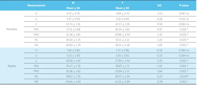
Table 1 - Comparison between mandibular and maxillary arches obtained for males and females.
* - statistically significant difference (P < 0.05). ns - statistically insignificant difference. The lines or horizontal and vertical distanc-es were exprdistanc-essed in millimeters with accuracy of 6 digits after the decimal point, thereby indicating satisfactory precision.
Data analysis
To assess intra-examiner error, 30% of the sam-ple was randomly selected, i.e., 21 pairs of cast mod-els 40 days after the first measurement. Student’s t-test assessed systematic error, with significance level set at 5%. Casual error was calculated
accord-ing to Dahlberg's formula: Error = √∑d2/2n, with
d = difference between the first and second measure-ments and n = number of repetitions. Systematic er-ror results were evaluated by a paired t-test.
Data is presented in tables and graphs as mean, standard deviation, minimum, median and
maxi-mum range, 25th percentile, 50th and 75th percentile,
respectively. Shapiro-Wilk test was used to check
data normality. All measurements met the normality criterion. A possible diference between males and fe-males was assessed by Student’s t-test. Measurements were determined as follows: For the medium arch,
mean values were used (P50th); for the small arch,
the 25th percentile was used (P25th), and for the large
arch, the 75th percentile (P75th) was used. For all
statis-tical tests, signiicance level was set at 5% (P < 0.05).
RESULTS
The sample consisted of 40% of males and 60% of females. Table I illustrates the diference between the mean value for male and female patients. Signiicant diferences were found for some measurements: The horizontal line of premolars and molars in the mandi-ble arch, and the values of canine, premolars and molar of the maxillary arch. Due to these diferences, other two tables were prepared to show the measurements of males (Table 2) and females (Table 3) individuals.
Measurement M F Dif. P value
Mean ± SD Mean ± SD
Mandible
CI 4.72 ± 0.70 4.60 ± 0.70 -0.12 0.497 ns
LI 3.37 ± 0.56 3.19 ± 0.49 -0.18 0.152 ns
C 22.74 ± 1.16 22.17 ± 1.28 -0.58 0.060 ns
PM1 27.51 ± 1.68 26.54 ± 1.60 -0.97 0.018 *
PM2 31.28 ± 1.65 29.96 ± 2.04 -1.32 0.006 *
M1 34.30 ± 1.75 33.11 ± 2.21 -1.19 0.020 *
M2 40.83 ± 1.79 39.14 ± 2.26 -1.69 0.001 *
Maxilla
CI 7.56 ± 0.85 7.37 ± 0.96 -0.19 0.390 ns
LI 5.23 ± 1.06 5.00 ± 0.61 -0.23 0.264 ns
C 29.28 ± 1.42 27.99 ± 1.54 -1.30 0.001 *
PM1 30.27 ± 1.76 28.87 ± 1.71 -1.40 0.001 *
PM2 35.58 ± 1.62 33.94 ± 2.17 -1.64 0.001 *
M1 38.67 ± 1.79 36.47 ± 2.34 -2.20 <0.001*
Table 2 - Measurements for male individuals.
Measurement Mean ± SD Median Minimum Maximum P25% P75%
Mandible
CI 4.7 ± 0.7 4.7 3.5 6.2 4.2 5.2
LI 3.4 ± 0.6 3.3 2.3 4.5 3.0 3.8
C 22.7 ± 1.2 22.7 19.7 25.0 22.1 23.7
PM1 27.5 ± 1.7 27.4 24.7 31.2 26.2 28.4
PM2 31.3 ± 1.7 31.2 27.8 34.6 30.0 32.8
M1 34.3 ± 1.8 34.2 31.3 37.9 32.7 35.8
M2 40.8 ± 1.8 41.3 36.5 43.4 39.2 42.2
Maxilla
CI 7.6 ± 0.8 7.6 5.6 9.1 7.1 8.1
LI 5.2 ± 1.1 5.1 3.9 9.3 4.5 5.7
C 29.3 ± 1.4 29.2 26.2 32.0 28.5 30.4
PM1 30.3 ± 1.8 30.2 27.0 35.1 29.2 31.2
PM2 35.6 ± 1.6 35.7 32.7 39.5 34.3 36.7
M1 38.7 ± 1.8 38.3 35.5 42.6 37.4 40.0
M2 43.6 ± 2.4 43.4 37.0 48.0 42.2 45.2
Table 3 - Measurements for female individuals.
Measurement Mean ± SD Median Minimum Maximum P25% P75%
Mandible
CI 4.6 ± 0.7 4.5 3.2 6.4 4.1 4.9
LI 3.2 ± 0.5 3.1 2.2 4.3 2.9 3.6
C 22.2 ± 1.3 22.0 19.9 25.0 21.2 23.0
PM1 26.5 ± 1.6 26.4 22.9 29.3 25.4 27.6
PM2 30.0 ± 2.0 29.6 24.9 34.0 28.8 31.7
M1 33.1 ± 2.2 33.0 28.8 37.4 31.9 34.7
M2 39.1 ± 2.3 38.8 35.3 44.6 37.6 40.5
Maxilla
CI 6.2 ± 0.8 6.1 4.8 8.3 5.6 6.7
LI 6.2 ± 0.7 6.1 4.9 7.9 5.6 6.6
C 28.0 ± 1.5 28.1 24.9 30.8 27.1 29.1
PM1 28.9 ± 1.7 28.6 25.7 32.4 27.6 29.9
PM2 33.9 ± 2.2 33.7 30.1 39.4 32.4 35.5
M1 36.5 ± 2.3 36.0 33.1 42.4 34.5 38.2
M2 41.4 ± 2.7 41.9 36.5 49.8 39.1 43.1
The first two lines refer to vertical measurements while the other lines show the horizontal
measure-ments. The mean 50th percentile is more reliable
than the median, and was used to determine the
medium size of the arches. P25th means that 25%
of the sample comprises small-sized arches, thereby
determining the size of small arches; whereas P75th
means that 25% of the sample comprises larger arches, thereby determining the size of larger arches.
Determining continuous lingual arches
Data obtained with statistics analysis were
import-ed into Delcam Power SHAPE™ 2010 sotware. Values
of P25th, means and P75th determined the shape and size
Figure 1 - Measurements of mandibular arch according to sex.
Figure 3 - Final outline of lingual arch.
Figure 4 - Sequence of continuous lingual arches (S, M, L) of male individuals.
Figure 2 - Measurements of maxillary arch according to sex. Figure 5 - Sequence of continuous lingual arches (S, M, L) of female individuals.
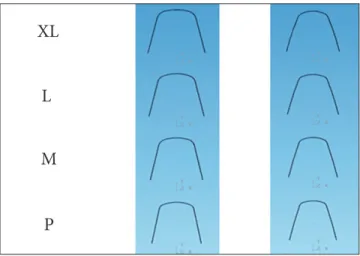
Thus, 12 different sizes (small, medium and large) of continuous maxillary and mandibular lin-gual arches were determined for female and male pa-tients, as shown in Figures 4 and 5.
DISCUSSION
In the literature6,7,9,18,19,20,25 there are several studies
in which diferent methods were used to obtain den-tal arch shape. The sample of 3D digitized images of cast models used in this study was also used by other
authors.16,21-24 The advantage of working with a 3D
digitized model is that it can be seen at the same time in three dimensions (horizontal, sagittal, and vertical), thereby yielding proportional results for all models.
Similarly to some authors16 and differently
from other studies that used only two coordinates
(x and y),13,19 the present study used x, y, and z axes
with a view to establishing landmarks, since the use M-mean
M-P25% M-P75% F-mean F-P25% F-P75%
30 20 10 0 -10 -20 -30
M-mean M-P25% M-P75% F-média F-P25% F-P75%
of two coordinates does not allow movement of mod-els due to lack of a third axis, the axis z — vertical.
Several authors use the cusp tips to determine the
shape of the arches,15,17,20 whereas others use the
ves-tibular middle points of the dental crown of anterior
and posterior teeth19 as well as lingual and occlusal
landmarks on the long axis of the teeth as reference.7
Lombardo et al13 used landmarks on the lingual
sur-face and selected points closer to the gingival third. Even though there are several ways to determine the shape of dental arches, this study was based on
Lom-bardo et al13 who advocated landmarks to be closer
to the cervical region of teeth, since it is the place where the difference between the lingual surfaces of
canines and premolars are smaller.10
To determine the configuration and size of
den-tal arches, the literature13,18,19,23 has used polynomial
functions or linear measurements.15,20,21,25 In this
study, linear measurements were used, given that
Del-cam Power SHAPE™ 2010 software provides accuracy of 6 digits after the decimal point. This accuracy was confirmed by Shapiro-Wilk test, which showed that all measurements met the criterion of normality.
Normality of data enabled comparison between males and females by means of Student’s t-test. This difference can be seen in Table 1. The litera-ture has not found differences between males and
females19,23,24,26, even though the sizes of male arches
are larger than those of female patients
anthropo-logically speaking.16 According to Lombardo et al,13
who did not find differences between males and fe-males, this probably occurs due to landmarks used on the lingual surface of teeth, since differences in vestibular-lingual diameters of teeth were not con-sidered, especially of first molars which have dif-ferent sizes between males and females. The pres-ent study also used landmarks on the lingual sur-face, but detected differences in arch shape between males and females, corroborating data obtained by
Ferrario et al16 and assigning sexual dimorphism to
the adopted measurements. A total of 14 linear mea-sures were taken — all within the normality
crite-rion — differently from Lombardo et al13 who used
only six linear measurements. Due to the abnormal
Accuracy of software measurements associated with the fact that the mirror method of the arch was
not previously used, as observed in some studies,13,19
allowed us to verify whether data had normal distri-bution for both male and female individuals.
Lom-bardo et al13 described and applied median
measure-ments different from the mean used in the present study. Similarly to this study, data were found to be statistically normal, as it used mean and not median to obtain the final measurements. Means are more ac-curate than medians and allow us to define
measure-ments of a medium-sized arch (50th percentile). The
small arch was determined by the minimum measures (25% percentile) while the large arch was determined by the maximum measures (75% percentile).
Figures 1 and 2 illustrate small, medium and large arches for female and male individuals. They also show the shapes for the mandible and maxilla dental arch, similar to a parabola-shaped arch slightly flat-tened on its anterior portion. Although the shape of the maxillary dental arch evidences slight bends in the canine region, continuous lingual arches were deter-mined because the indirect bonding of lingual brack-ets require a compensation of the lingual surfaces,
which are more irregular, by means of resin pads.2
Moreover, based not only on the fact that human dental arches are asymmetrical and the orthodontist
is who imposes symmetry,27 but also on the idea that
construction of symmetrical arches yields smaller
errors than if irregularities are obeyed,28
measure-ments could be adjusted (standard deviation) when-ever necessary to define the shape of continuous lingual arches.
Thus, despite using different methods, our study found similar values of continuous lingual arch shape in comparison to that registered by Lombardo
et al13 with a more square-shaped arch, or a
parabola-shaped arch more flattened on its anterior portion. Based on the results yielded herein, we deter-mined a diagram used for continuous lingual arches, assisting Lingual Orthodontics in building the set up and defining prefabricated arches.
indi-arch. Thus, in case of having to manufacture arches to meet both sexes, one could prepare a simplified diagram comprising only four arch shapes, as follows: arch S (designed only for women with small arch); arch M (designed for women with medium-sized arch and men with small-sized arch); arch L (designed for
men with medium-sized arch and women with large- sized arch); and, finally, arch XL (designed only for men with large-sized arch). Therefore, four sizes were established (S, M, L, and XL) for the maxilla and mandible, as shown in Figure 6.
CONCLUSION
The shape of mandibular and maxillary lingual arch is similar to a parabola-shaped arch slightly lattened on its anterior portion. The maxillary arch has slight bends in the canine region. Six arch sizes (small, medium and large) were determined, three for the maxilla and three for the mandible. Sexual dimorphism was found between sizes and lingual shape of maxillary and mandibular arch-es. Nevertheless, some arches were similar between males and females and, for this reason, enabled us to determine a smaller number of arches. As a result, four arch sizes were determined: S, M, L, and XL, all of which can be used in the maxilla and mandible. Thus, continuous lingual arches were determined and a diagram was developed for
the Lingual Straight Wire (LSW) technique.
Figure 6 - Diagram for the maxilla and mandible.
1. Alexander CM, Alexander RG, Gorman JC, Hilgers JJ, Kurz C, Scholz RP, et al. Lingual orthodontics: a status report part 1. J Clin Orthod. 1982;16(4):255-62. 2. Kurz C, Swarz ML, Andreiko C. Lingual orthodontics: a status report: part 2:
research and development. J Clin Orthod. 1982;16(7):735-40.
3. Fujita K. New orthodontic treatment with lingual bracket and mushroom archwire appliance. Am J Orthod. 1979;76(6):657-75.
4. Segner D. Some biomechanical considerations in treatment with the lingual technique. Lingual News. 2007;5(1).
5. Monini AC, Gandini Jr LG, Gandini MREAS, Figueiredo JFB. Biomechanical diferences between lingual and labial orthodontics. Rev Dental Press Ortod Ortop Facial. 2008;13(1):92-100.
6. Echarri P, Baca A. Ortodoncia lingual. Determinación de la forma del arco. Rev Iberoamericana Ortodoncia. 1998;17(1):1-8.
7. Tseng YC, Chang HP. A study of the dental arch form in lingual orthodontics. J Taiwan Assoc Orthod. 1998;10(2):3-14.
8. Creekmore T. Lingual Orthodontics: Its renaissance. Am J Orthod Dentofacial Orthop. 1989;96(12):120-37.
9. Fillion D. The resurgence of lingual orthodontics. Clin Impress. 1998;7(1):2-9. 10. Scuzzo G, Takemoto K, Takemoto Y, Takemoto A, Lombardo L. A new lingual
Straight-Wire technique. J Clin Orthod. 2010;44(2):114-23.
11. Takemoto K, Scuzzo G. The Straight-Wire concept in lingual Orthodontics. J Clin Orthod. 2001;35(1):46-52.
12. Kyung HM, Park HS, Sung JH, Bae SM, Kim IB. The Lingual Plain-Wire System with Micro-Implant Ancorage. J Clin Orthod. 2004;38(7):388-95.
13. Lombardo L, Saba L, Scuzzo G, Takemoto K, Oteo L, Palma JC, et al. A new concept of anatomic lingual arch form. Am J Orthod Dentofacial Orthop. 2010;138(3):260.e1-13; discussion 260-1.
14. Andrews LF. The six keys to normal occlusion. Am J Orthod. 1972;62(3):296-309. 15. Burris BG, Harris EF. Maxillary arch size and shape in american blacks and whites.
Angle Orthod. 2000;70(4):297-302.
16. Ferrario VF, Sforza C, Poggio CE, Serrao G, Colombo A. Three-dimensional dental arch curvature in human adolescents and adults. Am J Orthod Dentofacial Orthop. 1999;115(4):401-5.
REFERENCES
17. Taner TU, Ciger S, El H, Germeç D, Es A. Evaluation of dental arch width and form changes after orthodontic treatment and retention with a new computerized method. Am J Orthod Dentofacial Orthop. 2004;126(4):464-75; discussion 475-6. 18. Pepe SH. Polynominal and catenary curve its to human dental arches. J Dent
Res. 1975;54(6):1124-32.
19. Triviño T, Siqueira DF, Scanavini MA. A new concept of mandibular dental arch forms with normal occlusion. Am J Orthod Dentofacial Orthop. 2008;133(1):10-22.
20. Kook YA, Nojima K, Moon HB, McLaughlin RP, Sinclair PM. Comparison of arch forms between Korean and North American white populations. Am J Orthod Dentofacial Orthop. 2004;126(6):680-6.
21. Oda S, Arai K, Nakahara R. Commercially available archwire forms compared with normal dental arch forms in a Japanese population. Am J Orthod Dentofacial Orthop. 2010;137(4):520-7.
22. Paranhos LR, Andrews WA, Jóias RP, Bérzin, Daruge Jr E, Triviño T. Dental arch morphology in normal occlusions. Braz J Oral Sci. 2011;9(4):475-80. 23. Ferrario VF, Sforza C, Dellavia C, Colombo A, Ferrari RP. Three-dimensional hard
tissue palatal size and shape: a 10-year longitudinal evaluation in healthy adults. Int J Adult Orthod Orthognath Surg. 2002;17(1):51-8.
24. Camporesi M, Franchi L, Baccetti T, Antonini A. Thin-plate spline analysis of arch form in a Southern European population with in a ideal natural occlusion. Eur J Orthod. 2006;28(2):135-40. Epub 2005 Aug 22.
25. Nojima K, McLaughlin RP, Isshik Y, Sinclair PM. A comparative study of Caucasian and Japanese mandibular clinical arch forms. Angle Orthod. 2001;71(3):195-200. 26. Raberin M, Laumon B, Martin JL, Brunner F. Dimensions and form of dental
arches in subjects with normal occlusions. Am J Orthod. 1993;104(1):67-72. 27. Gottlieb EL. A Farewell to symmetry. J Clin Orthod. 1993;27(7):357-8. 28. Lundstrom A. Some asymmetries of the dental arches, jaws and skull, and their
etiological signiicance. Am J Orthod. 1961;47(2):81-106.