497 1 . Phd, Head of the Clinical Surgery Department - UFMS.
2 . Residente Doctor at the University Hospital (Cardiovascular Surgery Service) at UFMS.
3 . Masters, Assistant Professor of Cardiothoracic Surgery, Medical School, UFMS.
4 . Masters, Head of Cardiovascular Surgery at the Regional Hospital of Mato Grosso do Sul and Cardiovascular Surgeon at the Cardiovascular Surgery Service, University Hospital (UFMS).
Work performed at the Federal University of Mato Grosso do Sul, Campo Grande, MS, Brazil.
José Carlos Dorsa Vieira Pontes
1,
Guilherme Viotto Rodrigues da Silva
2,
Ricardo Adala Benfatti
3,
João Jackson Duarte
4Rev Bras Cir Cardiovasc 2011;26(3):497-9 CASE REPORT
Mixoma atrial esquerdo múltiplo. Relato de caso
Multiple left atrial myxoma. Case report
Abstract
Primary cardiac tumors are infrequent, with an incidence between 0.001% and 0.2%, mostly comprising benign histological characteristics in 75% of these cases. Myxomas account for approximately 50% of these neoplasms. As regards location, 75-80% of myxomas are in the left atrium, 18% in the right atrium, and more rarely in the ventricles. We report a case of a patient in functional class (FC) IV New York Heart Association (NYHA) and postoperative histological diagnosis of multilobular myxoma originating in the posterior left atrial wall. Clinical evaluation 3 months after surgery suggested NYHA functional class I and echocardiographic absence of intracardiac masses.
Descriptors: Myxoma. Heart neoplasms. Heart atria.
Correspondence address: José Carlos Dorsa Vieira Pontes.
Federal University of Mato Grosso do Sul/Department of Clinical Surgery
Senador Filinto Muller Avenue, Campo Grande, Mato Grosso do Sul, Brazil.
E-mail: [email protected]
Article received on March 18th, 2010 Article accepted on June 7th, 2010
Resumo
Os tumores primários cardíacos são infrequentes, apresentando incidência entre 0,001% a 0,2%, com características histológicas benignas em 75% dos casos. Os mixomas correspondem a aproximadamente 50% dessas neoplasias. Quanto à localização, 75 a 80% dos mixomas estão no átrio esquerdo, 18% no átrio direito, e mais raramente nos ventrículos. Relatamos o caso de um paciente em classe funcional (CF) IV New York Heart Association (NYHA) e diagnóstico anatomopatológico pós-operatório de mixoma multilobular originário na parede posterior atrial esquerda. À avaliação clínica no 3º mês pós-operatório se encontrava em CF I NYHA e a ecocardiográfica com ausência de massas intracardíacas.
Descritores: Mixoma. Neoplasias cardíacas. Átrios do coração.
INTRODUCTION
Primary cardiac tumors are rare conditions, with an incidence of 0.0017 to 0.19 in autopsy series, in which 60% of them are benign. Myxomas are the most common heart tumors, accounting for about 50% of benign primary cardiac tumors, with the majority located in the left atrium (LA), and 80% originate in the interatrial septum. Clinically, they often present themselves with signs and symptoms of mitral valve disease or thromboembolic events [1,2].
This is a patient with multiple left atrial myxoma, in case it is not mentioned in the literature, will be reported below.
CASE REPORT
Patient aged 42, male, complaining of progressive dyspnea, two months of evolution, New York Heart Association (NYHA) functional class IV, assisted at the cardiology clinic at the Federal University of Mato Grosso do Sul Some tests were required in the diagnostic
498
Pontes JCDV, et al. - Multiple left atrial myxoma. Case report Rev Bras Cir Cardiovasc 2011;26(3):497-9
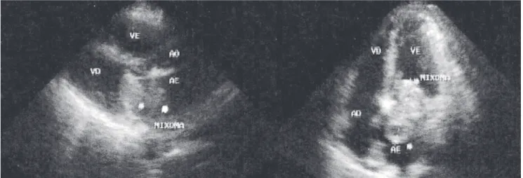
investigation: chest X-ray, electrocardiogram and transthoracic echocardiogram, which showed rectification of the atrial septum and dilated LA with the presence of two moving rounded masses, with the largest one measuring 5.3 x 3.2 cm, causing obstruction of the entrance channel of the left ventricle (LV), estimated to be 0.67 cm2 in diameter. They also emphasized on the LA (50mm); right ventricular diameter (37mm), 72% ejection fraction (EF) and right ventricle (RV) with increased dimensions. The masses apparently adhered to the mitral annulus and the LA free wall (Figure 1), suggesting myxoma, with surgical treatment being proposed.
After longitudinal median sternotomy, we proceeded to the cannulation of the ascending aorta and the superior and inferior vena cava, the establishment of extracorporeal circulation (EC) with moderate hypothermia at 27°C and
use of crystalloid cardioplegia (St. Thomas at 4ºC). After the right atriotomy that showed no tumor masses or thrombi, the transseptal incision was performed with visualization of three masses together, unlike the preoperative echocardiogram, originated at the mouth of the pulmonary veins and posterolateral wall of the left atrium.
The resection of the masses was performed with conventional electrocautery and, the material was referred to anatomopathological study, confirming the diagnosis of myxoma. The cavity was then reviewed, which showed no masses or thrombi, septoplasty, right atrial suture, EC outflow, and the patient was referred to the cardiac postoperative recovery and discharged on the fourth day after the procedure.
In the third month of postoperative follow-up, the patient was in NYHA functional class I, with normalization of the
Fig. 2 - Postoperative echocardiography with no evidence of tumor masses in the left atrium. AD: right atrium, AE: left atrium, Ao: aorta, VD: right ventricle, VE: left ventricle
499
echocardiographic patterns of left and right chambers, as well as an increase in EF to 78%, with the absence of intracavity thrombi in the LA and mitral valve within the normal parametrers (Figure 2).
DISCUSSION
Myxomas are the most common cardiac tumors, usually solitary and located in the LA. Bossert et al. [3] reported anomalous presentation of cardiac tumors in a study of 77 patients that underwent surgical treatment, with 59 of them presenting myxoma. Among them, 44 were located in the LA, 4 in the right atrium (RA), 10 cases were biatrial and 1 patient had myxoma in the LV.
In another study involving 49 patients with myxoma, 61.2% were located in the atrial septum, 26.5% in other parts of the LA, 6.1% originating from the mitral valve, 4.1% from the RA and 2% were biatrial. In one case, the tumor was found originating from the ventricular base of the mitral valve and the other one presenting multiple myxomas in both atria [4].
There are reports of multiple myxomas in the RV, originating from the ventricular base of the tricuspid valve and interventricular septum [5], as well as multiple myxomas located in the RA and RV, originating from the RA and RV free wall, below the tricuspid valve and ventricular apex [6]. Due to the location and morphology of the atrial myxoma less prevalent in the left atrial region and multilobular morphology, this present report demonstrates a rare and unusual morphological presentation of myxomas, with
REFERENCES
1. Reynen K. Cardiac myxomas. N Engl J Med. 1995;333(24):1610-7.
2. Vale MP, Freire Sobrinho A, Sales MV, Teixeira MM, Cabral KC. Mixoma gigante em átrio esquerdo: relato de caso. Rev Bras Cir Cardiovasc. 2008;23(2):276-8.
3. Bossert T, Gummert JF, Battellini R, Richter M, Barten M, Walther T, et al. Surgical experience with 77 primary cardiac tumors. Interact Cardiovasc Thorac Surg. 2005;4(4):311-5.
4. Keeling IM, Oberwalder P, Anelli-Monti M, Schuchlenz H, Demel U, Tilz GP, et al. Cardiac myxomas: 24 years of experience in 49 patients. Eur J Cardiothorac Surg. 2002;22(6):971-7.
5. Lobo Filho JG, Sales DLS, Borges AEPP, Leitão MC. Mixoma de átrio direito com prolapso para o ventrículo direito. Rev Bras Cir Cardiovasc. 2006;21(2):217-20.
6. Attar MN, Sharman DC, Al-Najjar Y, Moore RK, Millner RW, Khan SX. A rare case of multiple right heart myxomas. Int J Cardiol. 2007;118(2):e66-7.
classical clinical manifestations, satisfactory postoperative surgical outcome and total functional recovery of the patient in short-term postoperative period.