Rev Bras Anestesiol CLINICAL INFORMATION 2012; 62: 2: 262-268
262 Revista Brasileira de Anestesiologia
Vol. 62, No 2, March-April, 2012
Received from Instituto Nacional de Cardiologia/Ministério da Saúde (National Cardiology Institute/Health Ministry), Brazil.
1. Master in Health, Universidade Federal de Juiz de Fora (UFJF); Anesthesiologist at Insti-tuto Nacional de Cardiologia/ Ministério da Saúde (INC/MS), Professor of Anesthesiology at UNIPAC-JF
2. MBA in Hospital Management; Chief of the Departamento de Anestesiologia of INC/MS 3.PhD in Cardiovascular Surgery; Head of the Department of Cardiovascular Surgery, INC/MS 4. Diploma in Cardiovascular Surgery; Head of the Pediatric Cardiovascular Surgery Divi-sion, INC/MS
5. Medical Student, Universidade Federal do Rio de Janeiro (UFRJ)
6. Registered nurse, Universidade Federal de Juiz de Fora (UFJF); Master in Health (UFJF); Professor of Nursing, Faculdade Estácio de Sá
Submitted on July 29, 2010. Approved on June 19, 2011. Correspondence to:
Dr. Marcello Fonseca Salgado Filho Rua Alexandre Visentin, 100 Jardim do Sol
36061530 – Juiz de Fora, MG, Brazil E-mail: mfonsecasalgado@hotmail.com
CLINICAL INFORMATION
The Importance of Transesophageal Echocardiography in
Heart Harvesting for Cardiac Transplantation
Marcello Fonseca Salgado Filho
1, Arthur Siciliano
2, Alexandre Siciliano
3, Andrey José de Oliveira
4,
Júlia Salgado
5, Izabela Palitot
6Summary: Salgado Filho MF, Siciliano A, Siciliano A, Oliveira AJ, Salgado J, Palitot I – The Importance of Transesophageal Echocardiography in Heart Harvesting for Cardiac Transplantation.
Background and objectives: The use of transesophageal echocardiography (TEE) during heart harvesting for transplantation can guide the
heart assessment, as harvesting a marginal heart can jeopardize the cardiac transplantation.
Case report: Male, 30 years old, suffered a car crash that resulted in a severe traumatic brain injury (TBI) that evolved to brain death.The patient was intubated and ventilated with a fraction of inspired oxygen of 0.6, presetting Vt 500 mL, RR 14 bpm, PEEP of 3 mmHg, 99% O2 saturation, and normal blood gases. He was also hypovolemic, with urine output of 9,300 mL.day-1, sodium level of 157 mEq.L-1, hematocrit of 27%, and BP 90/60 mmHg maintained by infusion of norepinephrine 0.5 mcg.kg.min-1.The patient was clinically optimized and evaluated by TEE, which sho-wed normal size cardiac chambers, ejection fraction 66%, anatomical and functional heart valves with no changes, and foramen ovale integrity. Immediately after the confirmation of cardiac viability and clinical stabilization, the patient was taken to the operating room and the harvest began. The ischemic period lasted two hours and the heart was successfully transplanted.
Conclusions: In most heart transplant services, the cardiac assessment is made subjectively by the surgeon who often does not have the
anes-thesiologist support to clinically optimize the donor.At the Instituto Nacional de Cardiologia(INC/MS), the anesthesiologist is part of the harvesting team in order to perform intraoperative TEE, evaluating objectively the harvested heart. In doing so, it provides greater chances of heart transplan-tation success with lower costs for the Brazilian public health system.
Keywords: Echocardiography, Transesophageal; Heart Diseases; Transplantation.
©2012 Elsevier Editora Ltda. All rights reserved.
BACKGROUND AND OBJECTIVES
Heart transplantation is an effective treatment for patients with end-stage heart failure, but the number of donors is much smaller than the number of patients who are on the waiting list for a transplant 1.Between 10%-20% of patients die waiting for a heart transplant in the United Kingdom 2. In Brazil, despite
the lack of epidemiological studies on the subject, DATASUS 3
estimates that there are about 6.5 million patients with heart failure, and 1/3 of these patients are hospitalized. Of the hos-pitalized patients, about 6,000 are admitted by the Brazilian Unified Health System (SUS from Portuguese).
When a heart is evaluated for a possible transplant the
terms remain controversial 1. In an attempt to optimize donor
heart, the invasive hemodynamic monitoring (arterial punc-ture, central venous and pulmonary artery catheters) has been used, in addition to the subjective visual analysis of a
surgeon 1. However, the non-harvesting of a heart may
oc-cur considering the patient’s history that led to the brain stem death 1.
Transthoracic echocardiography (TTE) has been used to assess the donor heart since 1988 4 and over the years it has gained increasing popularity for being a noninvasive, portable and rapid assessment of cardiac function, which also diag-noses the presence of related cardiac pathologies, such as patent foramen ovale or valvular disease 3.
However, the use of transesophageal echocardiogra-phy (TEE) to evaluate the donor heart needs further
scien-tific work 1 because, as an adrenergic storm during the brain
stem death 5 and consequent cardiovascular overload occur,
a transient myocardial ischemia may happen. Brain injury
increases urine output, triggering sudden volume changes 6
with hemodynamic repercussions.When these situations are
associated with the TEE operator technical training, they can result in major constraints of the technique 7.
THE IMPORTANCE OF TRANSESOPHAGEAL ECHOCARDIOGRAPHY IN HEART HARVESTING FOR CARDIAC TRANSPLANTATION
Revista Brasileira de Anestesiologia 263
Vol. 62, No 2, March-April, 2012 CASE REPORT
Male, aged 30, 70 kg, 1.75 m, ASA physical status VI, suf-fered a car crash resulting in severe traumatic brain injury. Computed tomography showed cerebral intraparenchymal hemorrhage on the right with midline deviation, compression of the lateral ventricles and diffuse cerebral edema. The pa-tient developed brain stem death, which was confirmed by the team of RIOTRANSPLANTE following the World Health Orga-nization (WHO) guidelines 9.
During the preanesthetic evaluation the patient was intu-bated, ventilated with the aid of a respirator (Servo), which
parameters for fraction of inspired oxygen were 0.6, tidal vol-ume 500 mL, respiratory rate 14 bpm, PEEP 3 mmHg, oxygen saturation of 99%, and arterial blood gases within the normal range.
The patient had clinical signs of hypovolemia, with cen-tral venous pressure measuring 1 mm Hg, urine output was
9300 mL.day-1, hematocrit 27%, invasive blood pressure of
90/60 mmHg, and heart rate of 118 bpm. Noradrenaline was
being administered at a dose of 0.5 mcg.kg.min-1.
After emptying the stomach with a nasogastric tube (#18) and lubricating the oropharynx with lidocaine gel 2%, the TEE probe (Vivid I, GE, Finland, Helsinki, 2007) was introduced into the esophagus and the examination was carried out with 20 cuts, as recommended by the American Society of
Cardio-vascular Anesthesia 10.TEE evaluation showed normal size
cardiac cavities (Figure 1), left ventricular ejection fraction of
66% by Simpson’s technique 11 (Figure 2), and anatomical
heart valves without functional changes (Figure 3).The right
ventricle was hypovolemic (Figure 4) and the heart showed a hyperkinetic profile, ratifying the picture of hypovolemia.
With the infusion of 1,500 mL of Ringer’s solution#3,
guided by TEE to evaluate biventricular function, the heart rate decreased to 105 bpm, arterial pressure increased to 100x70 mmHg, central venous pressure increased to 5 mmHg,
and norepinephrine dose decreased to 0.3 mcg.kg.min-1.
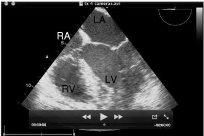
Figure 1 – Mid Esophageal Four Chamber View.
RV: rightventricle, RA: right atrium; LA: left atrium, LV: left ventricle.
Figure 2 – Mid Esophageal Four Chamber View.
The Markshows the left ventricular ejection fraction by Simpson’s technique (shaded area). RV: right ventricle; RA: right atrium; LA: left atrium; LVs: left ventricle during systole.
Figure 3 – Mid Esophageal Four Chamber View.
The Arrowshows the competent mitral valve during left ventricular systole on color Doppler. RV: right ventricle; RA: right atrium; LA: left atrium; LVs: left ventricle during systole.
Figure 4 – Short Axis View of Aortic Valve and Inlet and Outlet Pathways of Right Ventricle through the Mid Esophagus.
The Arrowshows the right ventricle during systole virtually collapsed by hypovolemia. RVs: right ventricle during systole; RA: right atrium; LA: left atrium; PA: pulmonary artery; and Ao V: aortic valve.
LA
RV
RA
LV
LA
RA
RV
LVs
LVs
LA
RA
RV
LA
Ao V
RA
SALGADO FILHO, SICILIANO, SICILIANO ET AL.
264 Revista Brasileira de Anestesiologia
Vol. 62, No 2, March-April, 2012
With the heart viability confirmed by TEE and by clinical sta-bilization, the patient was referred to the operating room and
the harvesting was initiated.The period of cardiac ischemia
lasted for two hours, and the heart was successfully trans-planted into a patient with Chagas’ cardiomyopathy, ASA IV, class 4 by New York Heart Association, and a left ventricular ejection fraction of 12%. The patient was discharged 25 days after transplantation.
DISCUSSION
Although the first heart transplant was described by Barnard in 1967, only in the 80s this surgical therapy gained worldwide popularity as an alternative technique for the treatment of ter-minal heart failure due to consistent advances in the manage-ment of the donor, to technical improvemanage-ment of surgeons, to
immunosuppressive drugs and to antibiotics 1.
With the increase in life expectancy, the number of patients waiting for a heart transplant is increasing steadily and, un-fortunately, many patients die waiting for a surgery 2.Based on the public health situation, many heart transplant centers worldwide, according to studies such as Livi et al. 13 and Kron et al. 14, are no longer so strict while choosing a donor. Hosen-pud et al. 15 showed that (in 2000) 11.5% of the transplanted hearts were from donors over 50 years of age.
During the last two decades, cardiovascular outcomes re-sulting from brain stem death have been studied intensively worldwide 2. Novitzky et al. 16, in their experimental study with baboons, have found that after brain stem death a cat-echolaminergic storm occurs with subsequent depletion of
these hormones – the Cushing reaction.Therefore there is
a direct myocardial dysfunction 6 and systemic inflammatory
reaction 5 that progresses to a cardiovascular collapse 5,6,16. Over 25% of the organs not used for cardiac transplantation are due to circulatory instability or direct myocardial dysfunc-tion 12.
Some studies have shown that 67.5% of the donor hearts have some degree of segmental dysfunction in the heart walls and 36% have global dysfunction of the left ventricular seg-mental function diagnosed by echocardiography during heart
harvesting 14,17. However, these abnormal hearts, once
trans-planted, showed improvement in cardiac function immediately after transplantation, with continued development up to 15 months after transplantation 14,17.
The Brazilian Unified Health System (SUS from Portu-guese), faced with the national reality of the public health
costs, recommends not harvesting marginal hearts 3,18 due
to the high surgical and post-operative costs.Therefore, the
hearts of donors over 50 years old; with cardiac malformation on echocardiography; left ventricular dysfunction on echocar-diography, significant coronary disease (for patients over 45 years of age a coronary angiography is mandatory); septice-mia; high doses or long-term use of vasoactive drugs (even after correction of hypovolemia); or HIV, hepatitis B and/or C infection detected by serology are all excluded 3,18.
In addition to myocardial dysfunction, diabetes insipidus is one of the metabolic changes found in brain death, which are characterized by polyuria, low urine osmolality, high serum osmolality, and hypernatremia 19. This physiological status as-sociated with decreased catecholamine levels and myocardi-al dysfunction triggers severe hemodynamic instability, which makes the clinical management of these patients a great chal-lenge to the anesthesiologist.
In this report, the patient had clinical (urine output of
5.5 mL.kg.h-1, low central venous pressure, hypotension, and
tachycardia) and laboratory (sodium 157 meq.dL-1, serum
os-molality of approximately 314 mOsmo.kg-1) signs of diabetes
insipidus. In addition, noradrenaline was being continuously administered. TEE examination was important because it showed good myocardial function (EF 60%), with no anatomi-cal and/or functional changes of the heart valves, and guided the volume optimization with crystalloid solution.
After the hemodynamic profile satisfactory response, with fluid replacement, improvement of clinical conditions, and de-creased norepinephrine, the patient was referred to the oper-ating room to have the heart harvested.
CONCLUSION
In most heart transplant services, the cardiac assessment is made subjectively by the surgeon who does not often have the anesthesiologist support to clinically optimize the donor. At the INC/MS, the anesthesiologist is part of the harvesting team in order to perform intraoperative TEE, objectively
eval-uating the harvested heart.There is then a greater chance of
268 Revista Brasileira de Anestesiologia Vol. 62, No 2, Março-Abril, 2012 SALGADO FILHO, SICILIANO, SICILIANO E COL.
REFERÊNCIAS/REFERENCES
1. Venkateswaran RV, Bonser RS, Steeds RP – The echocardiographic assessment of donor heart function prior to cardiac transplantation. Eur J Echocardiography, 2005;6:260-263.
2. Anyanwu AC, Rogers CA, Murday AJ – Intrathoracic organ transplan-tation in the United Kingdom 1995-99: results from the UK cardiotho-racic transplant audit. Heart, 2002;87:449-454.
3. Ministério da Saúde. Governo Federal. DATASUS. Disponível em: www.datasus.gov.br.
4. Gilbert EM, Krueger SK, Murray JL et al. – Echocardiographic evalua-tion of potential cardiac transplant donors. J Thorac Cardiovasc Surg, 1988;95:1003-1007.
5. Powner DJ, Hendrich A, Nyhuis A, Strate R – Changes in serum cat-echolamine levels in patients who are brain dead. J Heart Lung Trans-plant, 1992;11:1046-1053.
6. Rona G. Catecholamine cardiotoxicity. J Mol Cell Cardiol, 1985;17:291-306.
7. Lewandowski TJ, Aaronson KD, Pietroski RE, Pagani FD et al. – Discordance in interpretation of potential donor echos. J Heart Lung Transplant, 1998; 17(Suppl.1):S100.
8. English TA, Spratt P, Wallwork J et al. – Selection and procurement of hearts for transplantation. Br Med J,1984;288:1889-1891.
9. Wijdichs EFM – The diagnosis of brain desth. N Engl J Med, 2001;344:1215-1221.
10. Shanewise JS, Cheung AT, Aranson S et al. – ASE/SCA guidelines for performing a comprehensive intraoperative multiplane transesoph-ageal echocardiography examination: recommendations of the Ameri-can Society of Echocardiography Council for Intraoperative echocar-diography and he Society of Cardiovascular Anesthesiologist Task Force for certification in perioperative transesophageal Ecocardiogra-phy. AnesthAnalg, 1999;89:870-884.
11. Urbanowicz JH, Shaaban MJ, Cohen NH et al. – Comparison of transesophageal echocardiographic and scintigraphic estimates of left ventricular end-diastolic volume index and ejection fraction in pa-tients following coronary artery bypass grafting. Anesth, 1990;72:607-612.
12. Szabo G – Physiologic changes after brain death. J Heart Lung Trans-plant, 2004;23:223-226.
13. Livi U, Bortolutti U, Luciani Gb et al. – Donor shortage in heart trans-plantation. Is extension of donor ages limitsjustified? J Thorac Cardio-vasc Surgery, 1994;107:1346-55.
14. Kron IL, Tribble CG, Kern JA et al. – Successful transplantation of marginally acceptable thoracic organs. Ann Surg, 1993; 217: 518-524.
15. Hosenpud JD, Bennet LE, Keck BM et al. – The registry of the inter-national society for heart and lung transplantation: the official report – 2001. J Heart Lung Transplant, 2001;20:805-15.
16. Novitzky D, Wicomb WN, Cooper DKC et al. – Electrocardiographic, haemodynamic, and endocrine changes occurring during experimental brain death in the Chacma baboon. Heart Transplant, 1984;4:63-69. 17. Seiler C, Laske A, Galino A et al. – Echografic evalution of left
ven-tricular wall motion before and after transplantation. J Heart Lung Transplant, 1992;11:867-874.
18. Associação Brasileira de Transplantes de Órgãos – Disponível em http://www.abto.org.br.