RBCCV 44205-1525 DOI: 10.5935/1678-9741.20140015
Comparison of the solution of
tryptophan-alfacetoglutarate with
histidine-tryptophan-glutamate as cardioplegic agents in
isolated rat hearts: an immunohistochemical study
Comparação da solução de histidina-triptofano-alfacetoglutarato com histidina-triptofano-glutamato
como agentes cardioplégicos em corações isolados de ratos: estudo imuno-histoquímico
Marcos Aurélio Barboza de Oliveira
1, MD; Lívia Carvalho Ferreira
1; Débora Aparecida Pires de
Campos Zuccari
1; Antônio Carlos Brandi
2, MD; Carlos Alberto dos Santos
2, MD; Paulo Henrique
Husseni Botelho
2, MD;
Orlando Petrucci
3, MD, PhD; Domingo Marcolino Braile
1, MD, PhD
1. São José do Rio Preto Medical School (FAMERP), São José do Rio Preto, SP, Brazil.
2. Hospital de Base (HB), São José do Rio Preto, SP, Brazil.
3. State University of Campinas (UNICAMP), Faculty of Medical Sciences, Campinas, SP, Brazil.
Correspondence address:
Marcos Aurélio Barboza de Oliveira
Av. República do Líbano, 2700 – casa 80 – Jardim Tarraf II – São José do Rio Preto, SP, Brazil
Zip code: 15092-500
E-mail: [email protected]
This study was carried out at São José do Rio Preto Medical School (FAMERP), São José do Rio Preto, SP, Brazil.
No inancial support.
Article received on October 17th, 2013
Article accepted on November 21st , 2014
Abstract
Introduction: Cardiac arrest during heart surgery is a com-mon procedure and allows the surgeon to perform surgical pro-cedures in an environment free of blood and movement. Using a model of isolated rat heart, the authors compare a new car-dioplegic solution containing histidine-tryptophan-glutamate (group 2) with the histidine-tryptophan-alphacetoglutarate (group 1) routinely used by some cardiac surgeons.
Objective: To assess caspase, IL-8 and KI-67 in isolated rat hearts using immunohistochemistry.
Methods: 20 Wistar male rats were anesthetized and heparinized. The chest was opened, cardioctomy was performed and 40 ml/kg of the appropriate cardioplegic solution was infused. The hearts were kept for 2 hours at 4°C in the same solution, and thereafter, placed in the Langendorff apparatus for
30 minutes with Ringer-Locke solution. Immunohistochemistry analysis of caspase, IL-8, and KI-67 were performed.
Results: The concentration of caspase was lower in group 2 and Ki-67 was higher in group 2, both P<0.05. There was no statistical difference between the values of IL-8 between the groups.
Conclusion: Histidine-tryptophan-glutamate solution was better than histidine-tryptophan-alphacetoglutarate solution because it reduced caspase (apoptosis), increased KI-67 (cell proliferation), and showed no difference in IL-8 levels compared to group 1. This suggests that the histidine-tryptophan-glutamate solution was more eficient than the histidine-tryptophan-alphacetoglutarate for the preservation of hearts of rat cardiomyocytes.
to myocyte: the decrease in lactate and raising the pH in the mitochondrial matrix, even in ischaemia, avoiding intracellu-lar acidosis and edema, and contributing to the maintenance of intracellular adenosine triphosphate (ATP), protecting the myocyte of ischemia - reperfusion lesion.
In turn, the reduction of reperfusion injury cause decrease of caspase [10-12] and IL- 8, due to the reduction in cellular apoptosis and necrosis, respectively [13,14]. Nevertheless, the reduction of reperfusion injury may not be acting alone on behalf myocyte. Proliferative proteins such as KI- 67, could be re-coded, thus contributing to the reduction of the cell death and formation of new myocardial ibers [15,16]. This study assess HTG solution as a cardioplegic agent in isolated rat heart, considering immunohistochemical analysis of caspase markers, IL-8 and KI-67.
METHODS
After approval by the Ethics Committee on Animal Ex-perimentation of the Faculty of Medicine of São José do Rio Preto (autorization number 015/2012), 20 male Wistar rats (10 in each group) were used, weighing 280±29 grams.
All animals received care according to the recommenda-tions of the Committee on Care and Use of Laboratory An-INTRODUCTION
Induction of temporary arrest of the heart during cardiac surgery is a relatively common procedure that allows the sur-geon to perform procedures in an environment free of blood and movement [1-3]. One of the cardioplegic agents is histi-dine-tryptophan-ketoglutarate (HTK) solution.
The HTK was tested by Bretschneider et al. [ 4] in Ger-many, 1975. Its mechanism of action comes from the absence of calcium, which prevents its inlux into the cell by type “L” calcium channel in the plateau phase of the potential action, inhibiting the release of calcium from the sarcoplasmic retic-ulum over the myocyte, resulting in inactivation of myoila -ments [5,6].
This mechanism is complemented by cellular protection given by the constituents of this solution, whose main func-tions include: 1 - histidine: temperature-dependent buffer system, inhibitor of matrix metalloproteinases and cell im-permeant [7] 2 – tryptophan: acts in maintaining the integrity of cell membrane [8], and 3 - ketoglutarate: improves max-imum developed pressure and prevents increased creatine kinase MB fraction [8].
According Pisarenko et al. [9], the substitution of al-pha-Ketoglutaric acid by glutamate bring some advantages
Abbreviations, acronyms and symbols
ATP Adenosine triphosphate BSA bovine serum albumin MOD mean optical density
HTG histidine-tryptophan-glutamate HTK histidine-tryptophan-ketoglutarate IL Interleukin
IP Intraperitoneal AU Arbitrary units
Resumo
Introdução: A parada do coração durante a cirurgia cardíaca é procedimento comum e permite que o cirurgião realize os procedimentos cirúrgicos em ambiente isento de sangue e movimento. Os autores comparam, em modelo de coração isolado de rato, uma nova solução cardioplégica com triptofano-glutamato (grupo 2) com a histidina-triptofano-alfacetoglutarato (grupo 1) já utilizada de rotina por alguns cirurgiões cardíacos.
Objetivo: Avaliar por análise imuno-histoquímica a caspase, a IL-8 e KI-67 em corações isolados de ratos.
Métodos: 20 ratos machos de raça Wistar foram anestesiados e heparinizados. O tórax foi aberto, realizado cardiectomia e infundido 40 ml/kg de solução cardioplégica apropriada. Os corações foram mantidos por 2 horas na mesma solução a 4oC
e, após esse período, colocados em aparato de Langendorff por 30 minutos com solução de Ringer Locke. Foram feitas análises imuno-histoquímicas para caspase, IL-8 e KI-67.
Resultados: A concentração de caspase estava menor no grupo 2 e da KI-67 estava mais elevada no grupo 2, ambos com
P<0,05. Não houve diferença estatística entre os valores de IL-8 entre os grupos.
Conclusão: A solução com histidina-triptofano-glutamato foi melhor que a com histidina-triptofano-cetoglutarato, pois reduziu a caspase (apoptose), aumentou o KI-67 (proliferação celular) e não apresentou valores diferentes de IL-8 (inlamação e necrose) que no grupo 1. Isso sugere que a solução triptofano-glutamato foi mais eiciente que a histidina-triptofano-cetoglutarato na preservação dos cardiomiócitos dos corações de ratos.
imals - Institute of Laboratory Animal Resources (ILAR) - National Research Council, United States [17].
Experimental Protocol
The animals were anesthetized with an injection of 65 mg/kg intraperitoneal sodium pentobarbital and received IP systemic heparin (500 IU/kg). After opening the chest, car-diectomy was performed . Hearts received Ringer’s lactate solution to “wash” the coronary tree and then cardioplegic solution according to the corresponding group.
The hearts in this phase of the experiment were divided into 2 groups. In group 1, was used HTK solution at 4°C and in Group 2, solution of histidine-tryptophan-glutamate (HTG) at 4°C. Table 1 shows the composition of each solu-tion. In all cases, the infusion of cardioplegia was performed as a single dose 40 ml/kg at the aortic root, followed by im-mersion of the organ in the same solution for 2 hours at 4°C. After this time, the hearts were placed in a Langendorff system and perfused with oxygenated Locke Ringer buffer under normothermy and constant pressure of 100 cm H2O for gravitational method for 30 minutes. The drainage of the right ventricle was performed by opening the pulmonary ar-tery, and the right atrium was maintained intact in order to preserve the sinus node [18].
Three threads of epicardial pacemaker were inserted at equidistant points of the ventricles for electrocardiographic documentation of cardiac events. The time of onset of ven-tricular ibrillation and the irst heartbeat counted from the start of infusion of Ringer Locke solution was noted.
After 30 minutes of infusion of Ringer Locke, the ex-periment was discontinued. The hearts were removed from the Langendorff system and fragments of the cardiac apex, which were stored in sterile Falcon type tubes containing 10% formalin for subsequent histological and immunohisto-chemical preparation.
Histological and immunohistochemical technique preparation
Initially, the material was embedded in parafin, a proce -dure that provides resistance allow for cutting thickness of 3m and placed on silanized slides. The silanization of the blades consisted in preparing these with an adhesive that ixes the fragment to the blades, preventing their detachment during the immunohistochemical procedure. For this, they were im-mersed in acetone P.a. (2 minutes), 4% silane solution diluted with acetone (2 minutes) and again in acetone P.a. (4 to 5 dips). The drying of the slides was performed in an oven at 60ºC.
The block was attached to the microtome, the slice thick-ness was set to 3 µm and the cuts placed on silanized identi-ied and left in an oven at 60°C for 24 hours. The blade went through the process of deparafinization in xylene, followed by hydration in absolute alcohol I, II and III, inishing with six dives in tap water, incubated with 3% hydrogen peroxide for 30 minutes to block endogenous peroxidase.
Antigen retrieval was performed in the steamer with spe-ciic buffer for each antibody for 30 minutes (Table 2). Then the slides were covered up with a solution containing fetal bo-vine serum (BSA) and incubated with the primary antibody.
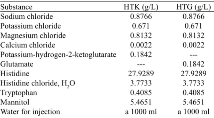
Table 1. Composition of solutions used.
Substance Sodium chloride Potassium chloride Magnesium chloride Calcium chloride Potassium-hydrogen-2-ketoglutarate Glutamate Histidine
Histidine chloride, H2O Tryptophan
Mannitol
Water for injection
HTK (g/L) 0.8766 0.671 0.8132 0.0022 0.1842 ---27.9289 3.7733 0.4085 5.4651 a 1000 ml
HTG (g/L) 0.8766 0.671 0.8132 0.0022 ---0.1842 27.9289 3.7733 0.4085 5.4651 a 1000 ml
HTK: Histidine-tryptophan ketoglutarate; HTG: histidine-tryptophan-glutamate
Table 2. List of antibodies used.
Antibody anti-Ki-67 anti-Caspase 3 anti-IL-8 Speciicity Monoclonal Polyclonal Monoclonal Dilution 1:200 1:1000 1:50 Buffer Citrato pH6 Citrato pH6 Citrato pH6 Lab Biocare Medical Abcam Santa Cruz
After this step, the slides were washed in PBS solution and incubated for 15 minutes with Starr Trek Universal HRP Detection kit (Biocare Medical®), which consisted in second-ary antibody biotinylated for 1 hour and streptavidin-peroxi-dase complex for 30 minutes, followed by washing with PBS for 15 minutes. The revelation was performed with substrate chromogen (DAB Betazoidchromogen) of the Starr Trek Universal HRP Detection kit (Biocare Medical®) for 2 to 5 minutes, and counterstained with Harrys hematoxylin for 40 seconds. The tissues were dehydrated in alcohol in ascending degree and bathed in xylene before mounting the slides on ERV-MOUNT amid (Erviegas®).
Negative control reactions were obtained by omitting the primary antibody. Tonsil tissue was used for Ki-67 reactions and Caspase 3 and as positive control breast tissue for IL-8 reaction.
Quantiication of immunohistochemical staining
(AU). The average optical density (AOD) was obtained with the aid of the following formula:
AOD = 255 – AU
This formula showed the intensity of immunostaining speciically in immunoreactive areas.
Statistical Analysis
The data were subjected to the Kolmogorov-Smirnov test and subsequently the parametric analysis by unpaired Stu-dent’s t test or non-parametric by Mann-Whitney test when appropriate, and Fisher’s exact test for categorical data. Re-sults were expressed only in mean ± standard deviation due to the fact all variables behave as continuous quantitative with Gaussian distribution. P values were presented, and those who were less than 0.05 were considered signiicant. The GraphPad Instat and Prism 6.0 softwares of statistical analysis, both for Windows® were used.
RESULTS
The average weight of the animals was 277.4 ± 24.6 g (group 1) and 288 ± 34.5 g (group 2 ), respectively, with no signiicant difference between groups (P=0.4396). Regarding the average volume of Ringer Locke collected from coronary sinus after 30 minutes (363.1 ± 177.3 ml and 277.4 ± 33.7 ml, respectively), there was no signiicant difference between groups (P=0.1923).
Findings during perfusion with cardioplegic solution and Ringer Locke
All hearts showed adequate perfusion of cardioplegia and Ringer Locke, evidenced by clear staining in the ven-tricular wall. The average heart rate after 5 minutes of perfu-sion (233±36 and 188±53.4 beats per minute, respectively), showed a signiicant difference (P=0.0086). The time of on-set of ventricular ibrillation (49 ± 28.2 and 45 ± 17 seconds, respectively) and time to irst heartbeat (153 ± 78 and 117 ± 96.8 seconds respectively) showed no signiicant difference (P=0.5869 and P=0.187, respectively).
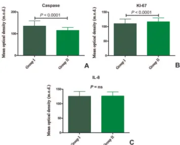
Immunohistochemical indings
After 2 hours of ischemia and 30 minutes of reperfu-sion, caspase activity was signiicantly lower in group 2 (P<0.0001), the activity of KI-67 was higher in group 2 (P<0.0001) and IL-8 was not different between groups (Figure 1).
DISCUSSION
Myocardial ischemia causes various cardiac effects, such as decreases force of contraction; increases diastolic pressure, indicating contraction of myoibrils in isovolumic conditions, causes a decline in phosphocreatine and ATP;
de-Fig. 1 - Histograms showing mean optical densities of: (A) caspase, (B) KI-67 and (C) IL-8. Group 1: solution with histidine-tryptophan-ketoglutarate, Group 2: solution with histidine-tryptophan-glutamate
creases contraction, and glutamate and aspartate; increases lactate, pyruvate, alanine and succinate [9]. According Pis-arenko et al. [9] the addition of glutamate in the perfusate keeps the intracellular ATP and decreases both lactate and pyruvate as that contribute to acidosis. These effects contrib-ute to improve cardiac function recovery after ischemia. Our results show similar behavior in the two solutions studied concerning the time and duration of ventricular ibrillation irst beat, however, it was better for group 2 concerning heart rate, which was lower, which can be correlated with lower acidosis, probably myocyte .
Another process that is intrinsically related to isch-emia-reperfusion injury is apoptosis [10,11], characterized by morphological changes such as chromatin condensation, fragmentation of nuclei and formation of “apoptotic bod -ies”. These changes are made by a family of proteases called caspases [12]. The degree of caspase activation is directly re-lated to the degree of apoptosis, which plays a critical factor in the recovery of cardiac function [13].
Our results demonstrate less caspase activity in group 2, suggesting a potential protective for myocardial function.
Authors’ roles and responsibilities
MABO Study design, execution of experiments, analysis of results and writing of the manuscript
LCF Assistance in immunohistochemical techniques DAPCZ Assistance in immunohistochemical techniques
ACB Participation in the preparation of the inal text
CAS Participation in the preparation of the inal text
PHHB Participation in drafting the inal text
OP Review of the version
DMB Study design, analysis of results and writing of the manuscript
REFERENCES
1. Chambers DJ. Mechanisms and alternative methods of achieving cardiac arrest. Ann Thorac Surg. 2003;75(2):S661-6.
2. Fannelop T, Dahle GO, Matre K, Moen CA, Mongstad A, Eliassen F, et al. Esmolol before 80 min of cardiac arrest with oxygenated cold blood cardioplegia alleviates systolic dysfunction. An experimental study in pigs. Eur J Cardiothorac Surg. 2008;33(1):9-17.
3. Braile DM, Ardito RV, Zaiantchick M, Santos JLV, Soares MJF. Cardioplegia sanguínea contínua normotérmica. Rev Bras Cir Cardiovasc. 1989;4(2):109-38.
4. Bretschneider HJ, Hübner G, Knoll D, Lohr B, Nordbeck H, Spieckermann PG. Myocardial resistance and tolerance to ischemia: physiological and biochemical basis. J Cardiovasc Surg (Torino). 1975;16(3):241-60.
5. Fallouh HB, Kentish JC, Chambers DJ. Targeting for cardioplegia: arresting agents and their safety. Curr Opin Pharmacol. 2009;9(2):220-6.
6. Chambers DJ, Hearse DJ. Developments in cardioprotection:
“polarized” arrest as an alternative to “depolarized” arrest. Ann
Thorac Surg. 1999;68(5):1960-6.
7. Antunovic M, Aleksic D. Preparation and testing of solutions for organ perfusion and preservation in transplantation. Vojnosanit Pregl. 2008;65(8):596-600.
8. Hachida M, Ookado A, Nonoyama M, Koyanagi H. Effect of HTK solution for myocardial preservation. J Cardiovasc Surg (Torino). 1996;37(3):269-74.
9. Pisarenko OI, Solomatina ES, Ivanov VE, Studneva IM, Kapelko VI, Smirnov VN. On the mechanism of enhanced ATP formation in hypoxic myocardium caused by glutamic acid. Basic Res Cardiol. 1985;80(2):126-34.
10. Xu YJ, Saini HK, Zhang M, Elimban V, Dhalla NS. MAPK activation and apoptotic alterations in hearts subjected to calcium paradox are attenuated by taurine. Cardiovasc Res. 2006;72(1):163-74.
11. Fischer UM, Cox CS Jr, Laine GA, Mehlhorn U, Bloch W, Allen SJ. Induction of cardioplegic arrest immediately activates the myocardial apoptosis signal pathway. Am J Physiol Heart Circ Physiol. 2007;292(3):H1630-3.
12. Pirnia F, Schneider E, Betticher DC, Borner MM. Mitomycin C induces apoptosis and caspase-8 and -9 processing through a caspase-3 and Fas-independent pathway. Cell Death Differ. 2002;9(9):905-14.
13. Lee S, Huang CS, Kawamura T, Shigemura N, Stolz DB, Billiar TR, et al. Superior myocardial preservation with HTK solution over Celsior in rat hearts with prolonged cold ischemia. Surgery. 2010;148(2):463-73.
protection provided by HTG solution was not different from that given by the HTK solution.
Mammalian hearts have low proliferative capacity after birth. One of the markers used to assess cell proliferation is the Ki-67 [15]. With this marker, Walsh et al . [16] demon-strated that 12% to 23% of fetal rat cardiomyocytes exhibit proliferative activity, from 1% to 8% up to the 7th day and vir-tually undetectable from the 14th day. In our study, there was a signiicant increase of the KI-67 in group 2, demonstrating early proliferative activity of HTG solution guaranteed with 2 hours of ischemia.
Associated with this, Walsh et al. [16] also draw the atten-tion to the KI-67 activity also be inversely related to apopto-sis, which is also conirmed in our study, in which caspase is lower in group 2 than in 1. However, the increased activity of this marker is worrisome because it has been associated with myxomas [19,20] and cardiac sarcomas [21].
Although the results obtained here are consistent with the literature, they are not deinitive regarding the replacement of alpha-Ketoglutaric acid by glutamate. Quantitative analy-sis with ATP and other nuclear markers for cell proliferation should be used to target a more comprehensive and safe con-clusion. Another relevant aspect is the concentration of glu-tamate. Would have the same protective effect on the heart if we change its concentration? Further studies are needed to answer these questions.
CONCLUSION
14. Anselmi A, Abbate A, Girola F, Nasso G, Biondi-Zoccai GG,
Possati G, et al. Myocardial ischemia, stunning, inlammation,
and apoptosis during cardiac surgery: a review of evidence. Eur J Cardiothorac Surg. 2004;25(3):304-11.
15. Lee Y. To proliferate or not to proliferate. Cardiovasc Res. 2010;86(3):347-8.
16. Walsh S, Pontén A, Fleischmann BK, Jovinge S. Cardiomyocyte cell cycle control and growth estimation in vivo: an analysis based on cardiomyocyte nuclei. Cardiovasc Res. 2010;86(3):365-73.
17. Committee on Care and Use of Laboratory Animals - Institute of Laboratory Animal Resources - Commission on Life Sciences - National Research Council. Guide for the care and use of laboratory animals. 8th ed. Washington: National Academies Press; 2010. 211p.
18. Lahaye Sle D, Gratas-Delamarche A, Malardé L, Vincent S, Zguira MS, Morel SL, et al. Intense exercise training induces adaptation
in expression and responsiveness of cardiac β-adrenoceptors in
diabetic rats. Cardiovasc Diabetol. 2010;9:72.
19. Kusumi T, Minakawa M, Fukui K, Saito S, Ohashi M, Sato F, et al. Cardiac tumor comprising two components including typical myxoma and atypical hypercellularity suggesting a malignant change. Cardiovasc Pathol. 2009;18(6):369-74.
20. Suvarna SK, Royds JA. The nature of the cardiac myxoma. Int J Cardiol. 1996;57(3):211-6.