Rev Bras Farmacogn 24(2014): 561-564
* Corresponding author.
E-mail: [email protected] (M. Taher).
0102-695X/$ - see front matter © 2014 Sociedade Brasileira de Farmacognosia. Published by Elsevier Editora Ltda. All rights reserved. http://dx.doi.org/10.1016/j.bjp.2014.10.003
Original article
Cytotoxic tirucallane triterpenes from the stem of
Luvunga scandens
Putri Nur Hidayah Al-Zikri
a, Muhammad Taher
a,*, Deny Susanti
b, Mohamad Fazlin Rezali
c,
Roger W. Read
d, Md. Hossain Sohrab
e, Choudhury Mahmood Hasan
f, Farediah Ahmad
gaDepartment of Pharmaceutical Technology, Faculty of Pharmacy, International Islamic University Malaysia, Pahang, Malaysia bDepartment of Chemistry, Faculty of Science, International Islamic University Malaysia, Pahang, Malaysia
cSIRIM Berhad, National Metrology Laboratory, Selangor, Malaysia
dSchool of Chemistry, the University of New South Wales, Sydney, Australia
ePharmaceutical Sciences Research Division, BCSIR Laboratories Dhaka, Dhaka, Bangladesh fManarat International University, Dhaka, Bangladesh
gDepartment of Chemistry, Faculty of Science, Universiti Teknologi Malaysia, Johor, Malaysia
Introduction
Development of new cytotoxic drugs is essential since the emerging resistance by cancer cells towards existing drugs. Medicinal plants are an ideal target for the discovery of potential bioactive compounds or lead structures for new cytotoxic drugs. Novel antitumor agents such as paclitaxel (Taxol) (Wani et al., 1971; Bills et al., 2002), vincristine and vinblastine (Hamburger et al., 1991; Tyler, 1986) were reported from plant sources.
The genus Luvunga (Rutaceae) is comprised by only about twelve species. Luvunga scandens (Roxb.) Buch-Ham ex Wight & Arn is mainly distributed in tropical and humid environments
throughout China, Cambodia, India, Laos, Indonesia, Philippines, Malaysia, Myanmar, Thailand and Vietnam. This plant is used for the treatment of several ailments by traditional folk medicine. A decoction of the stem of L. scandens is orally administered to treat malaria and fatigue (Eswani et al., 2010; Ong et al., 2011). A few reports of the phytochemical (Chopra, 2006), antifungal and insecticidal activity of L. scandens have been documented (Singh and Maurya, 2005; Garg and Jain, 1999). The present study was aimed to isolate and elucidate the structure of two compounds from the dichloromethane extract of the stem of Luvunga scandens by spectroscopic techniques, and the evaluation of their cytotoxicity.
A R T I C L E I N F O
Article history:
Received 8 August 2014 Accepted 2 October 2014
Keywords: Triterpenes Cytotoxicity Luvunga scandens Tirucallane
A B S T R A C T
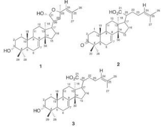
Two tirucallane triterpenes, namely flindissol (1) and 3-oxotirucalla-7,24-dien-21-oic-acid (2), were isolated from the dichloromethane extract of the stem of Luvunga scandens (Roxb.) Buch-Ham ex Wight & Arn, Rutaceae. This is the first report of their isolation from this plant. Their structures were constructed by high resolution mass and 2D NMR spectroscopic data. The cytotoxic potential of the two pure compounds 1 and 2 were determined by MTT assay against human breast adenocarcinoma cell line (MCF-7). Compounds 1 and 2 showed potent cytotoxicity against MCF-7 cell line with IC50 values of 13.8 µM and 27.5 µM, respec-tively. This result suggested their potential activity as antitumor agents.
562
Putri Nur Hidayah Al-Zikri et al. / Rev Bras Farmacogn 24(2014): 561-564Materials and methods
General experimental procedures
The NMR spectra were recorded on a Bruker 400 MHz NMR Spectrometer using CDCl3. The HRMS spectra were recorded using a Thermo LTQ-FT mass spectrometer (RMS 2 ppm with external calibration) in the Mark Wainwright Analytical Centre at The University of New South Wales, Sydney, Australia. IR spectra were measured on a Perkin Elmer infrared spectrophotometer. The UV spectra were obtained on a Shimadzu UV-1800 UV/VIS spectrophotometer. Column chromatography was performed on silica gel (70-230 and 230-400 mesh, Merck, Germany) and Sephadex LH-20 (40-70µm, GE healthcare). Organic solvents and TLC plates were purchased from Merck, Germany.
Dulbecco’s modified Eagle medium (DMEM), Fetal Bovine Serum (FBS), Penicillin-Streptomycin and TripLE Express were purchased from GIBCO. 3-(4,5-Dimethylthiazol-2-yl)-2,5-diphenyltetrazolium bromide (MTT) was purchased from Invitrogen (USA). Doxorubicine hydrochloride was obtained from Sigma Aldrich (USA).
Plant material
The stems of Luvunga scandens (Roxb) Buch-Ham ex Wight & Arn, Rutaceae, were collected from Bukit Pelindung, Kuantan, Pahang, Malaysia, in September 2012. Samples were further identified by Dr. Shamsul Khamis at Institute of Bioscience, Universiti Putra Malaysia. A voucher specimen (No. PIIU 0204) was deposited in the Herbarium of the Faculty of Pharmacy, International Islamic University Malaysia.
Extraction and isolation
The dried and powdered stem (730 g) of L. scandens was extracted by maceration using n-hexane, dichloromethane (DCM) and methanol (MeOH) as solvents. The concentrated DCM extract (133 g) was successively partitioned by n-hexane (1.8 l), n-hexane - DCM (1:1, 1.2 l), DCM (1.2 l), DCM-MeOH (1:1, 1.5 l), and MeOH (600 ml). The DCM-MeOH (1:1) fraction (67 g) was subjected to column chromatography (CC) for fractionation over silica gel (70-230 mesh) with a mobile phase of DCM-EtOAc (100 ml of each) of 100:0, 90:10, 80:20, 70:30, 60:40, 50:50 and 0:100 (v/v); and finally by EtOAc-MeOH (50:50). The fractions with similar TLC profile were combined to yield four fractions. Fraction 2 (62.2 g) was further extracted using the above-mentioned method to obtain compound 1 (32 mg) and 2 (17 mg).
Flindissol (1)
White amorphous powder; m.p. 184-187°C; UV (MeOH) λmax: 211 nm; IR (KBr) υmax: 3395, 2950, 2372, 1717, 1446, 979 cm-1; ESI-MS: [M + Na]+m/z 479.3482 (calculated for C
30H48O3Na, 479.3496); 1H NMR (400 MHz, CDCl3): d 0.80 (3H, s, CH3-18), 0.93 (3H, s, CH
3-19), 0.93 (3H, s, CH
3-29), 0.95 (3H, s, CH3-28), 0.99 (3H, s, CH3-30), 1.71 (3H, s, CH
3-27), 1.73 (3H, s, CH3-26), 3.49 (1H, bs, H-3), 4.71 (1H, m, H-23), 5.17 (1H, bs, OH-21), 5.19 (1H, d, J = 8.0 Hz, H-24), 5.27 (1H, bs, H-7). 13C NMR (100 MHz, CDCl
3): d 13.3 (C-19), 17.4 (C-11), 18.4
(C-27), 21.6 (C-29), 22.6 (C-18), 23.8 (C-6), 26.0 (C-26), 26.8 (C-28), 28.4 (C-30), 28.7 (C-2), 28.7 (C-16), 31.9 (C-12), 34.1 (C-15), 34.1 (C-22), 34.5 (C-10), 37.0 (C-4), 40.3 (C-1), 44.2 (C-13), 45.2 (C-17), 50.0 (C-20), 50.9 (C-9), 51.3 (C-14), 52.0 (C-5), 73.1 (C-23), 75.8 (C-3), 99.7 (C-21), 118.4 (C-7), 124.5 (C-24), 137.6 (C-25), 146.2 (C-8).
3-Oxotirucalla-7,24-dien-21-oic acid (2)
White amorphous powder; m.p. 238-241°C; UV (MeOH) λmax: 204 nm; IR (KBr) υmax: 3754, 3396, 2960, 1710, 1202 cm-1; ESI-MS: [M + H]+m/z 455.3510 (calculated for C
30H47O3, 455.3520); 1H NMR (400 MHz, CDCl3): d 0.92 (3H, s, CH
3-18), 1.00 (3H, s, CH3-19), 1.03 (3H, s, CH3-30), 1.07 (3H, s, CH
3-28), 1.13 (3H, s, CH3-29), 1.50 (1H, m, H-1), 1.50 (2H, m, H-11), 1.61 (3H, s, CH
3-26), 1.70 (3H, s, CH3-27), 1.77 (1H, m, H-5), 1.99 (1H, m, H-1), 1.99 (2H, m, H-23), 2.11 (2H, m, H-6), 2.11 (1H, m, H-17), 2.29 (1H, m, Heq-2), 2.29 (1H, m, H-9), 2.29 (1H, m, H-20), 2.75 (1H, ddd, J = 8.4, 8.4, 4.8 Hz, Hax-2), 5.11 (1H, bt, H-24), 5.34 (1H, bs, H-7). 13C NMR (100 MHz, CDCl
3): d 12.7 (C-19), 17.7 (C-26), 17.9 (C-11), 21.6 (C-18), 21.7 (C-29), 24.4 (C-6), 24.5 (C-28), 25.7 (C-27), 26.0 (C-23), 27.2 (C-16), 27.4 (C-30), 30.0 (C-12), 32.3 (C-22), 33.5 (C-15), 34.9 (C-2), 35.1 (C-10), 38.6 (C-1), 43.3 (C-13), 47.3 (C-20), 47.9 (C-4), 48.2 (C-9), 49.8 (C-17), 51.0 (C-14), 52.4 (C-5), 118.2 (C-7), 123.5 (C-24), 132.4 (C-25), 145.5 (C-8), 181.9 (C-21), 217.1 (C-3).
In vitro cytotoxicity assay
The cytotoxic activity of the isolated compounds against MCF-7 cells was measured by MTT assay according to Mosmann (1983) with some modifications. A total of 2 × 10⁵ cells/ml were plated in a 96-well plate overnight; then, the cells were treated in triplicate with various concentrations of the compounds. After 24 h of incubation, the cells were washed with phosphate buffer saline (PBS) and assayed by addition of 30 µl of MTT solution (5 mg/ml MTT in PBS). After 4 h of incubation, the formazan crystals were dissolved in 120 µl DMSO and left in the dark at room temperature for an additional 1 h. The absorbance measured at 570 nm and 630 nm used as reference wavelength was performed using a microplate reader (TECAN infinite M200). The wells with untreated cells were taken as the control, wells with medium with 0.5% (v/v) of EtOH were used as solvent control and wells with Doxorubicin were used as positive control. Results were expressed as a percentage of the average absorbance of sample treated cells with respect to untreated ones. The half maximal inhibitory concentration (IC50) was defined as the concentration of compound that caused a 50% reduction in cell viability against MCF-7 cell line. Viability of cells (%) was calculated using the following equation:
Viability of cells (%) = [(ASample-ABlank)/(AUntreated-ABlank)] × 100
Results and discussion
Compound 1 was obtained as a white amorphous powder. The 1H-NMR and 13C-NMR data together with the DEPT 135 spectrum proved the presence of 30 C-atom signals corresponding to seven Me, eight sp3 CH
Putri Nur Hidayah Al-Zikri et al. / Rev Bras Farmacogn 24(2014): 561-564
563
ESI-MS yielded a parent mass at m/z 479.3482 in positive ionization mode which, in conjunction with the data of other spectra, suggested the sodium adduct [M + Na]+ of a compound with a molecular formula of C30H48O3 (calculated mass 479.3496, [C30H48O3 + Na]+), accounting for 7 degrees of unsaturation. The 1H-NMR and 13C-NMR data of compound 1 were identical to those of the tirucallane triterpene flindissol, which has the unusual 3-a configuration of the A-ring hydroxyl group (Birch et al., 1963) and has been previously isolated from several species of the Rutaceae family (Guang-Yi et al., 1988; Bevan et al., 1967; Waterman, 1973). This is the first report of flindissol found in L. scandens.
by the ESI-MS mass spectrum with [M + H]+ at m/z 455.3510 for 2, which suggested the molecular formula C30H46O3 (calcd mass 455.3520, [C30H46O3 + H]+), accounting for 8 degrees of unsaturation. Accordingly, compound 2 was established as 3-oxotirucalla-7,24-dien-21-oic acid, a tirucallane triterpene first isolated from oleoresin of Aucoumea klaineana (Tessier et al., 1982) and later identified in Xanthoceras sorbifolia (Ma et al., 2000), Rhus javainica L. (Lee et al., 2005), Pancovia pedicellaris (Soh et al., 2009), and Feroniella lucida (Sriyatep et al., 2014), which displays inhibitory activity against HIV-1 protease (Ma et al., 2000; Nimmanpipug et al., 2009) and cytotoxicity (Lee et al., 2005).
Compounds 1 and 2 were assayed for their dose-response effect on the viability of human breast adenocarcinoma cell line (MCF-7) by MTT assay. Doxorubicin was used as positive control to compare the cytotoxicity of the pure 1 and 2. In order to determine the growth inhibition, the MCF-7 cells were incubated with various concentrations of the compounds for 24 h. The inhibition concentration required reduction of 50% of cell viability (IC50) was determined by comparison to the untreated control. Based on the results of the MTT assay (Table 1), both compounds isolated from stems of L. scandens inhibited the growth of human breast adenocarcinoma cells. A bar chart of the percentage of viability of cells was plotted against sample concentrations (Fig. 1). Compound 1 was comparatively more cytotoxic at a lower IC50 value (13.8 µM) than that of compound 2 (27.5 µM), and less than that of doxorubicin.
Components KI
1 13.8
2 27.5
Doxorubicin 6.21
Table 1
Cytotoxicity data (IC50 value in µM) of compounds 1 and 2 against MCF-7 cell line.
Figure 1 – Cytotoxic activity of compounds 1-2 against MCF-7 cell line. Untreated cells and Doxorubicin were used as negative and positive control respectively. The error bars indicate mean standard error of triplicate values. Asterisks represent statistically significant (p < 0.05) data compared to controls (untreated cells).
The NMR spectra of compound 2 were similar to those of 1, with exception to the additional one sp3 and two sp2 quaternary C-atoms and lack of three sp3 CH. The three sp3 CH signals (dC 75.8, 99.7 and 73.1) of compound 1 were bound to oxygen at C-3, C-21 and C-23, and their absence in compound 2 indicated a change in the 3-hydroxy substituent and a modification of the side-chain lactol. The appearance of two additional sp2 quaternary C-atoms in 2 suggests the presence of COOH (dC 181.9) and C=O (dC 217.1) moieties, which affirm the suggested structural modifications. In the 1H NMR spectrum, the presence of a 3-oxo moiety was confirmed by the coupling pattern ddd at dH 2.75 for the H-2 axial proton. This was further confirmed by tracing of cross peaks between the neighboring coupled protons at d 2.75 (Hax-2) and 1.99 and 1.50 (H-1, both m) in the COSY spectrum. Two olefinic proton signals, appeared at d 5.34 (H-7, bs) and 5.11 (H-24, bt), correlated in the HSQC spectrum (showing one-bond C-H correlations) to carbons at d 118.2 (C-7) and 123.5 (C-24), respectively.
564
Putri Nur Hidayah Al-Zikri et al. / Rev Bras Farmacogn 24(2014): 561-564Conclusions
It is concluded from spectroscopic evidence that two tirucallane triterpenes were isolated from the dichloromethane extract of the stems of Luvunga scandens. The tirucallane triterpenes flindissol (1) and 3-oxotirucalla-7,24-dien-21-oic acid (2) were isolated for the first time from this plant. The cytotoxicity of compounds 1 and 2, as determined by MTT assay against MCF-7 cell line, are consistent with the findings that medicinal plants are an ideal source of cytotoxic agents, thus, subsequent research towards the discovery of more cytotoxic agents should continue.
Authors’ contributions
PNHA and MT contributed in collecting plant material, extraction, purification, biological testing and writing of the manuscript. MT and DS designed the study and supervised the laboratory work. MFR contributed in running and advising NMR results. RWR contributed in MS analysis. RWR and FA gave critical reading of the manuscript. MT, RWR, MHS and CMH contributed in interpretation of NMR and MS data.
Conflicts of interest
The authors declare no conflicts of interest.
Acknowledgments
The research was partially supported by Science Fund (02-01-08-SF0110) from Ministry of Science, Technology and Innovation (MOSTI), Malaysia.
R E F E R E N C E S
Bevan, C.W.L., Ekong, D.E.U., Halsall, T.G., Toft, P., 1967. West African timbers. Part XX. The structure of turraeanthin, an oxygenated tetracyclic triterpene monoacetate. J. Chem. Soc. C. 820-828.
Bills, G., Dombrowski, A., Pelaez, F., Polishook, J., An, Z., 2002. Tropical Mycology: Micromycetes. Eds.: Walting, R., Frankland, J.C., Ainsworth, A.M., Issac, S., Robinson, C.H., vol. 2. CABI Publishing, New York, pp. 165-194.
Birch, A.J., Collins, D.J., Muhammad, S., Turnbull, J.P., 1963. The structure of flindissol. Some remarks on the elemi acids. J. Chem. Soc. 2762-2772.
Chopra, R.N., 2006. Indigenous Drugs of India. Bimal Kumar Dhur of Academic Publishers, p. 355.
Cotterrell, G.P., Halsall, T.G., Wriglesworth, M.J., 1970. The chemistry of triterpenes and related compounds. Part XLVII. Clarification of the nature of the tetracyclic triterpene acids of elemi resin. J. Chem. Soc. C. 739-743.
Eswani, N., Kudus, K.A., Nazre, M., Noor, A.G.A., Ali, M., 2010. Medicinal plant diversity and vegetation analysis of logged over hill forest of Tekai Tembeling Forest Reserve, Jerantut, Pahang. J. Agr. Sci. 2, 189-210.
Garg, S.C., Jain, R., 1999. Antifungal Activity of Luvunga scandens against some Keratinophilic fungi. Indian J. Pharm. Sci. 61, 248-249.
Guang-Yi, L., Gray, A.I., Waterman, P.G., 1988. Tirucallane and oleanane triterpenes from the resin of Aucoumea klaineana. Phytochemistry 27, 2283-2286.
Hamburger, M., Marston, A., Hostettmann, K., 1991. Search for new drugs of plant origin. Adv. Drug. Res. 20, 167-215. Lee, T.-H., Chiou, J.-L., Lee, C.-K., Kuo, Y.-H., 2005. Separation and
determination of chemical constituents in the roots of Rhus javanica L. var. roxburghiana. J. Chin. Chem. Soc. 52, 833-841. Ma, C.-M., Nakamura, N., Hattori, M., Kakuda, H., Qiao, J.-C.,
Yu, H.-L., 2000. Inhibitory effects on HIV-1 protease of constituents from the wood of Xanthoceras sorbifolia. J. Nat. Prod. 63, 238-242.
Mosmann, T., 1983. Rapid colorimetric assay for cellular growth and survival: application to proliferation and cytotoxicity assays. J. Immunol. Methods 65, 55-63.
Nimmanpipug, P., Lee, V.S., Wolschann, P., Hannongbua, S., 2009. Litchi chinensis-derived terpenoid as anti-HIV-1 protease agent: Structural design from molecular dynamics simulations. Molec. Simul. 35, 673-680.
Ong, H.C., Chua, S., Milow, P., 2011. Ethno-medicinal plants used by the Temuan villagers in Kampung Jeram Kedah, Negeri Sembilan, Malaysia. Ethno. Med., 5, 95-100.
Singh, G., Maurya, S., 2005. Antimicrobial, antifungal and insecticidal investigations on essential oils. An overview. Nat. Prod. Rad. 4, 179-192.
Soh, R.F., Bankeu, J.K., Lenta, B.N., Mba’ning, B.M., Ngouela, S., Tsamo, E., Sewald, N., 2009. Antibacterial allagic acid derivatives and other constituents from Pancovia pedicellaris. Zeit. Naturforsch., B: J. Chem. Sci. 64, 1070-1076.
Sriyatep, T., Chakthong, S., Leejae, S., Voravuthikunchai, S.P., 2014. Two lignans, one alkaloid, and flavanone from the twigs of Feroniella lucida. Tetrahedron 70, 1773-1779. Tessier, A.M., Delaveau, P., Piffault, N., 1982. Oleoresin of
Aucoumea klaineana. Planta Med. 44, 215-217.
Tyler, V.E., 1986. Plant drugs in the twenty-first century. Econ. Bot. 40, 279-288.
Wani, M.C., Taylor, H.L. Wall, M.E., Coggon, P., McPhail, A.T., 1971. Plant antitumor agents. VI. Isolation and structure of taxol, a novel antileukemic and antitumor agent from Taxus brevifolia. J. Am. Chem. Soc. 93, 2325-2327.