ISSN 0102-695X
http://dx.doi.org/10.1590/S0102-695X2012005000093 Received 2 Feb 2012 Accepted 21 Apr 2012 Available online 16 Aug 2012
may alter the urine excretion of Wistar rats
Monica S. C. Dalmacio,
*,1Sylvia M. N. Campos,
2,3Jorge J. J.
Carvalho,
4Mário J. S. Pereira,
4Ana Lúcia Nascimento,
4Patricia
O. Paschoal,
2,5Andrea P. Sobrinho,
2Monique M. B. Pedruzzi,
2,3Valeria Garrido,
2Silvana R. F. Moreno,
1,6Gerlinde A. P. B.
Teixeira,
2,3Gilberto P. Cardoso
11Programa de Pós-graduação em Ciências Médicas, Hospital Universitário Antônio Pedro, Universidade Federal Fluminense, Brazil,
2Departamento de Imunologia, Instituto de Biologia, Hospital Universitário Antônio Pedro, Universidade Federal Fluminense, Rio de Janeiro, Brazil,
3Programa de Pós-graduação em Patologia, Hospital Universitário Antônio Pedro, Universidade Federal Fluminense, Brazil,
4Departamento de Histologia e Embriologia, Universidade do Estado do Rio de Janeiro, Brazil,
5Programa de Pós-graduação em Pediatria, Hospital da Criança, Universidade do Estado de São Paulo, Brazil,
6Departamento de Patologia, Hospital Universitário Antônio Pedro, Universidade Federal Fluminense, Rio de Janeiro, Brazil.
Abstract: Wistar rats (n=20) were divided in two groups: G1 received 2 mg/kg of GBE (Ginkgo biloba extract 761), whereas G2 received the same volume of a sodium chloride solution (0.9%), both for 10 days. After a 7-day interval, the treatment was repeated for 8 days. Urine volume and food and water intake were measured daily during this protocol. Histological assessments were performed. No significant difference (p>0.05) was observed in food and water intake of animals during treatment with GBE. Animals who received GBE had a smaller urine volume and increase of weight with a significance difference (p<0.05) during the first and second exposure period. No histological alteration was observed in tissues, except for the kidney of the experimental group, which revealed a higher concentration of red cells in the glomerulus with a strong staining for Vascular Endothelial Growth Factor (VEGF). The introduction of GBE (therapeutic dose) in health rats may promote alterations in the physiology of the kidney, but no sufficient to modify the glomerulus architecture, including at ultra structural level (electron microscopy).
Keywords:
electron microscope
Ginkgo biloba
kidney
VEGF
Introduction
The Ginkgo biloba L. (Gb) tree is genus
Ginkgo is in the family Ginkgoaceae, is one of the world’s oldest living species, dating back more than 200 million years (Major, 1967).
The standardized dry extract from G. biloba
extract 761 (GBE) is obtained through an acetone-water extraction followed by subsequent purification steps with no addition of concentrates or isolated ingredients. The raw material for pharmacopeia grade GBE consists of the dried leaf of G. biloba, is characterized by: 22-27% flavones glycosides, determined as quercetin (1) and kaempferol (2), including isorhamnetin (3) (via HPLC) and calculated as flavones with a molar mass of MMr = 756.7 (quercetin glycosides) and Mr =740.7
(kaempferol glycosides); 5-7% terpene lactones, of which approximately 2.8-3.4% consists of ginkgolides A (4), B and C, as well as approximately 2.6-3.2% bilobalide (5); below 5 ppm ginkgolic acids (British Herbal Medicine, 1996).
Botanical identity coni rmation through a
thin-layer chromatography (TLC) test, as well as macroscopic and microscopic examinations of the medicinal plant are mandatory (British Herbal Medicine, 1996) and, the
British Herbal Pharmacopoeia requires that the dried leaf of GBE contains no less than 18% water-soluble extractive (British Herbal Medicine, 1996). It has described that both the acute and chronic toxicity of this extract is very low ( Blumenthal & Busse, 1998).
of the GBE should be taken twice or three times a day for cerebral insufficiency (Blumenthal & Busse, 1998), typical doses vary from 120 to 160 mg/day for others treatments (Valli & Giardina, 2002; Kudolo et al., 2005; Tang et al., 2007). Common side effects described are: nausea, dyspepsia, headache and allergic skin reactions. More serious adverse effects have been identified, including spontaneous subdural hematomas, intracerebral hemorrhage, as well as warfarin and trazodone interactions (Valli & Giardina 2002).
In humans, ginkgolide B is a potent inhibitor of PAF (Platelet-Activating Factor), a biologically active
phospholipids mediator involved in acute inlammatory
immunological responses which has been intimately linked to reperfusion injuries and cardiac dysfunction in
shock. Other components of GBE have been identiied
as inhibitors of phospholipase A2, thus inhibiting the production of PAF and eicosanoids (Valli & Giardina, 2002).
In the kidney, mesangial cells can synthesize and secrete a contract protein factor and PAF, which regulates the structure and the function of the glomerulus. Moreover, alteration of the function of this cell is central to the progression of glomerulus disease in numerous models of chronic renal failure (Wang et al., 2006). This suggests a role for PAF in the accumulation of mesangial matrix, which occurs during glomerulosclerosis while secretion of PAF by the epithelial cells inhibits proteoglycan production (Ruiz-Ortega et al., 1998).
Podocytes or visceral epithelial cells are highly differentiated glomerulus cells which cover GBM (Glomerulus Basement Membrane). They are unable
to replicate, forming a iltration slit structure that avoid
the protein to escape into urine. Considerable evidence in experimental animals indicates that disruption of podocytes contributes to the development of glomerulus sclerosis (Macedo et al., 2007).
Thus, the objective of this study was to evaluate the effects of a GBE (therapeutics dose): (i)
on the weight, on the food and water intake and the urinary excretion and (ii) on the organs morphology (liver, spleen and kidneys; targeting the organs most affected by potentially toxic or injury.
Materials and Methods
Chemicals and reagents
A commercial standardized mixture GBE, obtained (Farmacutis, Niteroi, RJ, Brazil), from Jiangsu Medicines and Health Products, lot No. GB002123, China, was used. In order to determine the concentration of water soluble chemical components, a solution of GBE was prepared by diluting 3.6 g of the powder contained in the GBE capsules in 9 mL of 9% physiologic NaCl. The percentage of extract of the capsule that is soluble in 0.9% saline was 24.66 mg. The soluble extract in saline was not filtered.
Having determined the concentration of the water soluble chemical components of GBE, and based on the clinical doses used for humans (120 mg/day), a fresh solution was prepared daily and immediately before its administration to the animals. Each animal received 2 mg/kg once a day according to the individual weight and the experimental design (Teske & Trentini, 1995; Moreno et al., 2007).
Experimental animals
Adult (3-5 months) female Wistar rats (n=20) weighing 320-380 g were obtained and maintained in metabolism cages. They were given free access to food and water in a temperature-controlled room (25 ºC) with a 12 h light/dark cycle, the percentage with relative humidity in the environment of experimental was the 60%.
Rats were randomly assigned to the experimental/ Gb and control groups (G1 and G2 respectively). During
the irst exposure period to GBE (1stE) animals of G1 O
O
H
HH O
O H H O H O O H 4 O O O O O O H H HO OH 5 O OH HO R OH O OH 1R=H 2R=OH
received a daily 2 mg/kg dose of GBE by the oral rout for ten days and we observe any kind of metabolism alterations. This period was followed by a 7-day rest to observe if occurs metabolism changes post treatment. The
animals inally received another cycle of a daily 2 mg/kg
dose of GBE by the oral rout for 8 days (2ndE) to conirm if
really happenings metabolism alterations in the treatment
with GBE. During both the irst and second exposure
periods, animals of G2 received an equivalent volume of 0.9% saline solution by the oral rout. This research received approval by the animal ethics committee at the School of Medicine of the Fluminense Federal University under the 0040/08 registration number.
Food and water intake and animal weight control
Food and water intake were monitored on a daily basis for the 25 days of this protocol for both groups. An excess of Purina® rat chow was weighed
and offered to the animals, and after 24 h of free consumption, the leftover diet was weighed and the result was expressed as milligrams of food per gram of body weight per day (mg/gbw/day). In the same manner, excess volume of water was offered to the animals during the 25 days of this protocol. After 24 h of free consumption, the leftover water was measured and the result was expressed as milliliters of water intake per day. The urinary volume was measured daily during the entire experimental period. All animals were weighed at the beginning and at the end of the experiment.
Histomorphology
After the necropsy, kidney, spleen and liver of all animals were fixed in 10% buffered formaldehyde and stained with hematoxylin and eosin (HE) to verify the presence of any histological alterations.
Immunohystochemistry
To perform immunohystochemistry, a standard
immunoperoxidase technique was used with formalin ixed parafin embedded tissue sections (5 µm thickness) and
mounted on silanized slides. To identify laminin distribution after blocking of endogenous peroxidase activity, slides were incubated with mouse antihuman laminin for 20 min, washed with PBS, incubated with biotinylated rabbit anti-mouse antibody for 30 min, and followed by the peroxidase conjugated streptavidin antibody at room temperature. Finally, slides were incubated with diaminobenzidine (DAB) at room temperature and then counterstained with haematoxylin.
To evaluate the VEGF (Vascular Endothelial
Growth Factor) and the Smooth Muscle α-actin (SMαA)
tissues sections were blocked with 3% bovine serum
albumin (BSA) in PBS, incubated with primary antibody for 2 h, and treated with LSAB2 system horseradish peroxidase (HRP) according to the manufacturers’ recommendations. Finally, the samples were incubated with diaminobenzidine (DAB) at room temperature and counterstained with haematoxylin.
Transmission electron microscopy
Fragments of kidney were ixed in 2.5%
glutaraldehyde diluted in 0.1 M sodium cacodylate buffer
c (pH 7.2). After ixation, the fragments were washed in the same buffer and then post-ixed for 60 min at room
temperature in 1% osmium tetroxide (OsO4) solution containing 0.8% potassium ferricyanide in 0.1 M
cacodylate. After ixation, the cells were washed in the
same buffer and thereafter, were dehydrated in a graded series of acetone and embedded in epoxy resin. Ultrathin sections were stained with uranyl acetate and lead citrate, and then examined with an EM 906 Zeiss transmission electron microscope.
Statistical analysis
Data is expressed as mean±SD. The program Graph-Pad InStat 4.01, by Graphpad Software Inc®, was used to determine the minimum signiicance difference
using ANOVA with Fisher’s or Tukey’s post-test. It was
considered a statistical signiicance when p<0.05.
Results
Weight analysis
All rats were weighed immediately before the
irst exposure of GBE and at the end of the second GBE
exposure. The mean weight of the experimental/Gb (G1) and control group (G2) was 333.46±12.22 g and 373.20±14.22 g respectively at the beginning of the protocol (Figure
1). Both groups showed a signiicant increase in body
weight, although the control group presented a smaller
signiicance (p=0.023) when compared to the experimental group (p=0.001). When analyzing the mean weight gain of
each group at the end of the assay, a signiicant difference
(p<0.05) was also observed. Animals of the experimental group gained a mean of 18.76 g while the control group gained a mean of 10.46 g.
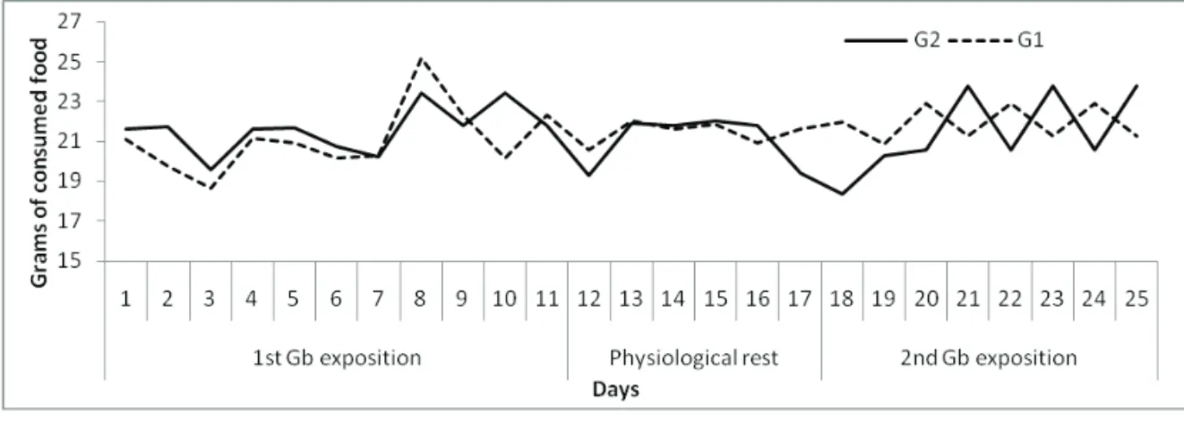
Food and water consumption
Evaluation of food and water intake of both G1 and G2 groups during the 25 days of the protocol (1stE/
Rest/2ndE) was performed. During the three stages of the protocol, it was not observed signiicant differences in
PR: 21.43±0.57 g; 2ndE: 21.91±0.88 g) and G2 (1stE:
21.59±1.17 g; PR: 21.03±1.31 g; 2ndE: 21.44±2.04 g)
(Figure 2).
Figure 1. Behavior of individual and mean body weight
throughout the experiment (…..) weights (─) mean of the
group. Weight increasing of 2.75% for the control group (G2) (p=0.023) and 5% for the experimental/Gb (G1) (p=0.001).
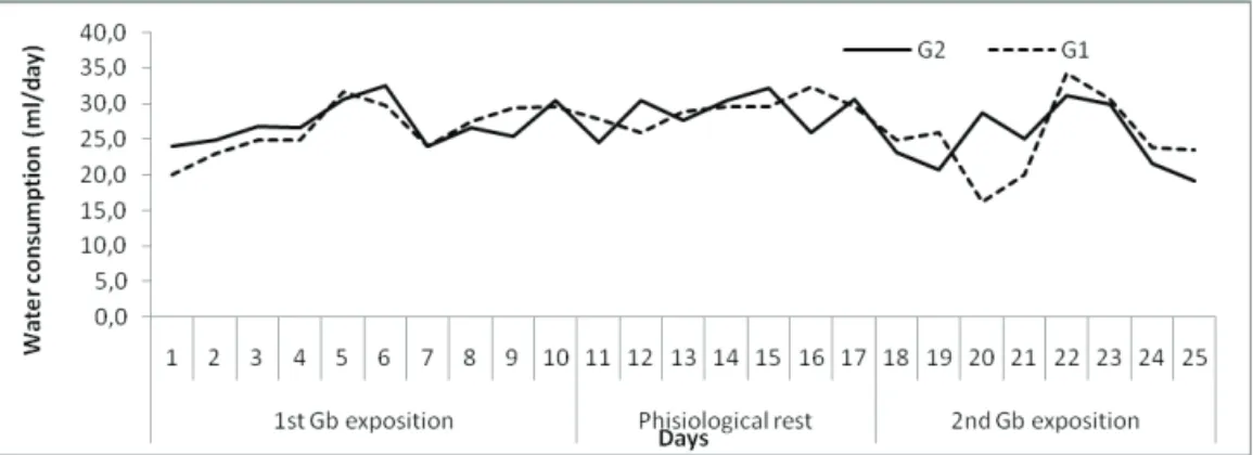
When evaluating the consumption of water during the same period, no differences between the groups were observed (Figure 3). The means for the three experimental periods were presented in Table 1.
Table 1. Mean food intake by five animals during the three experimental periods (1stE, PR and 2ndE).
First GBE Exposure G1 20.96±1.77*
G2 21.57±1.23
p value 0.092107
Physiological rest 21.55±0.61 21.13±1.23 0.081144
Second GBE exposure 21.91±0.88 21.44±2.04 0.492324
* mg/gbw/ day
Urinary excretion
When analyzing each period separately, our results demonstrate an interference of GBE on the urinary system of the rats. During the first and second periods of exposure, the mean urinary volume of the ten animals pertaining to G1 (experimental/Gb) is significantly smaller (0.228±0.068 mL/day) than that of the media of the animals of G2 (0.259±0.076 mL/ day) (p=0.0246) with a reduction the 11,97% when compared a control group. When the GBE treatment was interrupted an inversion occurs and animals of G1 (experimental/Gb) present a significantly larger urinary volume (p=0.000151) (0.17±0.010 mL/day) than that the animals of G2 (0.15±0.001 mL/day) with a smaller daily variation (p<0.01). With the reintroduction (2ndE)
of GBE, the urinary volume differs between the groups again (p=0.003162) with a reduction in urine volume in
Figure 2. Daily mean food intake of the media/animals of each group.
Immunohystochemistry
To check the integrity of blood vessels and the presence of angiogenesis in the kidney, immunohystochemistry assays to the basement membrane and smooth muscle were performed. Results
were negative for SMαA in the kidney samples of both
G1 and G2, and no vascular lesion or any other tissue alterations were present in the samples of the two groups (Figure 8).
Staining kidney sections of animals pertaining to G1 (experimental/Gb) and G2 (control) for lamimin showed the integrity of basal membrane with no differences at the glomeruli or tubules (Figure 9). However, staining was positive to VEGF in kidney sections of G1, but not of G2 (Figure 10). The darker areas seen in sections of G1 indicate a Vascular Endothelial Growth (light alteration), which was not observed in control animals.
Transmission electron microscopy
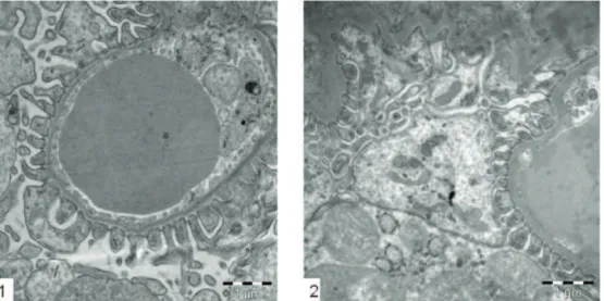
In treated animals with GBE, Glomerulus Basemen Membranes and Podocytes presented similar architecture to the control animals (Figure 11).
the experimental/Gb group.
The analysis of the kinetics of the urinary l ow shows a signii cant decrease in the urinary volume between the i rst and tenth day of the protocol for G1 group. On
the 11th day, the urinary volume of G1 (experimental/
Gb) presented a slight increase and maintained stable up to the 18th day, when the GBE was reintroduced (2ndE).
During this period, animals of G2 (control) presented a urinary volume similar to that of the end of the 1stE period.
During the second GBE exposure, a reduced of the mean urinary volume was observed in G1, whereas animals of G2 presented little variation, maintaining levels of urinary
l ow with a signii cant decrease (Figure 4).
Histomorphology
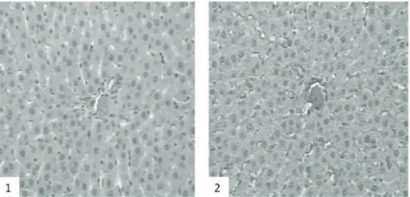
Comparing the microscopic morphology of the spleen of animals pertaining to G1 and G2, no alterations were observed in the white or red pulp, in the vascular system or in primary follicles (Figure 5). Likewise, no alteration was observed in the vascular (arteries and veins) or cellular (hepatocytes) structure of the liver (Figure 6). On the other hand, when examining the kidney, animals pertaining to G1 presented vascular congestion of the glomerulus and tubules (Figure 7).
Figure 4. Daily mean water consumption of the animals of each group.
Figure 6. Photomicrography of liver (20x). 1. Experimental/Gb animals (G1); 2. Control animals (G2). We cannot see any alterations in the hepatic cells or arterial structure in both groups.
Figure 7. Photomicrography of kidney (20x). 1. Experimental/Gb animals (G1); 2. Control animals (G2). We can see the concentration of red cells in the G1 are superior than the control group.
Figure 8. Photomicrography of kidney (20x). Immunohystochemistry smooth muscle α-actin. 1. Experimental/Gb animals (G1); 2.
Figure 9. Photomicrography of kidney (40x). Immunohystochemistry, laminin.
1. Experimental animals/Gb (G1); 2. Control animals (G2). No differences in the membrane structures in the both groups.
Figure 10. Photomicrography of kidney (20x). Immunohystochemistry VEGF (vascular endothelial growth factor). 1. Experimental/ Gb animals (G1); 2. Control animals (G2). In the control group we cannot observe the presence of VEGF as we can see in the
GBE group with the i xed color. There is a positive staining to the VEGF in experimental group (panel 2) when compared to the
control animals (panel 1).
Discussion and Conclusion
Although GBE has been reported in the traditional Chinese medicine as an adjuvant in weight reduction (Tian et al., 2004), our results do not confirm this finding. In our experiment, animals that received GBE for a total of eighteen days presented a significantly higher mean weight gain (18.76 g) when compared to the control group (10.46 g) during this period. Tian et al. (2004), examined 31 Chinese medicinal herbs used in weight reducing prescriptions of Traditional Chinese Medicine, and found that the inhibitory potencies of the active components of tuber fleece flower root, parasitic loran thus, green tea leaf and Ginkgo biloba leaf were similar to or greater than cerulenin and C75 (Pizer et al., 1998; Loftus et al., 2000; Kreider et al., 2010). It is possible to suggest that the treatment with GBE does not interfere in the weight gain or in the normal development of lean young animals.
Our data indicate that the kidneys of the animals submitted to the GBE treatment showed a more intense hematoxylin eosin (HE) coloring than the control animals, suggesting that some type of changes in histoarchitecture could be taking place. To elucidate these changes, it was performed the renal section to immunohystochemical assays, evaluating
SMαA, laminin and VEGF. No staining was observed for the first two antibodies (SMαA and laminin),
indicating that GBE either does not interfere. During
normal development, SMαA is expressed in mesangial
cells and is down regulated to a minimal expression in normal adult cells (Takeji et al., 2006). The only alteration observed in our histological analysis other than the more intense HE staining of kidneys pertaining to G1 (experimental/Gb) was the immunohistochemical staining for VEGF. Two processes may lead to blood vessel production: vasculogenesis and angiogenesis. Both the promotion and the reduction of angiogenesis have been reported with the use of GBE (Sun et al., 2009; Trompezinski et al., 2010).
Sun et al. (2009) reported that the GBE exerts protective effect on secondary cerebral ischemic injury after Sub Arachnoids Hemorrhage via the promotion of the expression of VEGF. On the other hand, Trompezinski et al. (2010) showed that GBE exerts a potent inhibition on VEGF levels in activated cells. GBE down regulates VEGF and IL-8 (interleukin 8) levels in a cumulative
manner in Tumor necrosis factor-α (TNFα)-stimulated
normal human keratinocytes. Thus, Trompezinski et al. (2010) suggests that GBE may contribute to moderate the
inlammatory processes in skin diseases associated with
angiogenesis.
Rodriguez de Turco et al. (1993) presented data indicating that GBE inhibits stress-induced polydipsia in rats due to the stress of daily handling and intubation;
whereas Porsolt et al. (1990) show that the repeated administration of GBE inhibits emotional hypophagia in mice due to the anxiolytic-like activity of this herb. In the study performed by Rodriguez de Turco et al. (1993) the GBE was dissolved in 5% ethanol and control animals received equivalent volumes of 5% ethanol alone. In our model, feeding or drinking was not altered by GBE
although weight gain was been larger signiicant for the
G1 (experimental/Gb) (p=0,001).
More insights into the regulation of the kidney function can provide not only a better understanding of pathophysiological processes, but also eventually, the development of new therapeutic approaches (Vallon et
al., 2006). On a recent search of the speciic literature
there were less than ten references which appeared upon a cross-search using "nephrotoxicity" and Ginkgo biloba
as keywords. All papers showed that this herb is beneicial
and protects the kidney from nephrotoxic substances such as uranium (Yapar et al., 2010), adriamycin (Abd-Ellah & Mariee, 2007), cisplatin (Fukaya & Kanno, 1999; Gulec et al., 2006), gentamicine (Naidu et al., 2000), and vancomycin (Celik et al., 2005). On the other hand, some herbal medications have potentially harmful side effects, as well as adverse interactions with prescription drugs (Qato, 2008). Despite the widespread concurrent use of conventional and alternative medicines, documented drug-herb interactions are sparse (Rai, 2004; Sparreboom et al., 2004; Zlotogorski & Littner, 2004; Yale & Glurich, 2005).
Renal excretion of fluid and electrolytes are under the control of a systemic neurohumoral system that plays a central role in body homeostasis (Atherton, 1972). Their fine control is regulated by specific factors and hormones such as arachidonic acid and its active metabolic products, which can modulate hemodynamic homeostasis (Feraille & Doucet, 2001). In the kidney, the eicosanoids, active products derived from arachidonic acid, not only regulate inflammatory processes, but also have the potential to regulate the kidney (Mullins et al., 2006). The PAF causes increased vascular permeability in several tissues and so how GBE (containing the ginkgolides) can cause reduced loss of water (Wang et al., 2006).
One of the properties anti-inlammatory in vivo of GBE is through the inhibition of Thromboxane-2 (TXB2) synthesis (Kudolo et al., 2004). The GBE also shows a potent antioxidant in both normo-and hypercholemic subjects, possibly by inhibiting cyclooxygenases-1 (COX-1) isoform activity. Collectively, these data suggest that GBE is comparable to acetylsalicylic acid in terms of the inhibition of TXB2 and COX-1 (Kudolo et al., 2004). Attention of the side effects of conventional AINE’S
(Non-steroidal anti-inlammatory drugs) including
of inducible nitric oxide synthase and cyclooxygenase-2 via suppression of nuclear factor-kappaB in RAW 264.7 cells induced by lipopolysaccharide stimulation. One of the molecules derived from Ginkgo biloba
leaves, a biflavone known as Ginkgetin, inhibits cyclooxygenases-2 and 5-lipoxygenase in mouse bone marrow-derived mast cells (Son et al., 2005; Park et al., 2006).
The importance of the cyclooxygenases pathways in the control of blood pressure is illustrated by the use of AINE’S in humans, which inhibit both cyclooxygenases 1 or 2 isoforms and cause Na+
retention and hypertension. COX-1 is constitutively expressed in a variety of tissues, including the kidney (Cheng & Harris, 2004). Probably, our data could be explained by the inhibitory effects of GBE on the COX and PAF pathway of arachidonic metabolism, but future researches are necessary, to prove this possibilities.
Another point that needs attention in the clinical setting is the fact that older populations are the main consumers of GBE and others prescription drugs. Despite concerns about drug safety, current information on both the use of prescription and over-the-counter medications, and dietary supplements is limited. Among prescription medication users, concurrent use of over-the-counter medications was 46% with a concurrent use of dietary supplements by 52% of the survey population (Qato et al., 2008). The fact of GBE promote reduction in the urinary volume it is possible to suggest that this extract may influence on the depuration time and to prolong the exposition time of drugs, consequently, affecting drug-herb interactions, but future researches are necessary, to prove this possibilities. The limitation of my study was no all questions were answers and we need future studies should look at measuring PAF, COX-1 and TXB2 to probably explain the reduced urinary volume. Avail what would have happened if we had treated the rats for a longer or shorter time period.
Concluding, as GBE may promote alterations in volume of the urine excretion. The introduction of GBE (therapeutic dose) in healthy rats may promote alterations in the physiology of the kidney, but no sufficient to modify the glomerulus architecture, including at ultra-structural level.
Reference
Abd-Ellah MF, Mariee AD 2007. Ginkgo biloba leaf extract (GBE 761) diminishes adriamycin-induced hyperlipidaemic nephrotoxicity in rats: association with nitric oxide production. Biotechnol Appl Bioc 46: 35-40.
Atherton JC 1972 . Renal physiology. Br J Anaesth 44: 236-245.
Blumenthal M, Busse W 1998: The complete German
commission E monographs: Therapeutic guide to herbal medicines. Boston: American Botanical Council.
British Herbal Medicine Association 1996. Scientific committee. British herbal pharmacopoeia. London. British Herbal Medicine Association.
Celik I, Cihangiroglu M, Ilhan N, Akpolat N, Akbuluty HH 2005. Protective effects of different antioxidants and amrinone on vancomycin-induced nephrotoxicity. Basic Clin Pharmacol 5: 325-332.
Cheng HF, Harris RC 2004. Cyclooxygenases, the kidney, and hypertension. Hypertension 3: 525-530.
Feraille E, Doucet A 2001. Sodium-potassium-adenosinetriphosphatase-dependent sodium transport in the kidney: hormonal control. Physiol Rev 1: 345-418. Fukaya H, Kanno H 1999. Experimental studies of the
protective effect of Ginkgo biloba extract (GBE) on cisplatin-induced toxicity in rats. Nippon Jibiinkoka Gakkai Kaiho 102: 907-917.
Gulec M, Iraz M, Yilmaz HR, Ozyurt H, Temel L 2006. The effects of Ginkgo biloba extract on tissue adenosine deaminase, xanthine oxidase, myeloperoxidase, malondialdehyde, and nitric oxide in cisplatin-induced nephrotoxicity. Toxicol Ind Health 3: 125-130. Kreider RB, Wilborn CD, Taylor L, Campbell B, Almada
AL, Collins R, Cooke M, Earnest CP, Greenwood M, Kalman DS, Kerksick CM, Kleiner SM, Leutholtz B, Lopez H, Lowery LM, Mendel R, Smith A, Spano M, Wildman R, Willoughby DS, Ziegenfuss TN, Antonio J 2010. ISSN exercise & sport nutrition review: research & recommendations. J Int Soc Sports Nutr 7: 1-44.
Kudolo GB, Delaney D, Blodgett J 2005. Short-term oral ingestion of Ginkgo biloba extract (GBE 761) reduces malondialdehyde levels in washed platelets of type 2 diabetic subjects. Diabetes Res Clin Pr 68: 29-38. Kudolo GB, Wang W, Barrientos J, Elrod R, Blodgett J
2004. The ingestion of Ginkgo biloba extract (GBE 761) inhibits arachidonic acid-mediated platelet aggregation and thromboxane B2 production in healthy volunteers. J Herb Pharmacother 4: 4-13. Loftus TM, Jaworsky DE, Frehywot GL, Townsend CA,
Ronnett GV, Lane MD, Kuhajda FP 2000. Reduced food intake and body weight in mice treated with fatty acid synthase inhibitors. Science 288: 2379-2381. Macedo CS, Lerco MM, Capelletti SM, Silva RJ, Pinheiro DO,
Spadella CT 2007. Reduction of podocytes number in late diabetic alloxan nephropathy: prevention by glycemic control. Acta Cir Bras 22: 337-341.
Major RT 1967. The ginkgo, the most ancient living tree. The resistance of Ginkgo biloba L. to pests accounts in part for the longevity of this species. Science 794: 1270-1273.
Experimental model to assess possible medicinal herb interaction with a radiobiocomplex: qualitative and quantitative analysis of kidney, liver and duodenum isolated from treated rats. Food Chem Toxicol 45: 19-23.
Mullins LJ, Bailey MA, Mullins JJ 2006. Hypertension, kidney, and transgenics: a fresh perspective. Physiol Rev 2: 709-746.
Naidu MU, Shifow AA, Kumar KV, Ratnakar KS 2000. Ginkgo biloba extract ameliorates gentamicin-induced nephrotoxicity in rats. Phytomedicine 3: 191-197. Park YM, Won JH, Yun KJ, Ryu JH, Han YN, Choi SK,
Lee KT 2006. Preventive effect of Ginkgo biloba extract (GBE) on the lipopolysaccharide-induced expressions of inducible nitric oxide synthase and cyclooxygenase-2 via suppression of nuclear factor-kappaB in RAW cells. Biol Pharm Bull 5: 985-990. Pizer ES, Chrest FJ, DiGiuseppe JA, Han WF 1998.
Pharmacological inhibitors of mammalian fatty acid synthase suppress DNA replication and induce apoptosis in tumor cell lines. Cancer Res 20: 4611-4615.
Porsolt RD, Martin P, Lenègre A, Fromage S, Drieu K. 1990. Effects of an extract of Ginkgo Biloba (GBE 761) on learned helplessness and other models of stress in rodents. Pharmacol Biochem Be 36: 963-971. Qato DM, Alexander GC, Conti RM, Johnson M, Schumm
P, Lindau ST 2008. Use of prescription and over-the-counter medications and dietary supplements among older adults in the United States. JAMA-J Am Med Assoc 24: 2867-78.
Rai J, 2004. Adverse interactions between low-dose aspirin/ warfarin and garlic/ginseng/Ginkgo biloba. Indian Heart 2: 176.
Rodriguez de Turco EB, Droy-Lefaix MT, Bazan NG 1993. GBE 761 inhibits stress-induced polydipsia in rats. Physiol Behav 53: 1001-1002.
Ruiz-Ortega M, Lorenzo O, Egidio J 1998. Angiotensin III up-regulates genes involved in kidney damage in mesangial cells and renal interstitial fibroblasts. Kidney Int Suppl 68: S41-45.
Son JK, Son MJ, Lee E, Moon TC, Son KH, Kim CH 2005. Ginkgetin, a biflavone from Ginko biloba leaves, inhibits cyclooxygenases-2 and 5-lipoxygenase in mouse bone marrow-derived mast cells. Biol Pharm Bull 12: 2181-2184.
Sparreboom A, Cox MC, Acharya MR, Figg WD 2004. Herbal remedies in the United States: potential adverse interactions with anticancer agents. J Clin Oncol 22: 2489-2503.
Sun BL, Hu DM, Yuan H, Ye WJ, Wang XC, Xia ZL 2009. Extract of Ginkgo biloba promotes the expression of
VEGF following subarachnoid hemorrhage in rats. Int J Neurosci 7: 995-1005.
Takeji M, Moriyama T, Oseto S, Kawada N, Hori M, Imai E 2006. Smooth muscle alpha-actin deficiency in myofibroblasts leads to enhanced renal tissue fibrosis. J Biol Chem 52: 40193-40200.
Tang J, Sun J, Zhang Y, Li L, Cui F, He Z 2007. Herb-drug interactions: Effect of Ginkgo biloba extract on the pharmacokinetics of theophylline in rats. Food Chem Toxicol 12: 2441-2445.
Teske M, Trentini AMM 1995. Herbarium: Compêndio de Fitoterapia Curitiba: Herbarium Laboratório Botânico.
Tian WX, Li LC, Wu XD, Chen CC 2004. Weight reduction by Chinese medicinal herbs may be related to inhibition of fatty acid synthase. Life Sci 19: 2389-2399. Trompezinski S, Bonneville M, Pernet I, Denis A, Schmitt D,
Viac J 2010. Gingko biloba extract reduces VEGF and CXCL-8/IL-8 levels in keratinocytes with cumulative effect with epigallocatechin-3-gallate. Arch Dermatol Res 3: 183-189.
Valli G, Giardina EG 2002. Benefits, adverse effects and drug interactions of herbal therapies with cardiovascular effects. J Am Coll Cardiol 7: 1083-1095.
Vallon V, Mühlbauer B, Osswald H 2006. Adenosine and kidney function. Physiol Rev 86: 901-940.
Wang JY, Yin XX, Wu YM, Tang DQ, Gao YY, Wan MR 2006. Ginkgo biloba extract suppresses hypertrophy and extracellular matrix accumulation in rat mesangial cells. Acta Pharmacol Sin 27: 1222-1230.
Yale SH, Glurich I 2005. Analysis of the inhibitory potential of Ginkgo biloba, Echinacea purpurea, and Serenoa repens on the metabolic activity of cytochrome P450 3A4, 2D6, and 2C9. J Altern Complem Med 11: 433-439.
Yapar K, Cavusoglu K, Oruç E, Yalçin E 2010. Protective role of Ginkgo biloba against hepatotoxicity and nephrotoxicity in uranium-treated mice. J Med Food 13: 179-188.
Zlotogorski HA, Littner M 2004. Potential risks, adverse effects and drug interactions associated with herbal medicine in dental patients. Refuat Hapeh Vehashinayim 21: 25-41.
*Correspondence
Monica Dalmacio Silveira Campos Câmara
Programa de Pós-graduação em Ciências Médicas, Hospital Universitário Antônio Pedro, Universidade Federal Fluminense
Rua Marquês do Paraná, 303, 4º andar, Centro, 24033-900 Niterói-RJ, Brazil