DOI: 10.1590/0004-282X20150154
ARTICLE VIEW AND REVIEW
When should MELAS
(Mitochondrial myopathy,
Encephalopathy, Lactic Acidosis, and
Stroke-like episodes
) be the diagnosis?
Quando o diagnóstico deveria ser MELAS (Miopatia mitocondrial, encefalopatia, acidose
lática, e episódios semelhantes a acidente vascular cerebral)?
Paulo José Lorenzoni, Lineu Cesar Werneck, Cláudia Suemi Kamoi Kay, Carlos Eduardo Soares Silvado, Rosana Herminia Scola
he irst description of cases with clinical features suggested MELAS was in 19751,2. he patients common point was the pres
-ence of mitochondrial myopathy associated with brain changes, such as mental retardation, seizures, myoclonus, ophthalmople
-gia, retinitis pigmentosa, blindness, calciication in basal ganglia and sudden hemiplegia suggestive of stroke1,2. In the following
years other cases with similar indings were added to the litera
-ture, and in 1984, Pavlakis et al., better characterize the patients who had normal early development, short stature, seizures, alter
-nating hemiparesis, hemianopsia and cortical blindness, through
the framework that called MELAS (Mitochondrial myopathy, Encephalopathy, Lactic Acidosis, and Stroke-like episodes)3.
In Brazil, the irst report of MELAS was made by Werneck et al. in 1987 who describe a boy who had recurrent episodes of seizures, headache and vomiting associated with focal neuro
-logical signs4. he brain computed tomography showed lesions
similar to ischemic stroke and calciication of the basal ganglia, ragged-red iber (RRF) in muscle biopsy, elevation of lactic acid and determination of enzyme activity consistent with the re
-spiratory chain complex IV deiciency also were found4.
Universidade Federal do Paraná, Hospital de Clínicas, Departamento de Clínica Médica, Serviço de Neurologia e Doenças Neuromusculares, Curitiba PR, Brazil.
Correspondence: Rosana Herminia Scola; Serviço de Doenças Neuromusculares, Hospital de Clínicas - UFPR; Rua General Carneiro, 181 / 3º andar; 80060-900 Curitiba PR, Brasil; E-mail: [email protected]
Conflict of interest: There is no conflict of interest to declare. Received 02 June 2015; Accepted 23 June 2015.
ABSTRACT
Mitochondrial myopathy, Encephalopathy, Lactic Acidosis, and Stroke-like episodes(MELAS) is a rare mitochondrial disorder. Diagnostic criteria for MELAS include typical manifestations of the disease: stroke-like episodes, encephalopathy, evidence of mitochondrial dysfunction (laboratorial or histological) and known mitochondrial DNA gene mutations. Clinical features of MELAS are not necessarily uniform in the early stages of the disease, and correlations between clinical manifestations and physiopathology have not been fully elucidated. It is estimated that point mutations in the tRNALeu(UUR) gene of the DNAmt, mainly A3243G, are responsible for more of 80% of MELAS cases. Morphological changes seen upon muscle biopsy in MELAS include a substantive proportion of ragged red fibers (RRF) and the presence of vessels with a strong reaction for succinate dehydrogenase. In this review, we discuss mainly diagnostic criterion, clinical and laboratory manifestations, brain images, histology and molecular findings as well as some differential diagnoses and current treatments.
Keywords: MELAS, mitochondria, myopathy, stroke, encephalopathy, genetics.
RESUMO
Miopatia mitocondrial, encefalopatia, acidose lática, e episódios semelhantes a acidente vascular cerebral (MELAS) é uma rara doença mitocondrial. Os critérios diagnósticos para MELAS incluem as manifestações típicas da doença: episódios semelhantes a acidente vascular cerebral, encefalopatia, evidência de disfunção mitocondrial (laboratorial ou histológica) e mutação conhecida em genes do DNA mitocondrial. Na fase inicial da doença, as manifestações clínicas podem não ser uniformes, e sua correlação com a fisiopatologia não está completamente elucidada. Estima-se que as mutações de ponto no gene tRNALeu(UUR) do DNAmt, principalmente a A3243G, sejam responsáveis por cerca de 80% dos casos de MELAS. As alterações morfológicas na biópsia muscular incluem uma grande proporção de fibras vermelhas rasgadas (RRF) e presença de vasos com forte reação para succinato desidrogenase. Nesta revisão, são discutidos os principais critérios diagnósticos, manifestações clínicas e laboratoriais, imagens cerebrais, padrões eletrofisiológicos, histológicos e alterações moleculares, bem como alguns dos diagnósticos diferenciais e tratamentos atuais.
WHAT IS THE DIAGNOSTIC CRITERION?
he cases previously published with a diagnosis suggestive of MELAS have been reviewed by Hirano et al. in 1992 with the purpose of obtaining diagnostic criteria for this group of pa
-tients5. After this literature review, the diagnostic criteria for
MELAS must include the following events: (1) signs of enceph
-alopathy, often with dementia and seizures, (2) episodes similar to stroke (stroke-like episodes) in young age, and (3) biochemi
-cal evidence of mitochondrial dysfunction such as lactic acido
-sis or RRF in muscle biopsy (Table 1)5. he diagnosis may also
be supported if at least two of the following was present: normal development, recurrent headache and vomiting (Table 1) 5.
Recently, the MELAS Study Group in Japan included oblig
-atory presentation of stroke-like episodes associated to evi
-dence of mitochondrial dysfunction and known mitochondrial gene mutations as diagnostic criterion.In Japan, the disease is also deined using the Japanese diagnostic criteria for MELAS (Table 2)6. Diferent of the Hirano et al. diagnostic criteria,
Japanese’s diagnostic criteria did not consider signs of enceph
-alopathy and includes mitochondrial gene mutations5,6.
WHAT IS THE PATHOGENESIS?
On clinical evaluation, stroke-like episodes of patients with MELAS are indistinguishable from those reported by patients with acute ischemic stroke, but the pathogene
-sis of these lesions in patients with MELAS is diferent and not fully elucidated. he two main hypotheses being consid
-ered are: (1) ischemic, with suggestion of a “mitochondrial
angiopathy” caused by mitochondrial dysfunction in smooth muscle cells of the small cerebral vessels leading to vascular occlusion with neuronal loss7,8,9,10; and (2) metabolic, due to a
“mitochondrial cytopathy”, that trigger energy failure of brain tissue causing neuronal damage11,12,13.
Currently, the mechanisms that trigger these episodes are correlated to a combination of these two hypotheses, in which both (neuronal and vascular dysfunction) are responsible for the pathogenesis of stroke-like episodes14. Iizuko and Sakai
in 2005 proposed a possible mechanism for the formation of stroke-like episodes, based on the main pathological indings related to these episodes (Figure 1): headache, seizures, fo
-cal hyperemia, vasogenic edema, lesion progression after the stroke-like episode and neuronal loss14.
WHAT ARE THE GENETIC ABNORMALITIES?
In 1990, Goto et al. and Kobayashi et al., described a point mutation of mtDNA afecting the gene encoding the leucine tRNA (UUR) (tRNALeu(UUR)) by the exchange at position 3243
of the nucleotide A by G (A3243G) in muscle of MELAS pa
-tients15,16. he tRNALeu(UUR), also known as MT-TL1, is located
between nucleotides 3230 and 3304 and is responsible for de
-coding of UUR codons (R = A or G)17. he A3243G mutation
afects the stability of the structure, methylation, aminoacyl
-ation and codon recognition of the tRNALeu(UUR), more sharp
-ly than other mutations in this gene14,17,18. his could reduce
the functional level of tRNALeu(UUR) that participates in the
process of mitochondrial protein synthesis14. After this irst
description other studies have shown that the A3243G muta
-tion was responsible for most cases of MELAS19,20,21.
In the years following the discovery of mutation A3243G, a second point mutation by substitution of T for C nucleotide at position 3271 (T3271C) in the tRNALeu(UUR) of the mtDNA
was also found in patients with MELAS22. Over the years, oth
-er point mutations in mtDNA w-ere found in patients with MELAS, revealing their genetic heterogeneity. However, the A3243G mutation still is responsible for remained about 80% of patients with MELAS and T3271C by approximately 7.5%, but up to 10% of patients with MELAS the mutations of mtDNA
Table 1. Hirano’s diagnostic criteria for MELAS (Hirano et al., 1992) 5.
Absolute criteria* Supportive criteria**
Encephalopathy (dementia and/or seizures) Normal development Stroke-like episodes in young age Recurrent headache Evidence of mitochondrial dysfunction
(lactic acidosis or ragged-red fibers in muscle biopsy)
Recurrent vomiting
*definitive diagnosis requires all absolute criteria; **diagnosis may also be more secure if at least two of three supportive criteria were present.
Table 2. Japanese’s diagnostic criteria for MELAS (Yatsuga et al., 2012) 6.
Category A. Clinical findings of stroke-like episodes Category B. Evidence of mitochondrial dysfunction
Headache with vomiting High lactate levels in plasma and/or cerebral spinal fluid or
deficiency of mitochondrial-related enzymes activities** Seizures
Hemiplegia Mitochondrial abnormalities in muscle biopsy***
Cortical blindness or hemianopsia Definitive gene mutation related to MELAS****
Acute focal lesion observed via brain imaging*
Definitive MELAS: two items of category A and two items of category B (four items or more) Supportive MELAS: one item of category A and two items of category B (at least three items)
remain unknown (Table 3)7,16. he mutations in DNAmt genes
coding to mitochondrial complex I, as ND5 gene, have been appointed as the second more frequent (Table 3)23. he pres
-ence of mutation by rearrangement of the deletion type, large or small, has rarely been described in mtDNA associated to MELAS phenotype. he presence of nuclear DNA gene muta
-tions has been also rarely associated to MELAS.
Although most MELAS patients have the A3243G point mutation, this mutation is not speciic for this group of pa
-tients, also found in patients with progressive external ophtalmoplegia (PEO), cardiomyopathy, sensorineural deaf
-ness and diabetes mellitus with maternal inheritance24.
Moreover, the clinical and laboratory evaluation of other fam
-ily members have found relatives with incomplete features for MELAS, usually with maternal inheritance pattern. However, genetic studies have showed that maternal relatives clinical
-ly asymptomatic but with the A3243G mutation, might have mitochondrial dysfunction with RRF in muscle biopsy5,19. In
addition, patients with MELAS concomitant with other mi
-tochondrial diseases such as myoclonic epilepsy associated with ragged red ibers (MERRF), Leigh syndrome and Leber’s hereditary optic neuropathy have been reported with associ
-ated mutation of the mtDNA. hese data contribute to state that mitochondrial diseases caused by mtDNA point muta
-tions have heterogeneous phenotype7.
he proportion of mutant mtDNA usually is diferent in tissues of MELAS patients associated to mutations of tRNALeu(UUR) 5,7,19,20. For these reasons the use of clinically af
-fected tissues, such as muscle, to perform the extraction of mtDNA for the molecular study could be recommended due to better results5,7,19,20,25. If clinically unafected tissues were
used, such as peripheral blood leukocytes, the pathogenic mutation may be undetectable5,7,19,20,25.
he methods used to detect each mutation may vary among laboratories (often PCR, PCR/RFLP or direct sequenc
-ing), but usually is recommended irst screening to targeted mutations to then perform the full sequence analysis.
WHAT ARE THE CLINICAL FEATURES?
Clinical symptoms are highly variable among patients with mitochondrial diseases. Some of these clinical indings may be absent in the early stage of the disease, while in advanced dis
-ease patients usually have more uniform clinical manifestations. Although the onset of clinical manifestation often occurs in childhood and early adulthood, a late onset in adults is not uncommon in patients with MELAS. Moreover, the age of stroke-like episodes may be diferent among afected mem
-bers of the same family26. Children psychomotor develop
-ment is usually normal in almost all patients with MELAS.
ion homeostasis changes? membrane instability?
pericapillary plasma extravasation
trigeminovascular activation
Headache
increase in anaerobic glycolysis
lactate production
Focal hyperemia Neuronal hyperexcitability
increasing energy demand
Epileptic seizure
increasing capillary permeability
Vasogenic edema
in the presence of mitochondrial capillary angiopathy
Energy imbalance
between demand and availability of ATP
Slowly progressive spread
of stroke-like lesions
Neuronal loss
with laminar (pseudo-laminar) distribution
CYTOPATHY
neurons astrocytes
Capillary ANGIOPATHY?
BBB breakdown
BBB: blood-brain barrier.
Stroke-like episodes are diagnostic criteria for MELAS and is expected that all patients have this clinical manifesta
-tion5,6. he stroke-like episode may occur alone or in associa
-tion with signs of the encephalopathy, such as seizures5,6.he
clinical outcome of stroke-like episodes is more benign, with improvement of symptoms in a few months, but the symp
-toms related encephalopathy, such as dementia and seizures, may progressively worsen6,12,14,27.
he presence of progressive dementia has also been found associated with changes in cerebral perfusion even in the absence of stroke-like episode, but with marked corti
-cal atrophy in the chronic phase of the disease characterized neuronal loss, similar to what occurs in vascular demen
-tia27,28. Partial seizures are commonly reported in MELAS
associated with mutations in the tRNALeu(UUR) gene, similar
other mitochondrial diseases as MERRF or PEO, but general
-ized seizures also occurs in MELAS29,30.
Headaches occur in the majority of afected individuals and are often severe during the acute phase of the stroke-like25,29,31.
Other multisystemic dysfunctions could be ind in MELAS patients: psychiatric manifestations, cerebellar atax
-ia, myoclonus, neuropathy, ocular motility abnormalities, exercise intolerance, pigmentary retinopathy, optic atrophy, deafness, short stature, diabetes mellitus, alteration of car
-diac conduction, cardiomyopathy, gastrointestinal changes, among others5,6,20,27,25,29. Cardiac involvement can occur in up
to 50% of patients with MELAS32,33. However, these disorders
are not the main clinical manifestation of the disease. MELAS patients should be followed at regular intervals to monitor disease progression and the appearance of new symp
-toms. Annual ophthalmologic, audiologic, cardiologic (electro
-cardiogram and echo-cardiogram), and endocrinologic (fasting blood sugar and TSH) assessments also are recommended25.
Children of woman with mtDNA mutation can inherit the mutation34. he risk to other family members depends on
the genetic status of the mother25,34. However, it is appropri
-ate to evalu-ate other family members because the phenotype
manifestation results from a combination of factors, due to the diferent inherit percentages of mutant mtDNA, and therefore, can have a wide range of clinical symptoms from asymptomatic to full manifestation.
WHAT ARE THE IMAGING FEATURES?
Brain conventional radiological studies, such as CT or MRI, in patients with MELAS reveal changes in gray matter more than in white matter, predominate in the occipital, pa
-rietal and temporal lobes, which simulate ischemic stroke (Figure 2)14,35,36. Most injuries occur in the cortical region of
the cerebral hemispheres and more rarely in the cerebellum or basal ganglia32,36. hese brain lesions can be unilateral or
bilateral (Figure 2). However, these areas do not occur in a speciic vascular territory and angiographic studies show that the vessels in the afected regions have blood low and sometimes are dilated5,32,36. Brain MRI revealed that the lesion
in the acute phase begins in the temporal region on focal form, but in 2 to 3 weeks may progress to the parietal and occipital regions, in one third of patients37. his shows that
even after the stroke episode the disease process continues to progress37. he changing pattern of lactic acid concen
-tration between these regions during the clinical outcome of stroke-like episodes, was also observed in patients with
MELAS38. hese indings may mean that the vulnerability of
neurons to mitochondrial dysfunction may be higher in these regions. he exact mechanism of the disease predilection for certain locations in the central nervous system has not been elucidated, but the possibility of heteroplasmy in brain tissue was ruled-out by genetic studies when comparing tis
-sue from diferent brain areas11,14,39. Studies using spectrosco
-py show increased lactic acid in acute lesions while the use of magnetic resonance difusion can reveal increased coef
-icient of difusion35,40,41. Both methods being more sensitive
than conventional in the acute phase of the injury35,40,41. he
MRI images are compatible with subcortical laminar necro
-sis in the subacute phase of episodes36,37.
he presence of multiple focal areas of cortical necro
-sis associated with difuse cortical atrophy in both cerebral hemispheres and cerebellum, are the most frequent patho
-logical indings in the brain of patients with MELAS, while the brainstem is rarely afected32,36,42. he presence of cortical
atrophy and calciications in the basal ganglia are also found in the progression of some patients36,42.
WHAT ARE THE LABORATORIAL AND BIOCHEMICAL FEATURES?
he concentration of cerebrospinal luid (CSF) protein may be elevated but rarely surpasses 100 mg/dL25,29. he lactic acid
level in blood and CSF is elevated during, or shortly after, the
Table 3. Summary of mutations in mitochondrial DNA in the
two most frequent genes associated with MELAS.
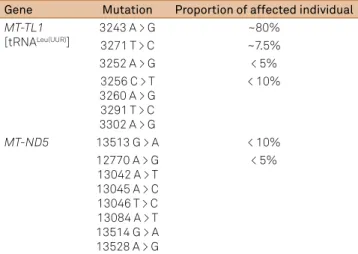
Gene Mutation Proportion of affected individual
MT-TL1
[tRNALeu(UUR)]
3243 A > G ~80%
3271 T > C ~7.5%
3252 A > G < 5% 3256 C > T
3260 A > G 3291 T > C 3302 A > G
< 10%
MT-ND5 13513 G > A < 10% 12770 A > G
13042 A > T 13045 A > C 13046 T > C 13084 A > T 13514 G > A 13528 A > G
stroke-like episodes in virtually all patients with MELAS20,25.
he level of ventricular lactic acid measured by spectroscopy, is usually increased in most patients with MELAS and their families, and this increase is associated with the severity of neurological symptoms43. Other tests may be used in MELAS
investigation, such as serum levels of creatine kinase (CK), but their indings are not speciic for MELAS, even though helps especially in the diferential diagnosis with other diseases29.
Serum CK may be slightly increased in some patients, usually during or after stroke-like episodes, helping to demonstrate the muscular involvement in these patients, although non
-speciic to demonstrate mitochondrial myopathy20,29.
Biochemical studies have showed that several complex of mitochondrial respiratory chain may be deicient, isolat
-ed or in combination, but the complex I appears to be more involved, typically associated with changes to other respi
-ratory chain complexes, while the complex II seems least afected5,19,20,32,44. his can also be observed in MELAS cases
with A3243G mutation who present RRF with normal cyto
-chrome c oxidase (COX) staining in muscle biopsy7. In con
-trast to patients with PEO with A3243G mutation, who show a high incidence of RRF with deiciency of COX activity7. he
relationship between the defects of complex I of the respira
-tory chain and MELAS phenotype is also suggested by the identiication of mutations of the ND genes of mtDNA, cod
-ing subunits of complex I, in patients with MELAS. Complex II seems less involved in patients with MELAS, possibly by being encoded by nDNA5,19. he biochemical analysis of respi
-ratory chain complexes may be normal in some cases.
WHAT ARE THE HISTOLOGICAL FEATURES?
Initially the muscle biopsy may show only muscle ibers with subsarcolemmal accumulation of mitochondria without typical RRF, but all patients with MELAS have RRF in the course of the disease, and its frequency is usually higher in the histochemical reaction for succinic dehydrogenase (SDH) staining than by modiied Gomori Trichrome (TGM), similar to what occurs in other mitochondrial diseases (Figure 3)7,20,29.
he degree of heteroplasmy (proportion of normal and mutant
mtDNA in each tissue) is also an important factor inluencing the variability of the muscle biopsy indings45,46.
However, the two morphological abnormalities in mus
-cle biopsy that can help distinguish MELAS from other mi
-tochondrial diseases are: a large proportion of RRF with nor
-mal activity of COX and the presence of vessels with strong reaction for SDH (Figure 3) 29,32,47,48. he quantitative analy
-sis showed that 80-90% of the muscle ibers have greater amount of mtDNA (normal and mutant), and the ratio of mtDNA mutant RRF is extremely high in comparison with the other ibers which do not form RRF45,46. However, as men
-tioned earlier, the presence of COX negative ibers in patients with MELAS is lower than that found in other mitochon
-drial diseases, as MERRF or PEO7,20,30. Some authors report
that the COX activity in the muscle ibers is variable, and the presence of COX negative areas is segmentally along the same iber muscle in patients with MELAS, similar to what occurs in other mitochondrial disorders, suggesting that the alteration of complex IV is not the main change in this group of patients7,48,49,50. hus, it may occur in a single muscle
iber portions well demarcated with COX positive and COX negative49,50,51. hese muscle ibers might have the ratio of
mtDNA normal and mutant in those regions inluence the functional impairment of complex IV, even in patients with mutations without involvement of genes encoding the COX subunits, for example, with A3243G mutation50,51,52.
he presence of vessels in the muscle biopsy, usually arte
-rioles, with strong reaction to SDH (SDH+) is a common ind
-ing in patients with MELAS or MERRF, but rarely found in pa
-tients with PEO7,20,29,30,53. hese vessels may also occur in other
tissues such as brain and gastrointenstinal tract7,20,53. Electron
microscopy has been observed that these vessels have mito
-chondria in number and size similar to what occurs in the central nervous system11,53. Similarly to what occurs in RRF,
the study of these vessels with strong reaction to SDH shows that the ratio of mtDNA mutant is extremely high in these vessels when compared to vessels with normal reaction to SDH45,46. his type of vascular involvement in patients with
MELAS has no meaning pathogenic fully known, however, some researchers still believe that this change in the blood vessels indicates that MELAS is a systemic angiopathy32,45,46.
A
B
Figure 2. Imaging features in brain MRI showing unilateral (A) or bilateral (B) lesions in MELAS patients (with permission from Arq
For some authors, even in the absence of RRF in muscle biopsy of patients with suspected, MELAS can be supported by the histological diagnosis when is found a strong reaction of these vessels in SDH7,20.
hus, the muscle biopsy of MELAS patients, especially those with the A3243G mutation, usually has vessels with strong re
-action for SDH and RRF with normal COX activity29,45,46,50.
WHAT ARE THE MAIN DIFFERENTIAL DIAGNOSES?
Many diferential diagnoses have been published, but three categories should be especially considerate in investi
-gation of MELAS patients:
First, MELAS should be considered in the diferential di
-agnosis of all acute stroke in young people along with heart disease, carotid or vertebral diseases, sickle cell disease, vasculopathies, lipoprotein dyscrasias, venous thrombosis, Moyamoya disease, complicated migraine (as familial hemi
-plegic migraine), Fabry disease, homocystinuria caused by cystathionine beta-synthase deiciency, and other25. Previous
studies not support screening for mtDNA mutations to diagnose oligosymptomatic forms of MELAS in cryptogen
-ic strokes in the absence of other features of the disease31.
Besides appropriate speciic tests, a maternal history of oth
-er problems suggesting mitochondrial dysfunction (short stature, migraine, hearing loss, diabetes mellitus, cardiac involvement), clinical manifestation (improvement of the stroke symptoms but worsening, or start, seizures and/or en
-cephalopathy) and brain imaging (stroke not corresponding to a vascular distribution or change in stroke pattern after acute ictus) can help orient the clinician toward the cor
-rect diagnosis and guide mitochondrial histological and/or genetic testing.
Second, stroke-like episodes can also be rarely associat
-ed with a variety of other mitochondrial disorders including PEO, Kearn-Sayre syndrome, MERRF, Leigh syndrome, op
-tic neuropathy, maternally inherited diabetes mellitus with or without deafness, cardiomyopathy, deafness, and other. In addition, some patients also present MELAS with mutations in the nuclear DNA genes, as POLG, and typical phenotype can sufer changes. he family history, clinical features and laboratorial data can help orient the clinician toward the correct diagnosis.
hird, the inborn errors of metabolism which causes pro
-gressive encephalopathies, especially when onset is in child
-hood and early adult-hood, as X-linked adrenoleukodystro
-phy, metachromatic leukodystro-phy, Krabbe disease, GM1 gangliosidosis, GM2 gangliosidosis, Fabry disease, Niemann Pick type C, amino acidopathies, organic acid disorders, among other diseases54. Because of the multiplicity of condi
-tions, many diferent diagnostic tests are used for screening. An abnormal result is often followed by a subsequent “deini
-tive test” to conirm the suspected diagnosis.
WHAT IS THE TREATMENT?
Similar to occurs in other mitochondrial diseases, there is no speciic treatment for MELAS. herapeutic compounds may ameliorate symptoms in individual cases; however, the available therapeutic interventions are not able to afect the essential progression of this disease.
Many therapeutic strategies have been adopted based in the result of isolated case reports or limited clinical studies that have included a heterogeneous population of patients with MELAS or other mitochondrial disorders. he thera
-peutic compounds were used in the treatment of MELAS to
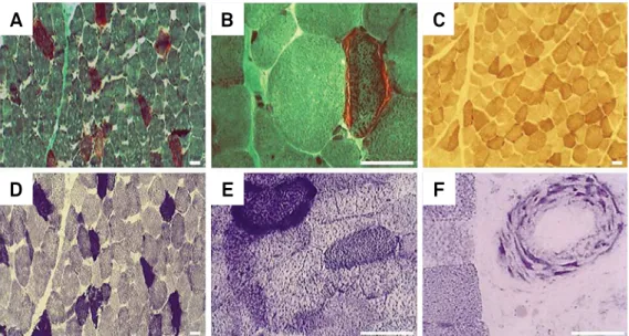
Figure 3. Classic features in the muscle biopsy of MELAS: ragged-red fibers (RRF) on modified Gomori trichrome stain (A, B);
muscle fibers with normal activity of the cytochrome c oxidase (COX) (C); RRF on succinic dehydrogenase (SDH) stain (D, E); and SDH strongly reactive blood vessel (F). Bar = 50 µm.
C
D
E
F
improve respiratory chain or to reduce the levels of reactive oxygen species arising from disrupted mitochondrial metab
-olism. Some of the most frequently prescribed agents include ubidecarenone (coenzyme Q10, CoQ), idebenone, edaravone, levoarginine (L-arginine), complex B vitamins, vitamin C, vi
-tamin E, and levocarnitine (L-carnitine) in various combina
-tions and doses25,55,56,57,58. he concomitant administration of
these diferent medications can be useful and has been ben
-eit to some MELAS patients.
here is no standardized treatment of stroke-like episodes but there is increasing evidence that these patients beneit from the administration of L-arginine and consequent anti
-epileptic treatment if the stroke-like episodes are associated with epileptic activity25,55. Although the underlying mecha
-nisms are not completely understood, in acute phase of the stroke-like episodes in MELAS, seems that L-arginine ther
-apy improve microcirculation and endothelial dysfunction, and improve almost all symptoms associated with stroke-like, with the exception of migraine headaches and visual ields55,56,57,59. According to these studies L-arginine is applied
in a dosage until 0.5 g/kg body weight intravenously during the acute phase, followed by oral administration thereaf
-ter55,56,57. Occasionally, L-arginine is also given together with
other drugs55.Symptomatic drug treatment of stroke-like
episode include antiepileptic treatment if it was accompa
-nied by seizures, analgesic treatment if it was accompa-nied by headache, or anti-psychotic or sedative therapy if it was dominated by confusion, agitation, anxiety, hyperactivity, or psychosis57. Physical therapy should be implemented in indi
-viduals after stroke-like25.
MELAS management also includes additional therapy for its complications, such as, cardiac disease (standard pharma
-cologic therapy), diabetes mellitus (dietary modiication, oral hypoglycemic agents and/or insulin therapy), deafness (hear
-ing devices and cochlear implantation) or epilepsy (tradition
-al antiepileptic treatment)25,58,60. Because febrile illnesses may
trigger acute exacerbations, MELAS patients should receive standard childhood vaccinations, lu vaccine, and pneumococ
-cal vaccine25,55. In addition, toxins or drugs that had potential
to cause mitochondrial dysfunction or lesion, such as amino
-glycoside antibiotics, linezolid, aspirin, Zidovudine, cigarettes or alcohol consumption, should be recognized and avoided55,58.
Valproic acid for seizure treatment (high risk by carnitine uptake inhibition) and dichloroacetate for acute stroke-like episodes (high risk for peripheral neuropathy) are not recom
-mended58,60. Exercise (endurance and resistance training) is
helpful in MELAS as well other mitochondrial diseases58.
It is appropriate to ofer genetic counseling (including dis
-cussion of potential risks to children and reproductive options) to young women in fertile age who are afected or at risk58.
Prenatal testing and preimplantation genetic diagnosis may be an option for some families in which the disease-causing mutations have been identiied but their interpretation is complex yet25,34,58. he transfer of nuclear DNA from fertilized
oocytes or zygotes harboring a mtDNA mutation to an enu
-cleated recipient cells could theoretically prevent transmis
-sion of mtDNA diseases and this promising therapy is under investigation34,58. In the pregnancy, MELAS women should be
monitored, mainly for diabetes mellitus and respiratory failure, which may require speciic therapeutic interventions25.
References
1. Koenigsberger MR, Pellock JM, DiMauro S, Eastwood A.. Juvenile mitochondrial myopathy, short stature and lactic acidosis: a clinical, biochemical, and ultrastructural study (Abstract #80). In: Fifth Annual Meeting of the Child Neurological Society; 1976 October 28-30; Monterey, CA.
2. Shapira Y, Cederbaum SD, Cancilla PA, Nielsen D, Lippe BM. Familial poliodystrophy, mitochondrial myopathy, and lactate acidemia. Neurology. 1975;25(7):614-21. doi:10.1212/WNL.25.7.614 3. Pavlakis SG, Phillips PC, DiMauro S, De Vivo DC, Rowland LP.
Mitochondrial myopathy, encephalopathy, lactic acidosis, and strokelike episodes: a distinctive clinical syndrome. Ann Neurol.1984;16(4):481-8. doi:10.1002/ana.410160409 4. Werneck LC, Abdalla H, Lohr A. [MELAS (mitochondrial
encephalopathy, lactic acidosis and stroke like episodes): case report]. Arq Neuropsiquiatr. 1987;45(3) 288-94. Portuguese. doi:10.1590/S0004-282X1987000300009
5. Hirano M, Ricci E, Koenigsberger MR, Defendini R, Pavlakis SG, DeVivo DC et al.. Melas: an original case and clinical criteria for diagnosis. Neuromuscul Disord.1992;2(2):125-35. doi:10.1016/0960-8966(92)90045-8
6. Yatsuga S, Povalko N, Nishioka J, Katayama K, Kakimoto N, Matsuishi T et al.. MELAS: a nationwide prospective cohort study of 96 patients in Japan. Biochim Biophys Acta. 2012;1820(5):619-24. doi:10.1016/j.bbagen.2011.03.015
7. Goto Y. Clinical features of MELAS and mitochondrial DNA mutations. Muscle Nerve. 1995;18(Suppl 3):S107-12. doi:10.1002/mus.880181422
8. Mizukami K, Sasaki M, Suzuki T, Shiraishi H, Koizumi J, Ohkoshi N et al.. Central nervous system changes in mitochondrial encephalomyopathy: light and electron microscopic study. Acta Neuropathol. 1992;83(4):449-52. doi:10.1007/BF00713541 9. Ohama E, Ohara S, Ikuta F, Tanaka K, Nishizawa M, Miyatake T.
Mitochondrial angiopathy in cerebral blood vessels of mitochondrial encephalomyopathy. Acta Neuropathol. 1987;74(3):226-33. doi:10.1007/BF00688185
10. Sakuta R, Nonaka I. Vascular involvement in mitochondrial myopathy. Ann Neurol. 1989;25(6):594-601. doi:10.1002/ana.410250611 11. Gilchrist JM, Sikirica M, Stopa E, Shanske S. Adult-onset
MELAS. Evidence for involvement of neurons as well as cerebral vasculature in strokelike episodes. Stroke. 1996;27(8):1420-3. doi:10.1161/01.STR.27.8.1420
12. Iizuka T, Sakai F, Suzuki N, Hata T, Tsukahara S, Fukuda M et al.. Neuronal hyperexcitability in stroke-like episodes of MELAS syndrome. Neurology. 2002;59(6):816-24. doi:10.1212/WNL.59.6.816 13. Sparaco M, Bonilla E, DiMauro S, Powers JM. Neuropathology
14. Iizuka T, Sakai F. Pathogenesis of stroke-like episodes in MELAS: analysis neurovascular cellular mechanisms. Curr Neurovasc Res. 2005;2(1):29-45. doi:10.2174/1567202052773544
15. Goto YI, Nonaka I, Horai S. A mutation in the tRNA(Leu)(UUR gene associated
with the MELAS subgroup of mitochondrial encephalomyopathies. Nature. 1990;348(6302):651-3. doi:10.1038/348651a0
16. Kobayashi Y, Momoi MY, Tominaga K, Momoi T, Nihei K, Yanagisawa M et al.. A point mutation in the mitochondrial tRNA(Leu)(UUR) gene
in MELAS (mitochondrial myopathy, encephalopathy, lactic acidosis and stroke-like episodes). Biochem Biophys Res Comm. 1990;173(3):816-22. doi:10.1016/S0006-291X(05)80860-5 17. Finsterer J. Genetic, pathogenetic, and phenotypic implications of
the mitochondrial A3243G tRNALeu(UUR) mutation. Acta Neurol Scand. 2007;116(1):1-14. doi:10.1111/j.1600-0404.2007.00836.x 18. Hao R, Yao YN, Zheng YG, Xu MG, Wang ED. Reduction of mitochondrial
tRNALeu(UUR) aminoacylation by some MELAS-associated mutations.
FEBS Lett. 2004;578(1-2):135-9. doi:10.1016/j.febslet.2004.11.004 19. Ciafaloni E, Ricci E, Shanske S, Moraes CT, Silvestri G, Hirano M et al..
MELAS: clinical features, biochemistry and molecular genetics. Ann Neurol. 1992;31(4):391-8. doi:10.1002/ana.410310408
20. Goto Y, Horai S, Matsuoka T, Koga Y, Nihei K, Kobayashi M et al.. Mitochondrial myopathy, encephalopathy, lactic acidosis and stroke-like episodes (MELAS): a correlative study of the clinical features and mitochondrial DNA mutation. Neurology. 1992;42(3):545-50. doi:10.1212/WNL.42.3.545
21. Hammans SR, Sweeney MG, Brockington M, Morgan-Hughes JA, Harding AE. Mitochondrial encephalopathies: molecular genetic diagnosis from blood samples. Lancet. 1991;337(8753):1311-3. doi:10.1016/0140-6736(91)92981-7
22. Goto Y, Nonaka I, Horai S. A new mtDNA mutation associated with mitochondrial myopathy, encephalopathy, lactic acidosis and stroke-like episodes (MELAS). Biochim Biophys Acta. 1991;1097(3): 238-40. doi:10.1016/0925-4439(91)90042-8 23. Zhao D, Hong D, Zhang W, et al.. Mutations in mitochondrially
encoded complex I enzyme as the second common cause in a cohort of Chinese patients with mitochondrial myopathy, encephalopathy, lactic acidosis and stroke-like episodes. J Hum Genet. 2011;56(11):759-64. doi:10.1038/jhg.2011.96
24. Zeviani M, Di Donato S. Mitochondrial disorders. Brain. 2004;127(10):2153-72. doi:10.1093/brain/awh259 25. DiMauro S, Hirano M. MELAS. In: Pagon RA, Adam MP,
Ardinger HH, Wallace SE, Amemiya A, Bean LJH et al., editors. GeneReviews® [Internet]. Seattle (WA): University of
Washington, Seattle; 2013 [citado 21 nov 2013]. Disponível em: http://www.ncbi.nlm.nih.gov/books/NBK1233/
26. Bataillard M, Chatzoglou E, Rumbach L, Sternberg D, Tournade A, Laforêt P et al.. Atypical MELAS syndrome associated with a new mitochondrial tRNA glutamine point mutation. Neurology. 2001;56(3):405-7. doi:10.1212/WNL.56.3.405
27. Kaufmann P, Engelstad K, Wei Y, Kulikova R, Oskoui M, Sproule DM et al.. Natural history of MELAS associated with mitochondrial DNA m.3243A>G genotype. Neurology. 2011;77(22):1965-71. doi:10.1212/WNL.0b013e31823a0c7f
28. Nishioka J, Akita Y, Yatsuga S, Katayama K, Matsuishi T, Ishibashi M et al.. Inappropriate intracranial hemodynamics in the natural course of MELAS. Brain Dev. 2008;30(2):100-5. doi:10.1016/j.braindev.2007.06.008
29. Lorenzoni PJ, Scola RH, Kay CS, Arndt RC, Freund AA, Bruck I et al.. MELAS: clinical features, muscle biopsy and molecular genetics. Arq Neuropsiquiatr.. 2009;67(3A):668-76. doi:10.1590/S0004-282X2009000400018
30. Lorenzoni PJ, Scola RH, Kay CS, Arndt RC, Silvado CE, Werneck LC. MERRF: clinical features, muscle biopsy and molecular genetics in Brazilian patients. Mitochondrion. 2011;11(3):528-32. doi:10.1016/j.mito.2011.01.003
31. Conforto AB, Yamamoto FI, Oba-Shinjo SM, Pinto JG, Hoshino M, Scaff M et al.. Screening for MELAS mutations in young patients with stroke of undetermined origin. Arq Neuropsiquiatr. 2007;65(2B):371-6. doi:10.1590/S0004-282X2007000300001
32. Hirano M, Pavlakis SG. Mitochondrial myopathy, encephalopathy, lactic acidosis, and strokelike episodes (MELAS): current concepts. J Child Neurol. 1994;9(1):4-13. doi:10.1177/088307389400900102 33. Okajima Y, Tanabe Y, Takayanagi M, Aotsuka H. A follow up
study of myocardial involvement in patients with mitochondrial encephalomyopathy, lactic acidosis, and stroke-like episodes (MELAS). Heart. 1998;80(3):292-5. doi:10.1136/hrt.80.3.292 34. Richardson J, Irving L, Hyslop LA, Choudhary M, Murdoch A, Turnbull
DM et al.. Concise reviews: assisted reproductive technologies to prevent transmission of mitochondrial DNA disease. Stem Cells. 2015;33(3):639-45. doi:10.1002/stem.1887
35. Haas R, Dietrich R. Neuroimaging of mitochondrial disorders. Mitochondrion. 2004;4(5-6):471-90. doi:10.1016/j.mito.2004.07.008 36. Tschampa HJ, Urbach H, Greschus S, Kunz WS, Kornblum C.
Neuroimaging characteristics in mitochondrial encephalopathies associated with the m.3243A>G MTTL1 mutation. J Neurol. 2013;260(4):1071-80. doi:10.1007/s00415-012-6763-4 37. Iizuka T, Sakai F, Kan S, Suzuki N. Slowly progressive spread of
the stroke-like lesions in MELAS. Neurology. 2003;61(9):1238-44. doi:10.1212/01.WNL.0000091888.26232.FE
38. Kamada K, Takeuchi F, Houkin K, Kitagawa M, Kuriki S, Ogata A et al.. Reversible brain dysfunction in MELAS: MEG, and (1)H MRS analysis. J Neurol Neurosurg Psychiatry. 2001;70(5):675-8. doi:10.1136/jnnp.70.5.675
39. Betts J, Jaros E, Perry RH, Schaefer AM, Taylor RW, Abdel-All Z et al.. Molecular neuropathology of MELAS: level of heteroplasmy in individual neurones and evidence of extensive vascular involvement. Neuropathol Appl Neurobiol. 2006;32(4):359-73. doi:10.1111/j.1365-2990.2006.00731.x
40. Abe K, Yoshimura H, Tanaka H, Fujita N, Hikita T, Sakoda S. Comparison of conventional and diffusion-weighted MRI and proton MR spectroscopy in patients with mitochondrial encephalomyopathy, lactic acidosis, and stroke-like events. Neuroradiology. 2004;46(2):113-7. doi:10.1007/s00234-003-1138-2 41. Oppenheim C, Galanaud D, Samson Y, Sahel M, Dormont D, Wechsler
B et al.. Can diffusion weighted magnetic resonance imaging help differentiate stroke from stroke-like events in MELAS? J Neurol Neurosurg Psychiatry. 2000;69(2):248-50. doi:10.1136/jnnp.69.2.248 42. Tanahashi C, Nakayama A, Yoshida M, Ito M, Mori N, Hashizume Y. MELAS with the mitochondrial DNA 3243 point mutation: a neuropathological study. Acta Neuropathol. 2000;99(1):31-8. doi:10.1007/PL00007403 43. Kaufmann P, Shungu DC, Sano MC, Jhung S, Engelstad K, Mitsis
E et al.. Cerebral lactic acidosis correlates with neurological impairment in MELAS. Neurology. 2004;62(8):1297-302. doi:10.1212/01.WNL.0000120557.83907.A8
44. Kobayashi M, Morishita H, Sugiyama N, Yokochi K, Nakano M, Wada Y et al.. Mitochondrial myopathy, encephalopathy, lactic acidosis and stroke-like episodes syndrome and NADH-CoQ reductase deficiency. J Inherit Metab Dis. 1986;9(3):301-4. doi:10.1007/BF01799670 45. Tokunaga M, Mita S, Sakuta R, Nonaka I, Araki S. Increased mitochondrial
DNA in blood vessels and ragged-red fibers in mitochondrial myopathy, encephalopathy, lactic acidosis, and stroke-like episodes (MELAS). Ann Neurol. 1993;33(3):275-80. doi:10.1002/ana.410330308
46. Tokunaga M, Mita S, Murakami T, Kumamoto T, Uchino M, Nonaka I et al.. Single muscle fiber analysis of mitochondrial myopathy, encephalopathy, lactic acidosis, and stroke-like episodes (MELAS). Ann Neurol 1994;35(4):413-9. doi:10.1002/ana.410350407 47. Rossmanith W, Freilinger M, Roka J, Raffelsberger T, Moser-Thier
48. Tanji K, Kaufmann P, Naini AB, Lu J, Parsons TC, Wang D et al.. A novel tRNA(Val) mitochondrial DNA mutation causing MELAS. J Neurol Sci. 2008;270(1-2):23-7. doi:10.1016/j.jns.2008.01.016
49. Matsuoka T, Goto Y, Hasegawa H, Nonaka I. Segmental cytochrome c-oxidase deficiency in CPEO: teased muscle fiber analysis. Muscle Nerve. 1992;15(2):209-13. doi:10.1002/mus.880150213
50. Petruzzella V, Moraes CT, Sano MC, Bonilla E, DiMauro S, Schon EA. Extremely high levels of mutant mtDNAs co-localize with cytochrome c oxidase-negative ragged-red fibers in patients harboring a point mutation at nt 3243. Hum Mol Genet. 1994;3(3):449-54. doi:10.1093/hmg/3.3.449
51. Moraes CT, Ricci E, Petruzzella V, Shanske S, DiMauro S, Schon EA et al.. Molecular analysis of the muscle pathology associated with mitochondrial DNA deletions. Nature Genet. 1992;1(5):359-67. doi:10.1038/ng0892-359
52. Sciacco M, Bonilla E, Schon EA, DiMauro S, Moraes CT. Distribution of wild-type and common deletion forms of mtDNA in normal and respiration-deficient muscle fibers from patients with mitochondrial myopathy. Hum Mol Genet. 1994;3(1):13-9. doi:10.1093/hmg/3.1.13 53. Hasegawa H, Matsuoka T, Goto Y, Nonaka I. Strongly succinate
dehydrogenase-reactive blood vessels in muscles from patients with mitochondrial myopathy, encephalopathy, lactic acidosis, and stroke-like episodes. Ann Neurol. 1991;29(6):601-5. doi:10.1002/ana.410290606
54. Vanderver A. Tools for diagnosis of leukodystrophies and other disorders presenting with white matter disease. Curr Neurol Neurosci Rep. 2005;5(2):110-8. doi:10.1007/s11910-005-0008-1 55. Finsterer J. Management of mitochondrial
stroke-like-episodes. Eur J Neurol. 2009;16(11):1178-84. doi:10.1111/j.1468-1331.2009.02789.x
56. Koga Y, Povalko N, Nishioka J, Katayama K, Yatsuga S, Matsuishi T. Molecular pathology of MELAS and L-arginine effects. Biochim Biophys Acta. 2012;1820(5):608-14. doi:10.1016/j.bbagen.2011.09.005
57. Finsterer J. Stroke and stroke-like episodes in muscle disease. Open Neurol J. 2012;6(1):26-36. doi:10.2174/1874205X01206010026 58. Horvath R, Gorman G, Chinnery PF. How can we treat mitochondrial
encephalomyopathies? Approaches to therapy. Neurotherapeutics 2008;5(4):558-68. doi:10.1016/j.nurt.2008.07.002
59. Koga Y, Akita Y, Nishioka J, Yatsuga S, Povalko N, Katayama K et al.. MELAS and L-arginine therapy. Mitochondrion. 2007;7(1-2):133-9. doi:10.1016/j.mito.2006.11.006