Efects of eliminating tension by
means of epineural stitches
A comparative electrophysiological and
histomorphometrical study using diferent
suture techniques in an animal model
Jorge Bustamante1, Mariano Socolovsky2, Roberto S. Martins3,
Juan Emmerich4, Maria Gabriela Pennini4,
Natalia Lausada1, Luis Domitrovic2
ABSTRACT
Epineural stitches are a means to avoid tension in a nerve suture. We evaluate this technique, relative to interposed grafts and simple neurorraphy, in a rat model. Method: Twenty rats were allocated to four groups. For Group 1, sectioning of the sciatic nerve was performed, a segment 4 mm long discarded, and epineural suture with distal anchoring stitches were placed resulting in slight tension neurorraphy. For Group 2, a simple neurorraphy was performed. For Group 3, a 4 mm long graft was employed and Group 4 served as control. Ninety days after, reoperation, latency of motor action potentials recording and axonal counts were performed. Inter-group comparison was done by means of ANOVA and the non-parametric Kruskal-Wallis test. Results: The mean motor latency for the simple suture (2.27±0.77 ms) was lower than for the other two surgical groups, but lower than among controls (1.69±0.56 ms). Similar values were founding in both group 1 (2.66±0.71 ms) and group 3 (2.64±0.6 ms). When fibers diameters were compared a significant difference was identified between groups 2 and 3 (p=0.048). Conclusion: Good results can be obtained when suturing a nerve employ with epineural anchoring stitches. However, more studies are needed before extrapolating results to human nerve sutures.
Key words: neurorraphy, tension, autologous graft, nerve suture, peripheral nerve.
Avaliação dos efeitos da eliminação da tensão através de ancoramento epineural: estudo experimental comparando resultados eletrofisiológicos e histomorfométricos após diferentes técnicas de reparo no nervo
RESUMO
A aproximação através de pontos epineurais é uma forma de se reduzir a tensão numa neurorrafia. Neste estudo esta técnica é avaliada através da sua comparação com a interposição de enxertos e neurorrafia simples num modelo experimental utilizando o rato. Método: Vinte ratos foram utilizados e divididos em 4 grupos. No Grupo 1, após a ressecção de 4 mm, os cotos do nervo foram aproximados através de pontos de ancoramento epineurais e suturados com tensão. No Grupo 2, uma neurorrafia simples foi realizada após secção do nervo. No Grupo 3, um enxerto de 4 mm foi utilizado para o reparo e o Grupo 4 foi utilizado como controle. Noventa dias após, os nervos foram novamente expostos e a medida da latência do potencial de ação motor e a contagem axonal foram realizados. A comparação entre os grupos foi realizada através da comparação entre as médias (ANOVA) e com o teste não-paramétrico de Kruskal-Wallis. Resultados:
Correspondence
Mariano Socolovsky University of Buenos Aires School of Medicine La Pampa 1175 Torre 2 5A Buenos Aires 1428, Argentina E-mail: [email protected]
Received 19 May 2010
Received in final form 16 September 2010 Accepted 23 September 2010
1Laboratory of the Program of Organ and Tissue Transplants; School of Medical Sciences; National University of La Plata,
Argentina; 2Peripheral Nerve Section, Department of Neurosurgery, Hospital de Clínicas, University of Buenos Aires,
Argentina; 3Peripheral Nerve Unit, Discipline of Neurosurgery, Hospital das Clínicas, University of São Paulo and Hospital do
A média da latência motora na sutura simples (2,27±0,77 ms) foi menor em relação aos outros dois grupos onde o nervo foi seccionado e reparado e maior que o grupo controle (1,69±0,56 ms). Resultados semelhantes foram identificados nos grupos 1 (2,66±0,71 ms) e 3 (2,64±0,6 ms). Uma diferença significativa diâmetros das fibras foi identificada quando comparados os grupos 2 e 3 (p=0,048). Conclusão: Resultados equiparáveis aos obtidos com enxerto podem ser obtidos quando a neurorrafia é realizada com pontos epineurais de ancoramento com tensão, mas estudos adicionais são necessários antes desses resultados serem extrapolados para o reparo de nervo em seres humanos.
Palavras-chave: neurorrafia, tensão, enxerto autólogo, sutura nervosa, nervo periférico.
A universally-accepted concept referring to the su-ture of a nerve is that if one draws both ends together under tension at the site of union, the resultant tension jeopardizes the final outcome1. In the majority of
pe-ripheral nerve sections of traumatic origin, nerve tissue is lost; consequently, a gap is created that prevents the union of the two ends without tension. his gap is in-creased by the natural elasticity of the nerve stumps, which tend to retract and further separate over time. In-numerable mechanisms have been developed and tested over the last 100 years, in attempts to avoid this ten-sion, including lexion of articulations2, transpositions,
the use of nerve grafts3-5 and modern neurotubes6-8.
Ar-ticular lexion is restricted to certain nerves, and only when injured at a site where they transverse a joint. A common complication of this technique is articular ri-gidity5. Transpositions are commonly used, but also
lim-ited to speciic nerves at certain locations along their path as the ulnar nerve at the elbow. Neurotubes have proven successful, but only when the gap is short (up to 3 cm)6,7. Moreover, the high costs of neurotubes limit their
utility. he gold standard for uniting two ends of a nerve when there is loss of substance between them is an inter-posing neurorraphy with an autologous graft. At present, this method has shown to be the most appropriate4.
However, the use of grafts is not without complications, as is the loss of sensitivity in the territory innervated by the donor nerve, generally the sural nerve, thereby in-creasing operating time, pain due to neuromas, infection risk, etc. But perhaps the most important point that must be taken into account when evaluating sutures using a graft, is that its results are always inferior, in terms of re-innervation, to those involving direct suturing without tension or termino-terminal neurorraphy. In accor-dance with Saint Venant’s principle, the epineurium can be used as a splint to distribute tension at a repair site9.
If a small number of anchoring sutures are placed one nerve diameter away from the main repair site, this re-lieves tension and allows for placement of the remaining small stitches designated to maintain stump alignment. With this, ibrosis can be eliminated and axonal passage through the neurorraphy facilitated.
he objective of the present study is to compare, elec-trophysiologically and morphometrically the results of nerve sutures employing distal anchoring stitches in a rat model against the results obtained employing grafts and sutures without tension, as well as against non-in-jured controls without sutures.
METHOD
previ-ously placing one or two epineural stitches at a distance of 3 mm from the ends of both stumps (Fig 1). A knot was made with the objective of drawing the two ends nearer to each other and undertaking a termino-ter-minal neurorraphy without tension at the site of the su-ture (Group 1) (Fig 2A).
On the left side of the same 10 rats, the procedure was repeated; but instead of resecting 4 mm of the sciatic nerve, a direct termino-terminal suture under microscopy was performed with very little or no tension, coapting the extremes of the sectioned nerve (Group 2) (Fig 2B).
A second lot of 10 Wistar rats underwent interven-tion on the right side in much the same way as for Group 1, resecting a 4 mm long segment of the sciatic nerve, but then rotating it in the opposite direction, and later suturing it under a microscope, using a graft in an in-terposed form (Group 3) (Fig 2C). he left side of the second lot of rats was left intact, and later used as a con-trol group (Group 4) (Fig 2D).
Animals were maintained in the lab under optimal conditions for food, light and sleep, to ultimately be re-operated upon 90 days after the irst surgery. At the time
of the second procedure, again under general anesthesia, the operated zone for Groups 1, 2 and 3 was explored; whereas the sciatic nerve was exposed for the irst time in Group 4 limbs. Electrophysiological measurements and microscopic studies were performed, as described below. All animals ultimately were euthanized by means of an intra-peritoneal overdose of thiopental.
Electrophysiological measurements
Electrophysiological studies were performed to measure the latency of the motor action potentials (MAP)10. For this evaluation, we designed two ground
electrodes; one electrode to administer the stimulus; and a fourth for recording. One of the ground electrodes was constructed of stainless steel wire, 316L gauge and 0.40 mm in diameter, with one end shaped in an helicoidal fashion, so positioned as to surround the nerve, thereby increasing the area of contact. he other ground elec-trode consisted of a monopolar needle with a diameter of Fig 1. Scheme for anchoring epineural stitches. [A] Gap. [B]
pas-sage of inextensible suture string (for example, nylon 6.0) through epineurium at a site at least 5 mm distal to the suture. Once both stumps are drawn closer together by means of anchorage stitches, a microsuture with 10.0 stitches is performed by means of the common technique.
0.40 mm, length of 25 mm, and gauge (G) 2611. he
elec-trodes for stimulation were made of wire formed from a voltaic arc of stainless steel 316L, 0.5 mm in diam-eter, with a distance of one mm between the anode and cathode. For recording, a coaxial needle, 0.30 mm in di-ameter, 25 mm long and 30G, as well as a recording area of 0.019 square mm was employed. Records were made by means of a portable electroneuromyography device with two channels (Medtronic, Denmark).
After exposing the sciatic nerve, the ground elec-trodes were installed, the helicoidal ground electrode placed half the distance between the electrodes for stim-ulation and recording. he recording electrode was in-stalled in the gastrocnemius muscle by means of a per-cutaneous puncture using a coaxial needle. he distance between the stimulating and recording electrodes was always 30 mm. Short duration supra-maximal stimula-tion (1 millisecond) was applied, so as to generate an action potential. To determine the supra-maximal cur-rent of stimulation, the stimulation values were increased gradually, at intervals of 0.1 milli-amperes (mA), until the scope of the last two potentials remained unaltered. From this potential record, called MAP, the latency were estimated and called LATM. All electrophysiological procedures were undertaken by the electrophysiologist on our research team. Obtained results were analyzed by a diferent researcher (RSM).
Histomorphometric measurements
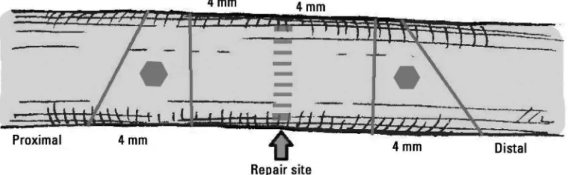
Immediately after the electrophysiological evaluation, both sciatic nerves were ixed in vivo with instillation of Karnovsky ixative solution (paraformaldehyde, 2.5% glu-taraldehyde and 0.1M bufer at pH=7.32%). After 3 min-utes, a 2 cm segment of nerve was sectioned, including the suture site; in the suture with graft group, a 3 cm seg-ment was cut. Both 5 mm distal and proximal to the su-ture, sectioning with a scalpel, 2 segments 4mm in length were obtained (Fig 3). In the graft group, this entire graft area was considered the ‘repair zone’, instead of just the
suture lines, as in Groups 1 and 2. hese segments were stored in labeled bottles containing the same ixative so-lution. Each segment then was post-ixed in 2% osmium tetroxide solution, dehydrated in growing solutions of ethyl alcohol, clareated with ethylene oxide, iniltrated, and inally added to an epoxy resin, which allowed us to obtain 1mm transversal cuts, toluidine blue-included, suitable for microscopic analysis.
To evaluate the number of myelinized axons, 5 pho-tographs of each cut were obtained with an 8.3 mega-pixel digital camera, with a 100× zoom. he inal number express the addition of counting axons from these ive samples. he samples were analyzed using the program Sigma Scan Pro 5.0 (SPSS Inc., Chicago, IL) for axon counts. After measuring the proximal and distal seg-ments, it was possible to generate an index called the
regeneration index (RI) by dividing the total number of regenerated axons in the distal segment by the total number of regenerated axons in the proximal segment. his index was used to evaluate the number of axons that had crossed the repair site. he iber diameter distal to repair in the operated groups was also evaluated using the same program. After all these studies, the animals were sacriiced via the intra-peritoneal administration of an over-dose of pentobarbital.
Statistical analysis
he results were expressed as mean±standard devi-ation. Analysis of variance (ANOVA) with Tukey’s post hoc test was used to compare values of motor latency. he non-parametric Kruskal-Wallis test was used to compare RI results. A p value of 0.05 was considered signiicant.
RESULTS
All 20 animals who survived beyond 24 hours exhib-ited an excellent post-operative course, regaining mo-bility of their limbs within less than 24 hours. With re-spect to surgical wounds, while the stitches of cutaneous sutures were self-extracted, only one female presented Fig 3. Segments of the nerve obtained after Karnovsky ixation solution application. The proximal and distal
with dehiscence, a few millimeters in length; since the length of dehiscence was so short, it was decided not to re-operate on her. No infection at the surgical site or self-mutilation behaviors was reported. Over the duration of the experiment, the animals increased their average body weight by between 7 and 10%.
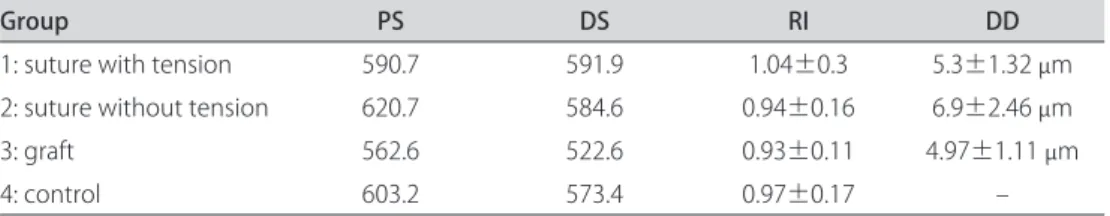
Electromyographic measurements were obtained in all animals, tabulated and analyzed, so as to calcu-late the conduction velocity of motor action potentials. In decreasing order, we noticed the best results con-cerning motor latency after simple sutures (2.27±0.77 ms), followed by sutures under tension (2.66±0.61 ms), and followed by nerve grafts (2.64±0.6 ms). The con-duction velocity for the control group was 1.69±0.56 ms (Table 1). he comparison between groups showed statistical diference when comparing group 1 (suture with epineural stitches) and 4 (control) with p=0.013, and group 3 (graft) and control (p=0.024). No statis-tical diference was founded when comparing group 1 and 2 (simple suture) with p=0.732, when comparing group 1 and 3 (p=0.982), when comparing group 2 and 3 (p=0.505) and when comparing group 2 and 4 (p=0.252). Table 2 presents the results of axon counts. The means for the RI were 1.04±0.3, 0.94±0.16, 0.93±0.11 and 0.97±0.17 for Groups 1, 2, 3 and 4, respectively, with no signiicant diference identiied between the four groups (Kruskal-Wallis test, p=0.91). he iber diameter distal to the repair site was 5.3±1.32, 6.9±2,46 and 4,97±1.11 µm in groups 1, 2 and 3, respectively. A signiicant dif-ference was identiied between groups 2 and 3 (p=0.048).
DISCUSSION
The mechanism by which tension interferes with axoplasmatic passage through a suture site is ibrosis, which occurs at both stumps and prevents the passage of growing axons towards the distal end. Anchoring epi-neural traction stitches could be a mechanism by which one could avoid this ibrosis and retraction. Of course, this method only can be employed in nerves with a lim-ited loss of tissue resulting in short intervals, since it is impossible to draw two stumps that are separated by a large gap into close proximity without generating a se-vere lesion due to traction at both ends, or lacerations
involving the epineurium, which would deinitely pre-vent connection by this means. Furthermore, it has been shown that both stumps of a sectioned nerve continue to retract over the irst couple of weeks after section, until they ind a point of neutral elasticity12. his could be
an-other mechanism behind the failure of a suture under tension. After suturing under tension, both ends con-tinue to separate until they are so far away that regener-ation is prevented.
Interpreting our other indings, it was apparent that any type of repair of a nerve alters neurophysiological conduction relative to a nerve that has undergone no ma-nipulation. Moreover, this is further proof that if a nerve suture must be performed, it is better to leave it without tension at the same level, such being the ideal way to re-pair an injured nerve (Group 1). In cases where there is a loss of tissue from a nerve due to trauma, according to what has been extensively published in the literature to date, the tension generated by drawing the two severed ends together for suturing appears to be detrimental to procedural success, since the absence of re-innervation is veriied5. An alternative way to correct such an
im-perfection is to anchor epineural stitches at a distance, so as to draw the two stumps closer together and avoid concentrated tension at the nerve suture site itself, dis-placing all tension to parts further away from the nerve. his suture technique is no novelty, having already been described in several papers13,14.
he results of the experiment described in this paper showed that regarding motor latency, there was no sta-tistical diference when employing this technique (epi-neural suture under tension) when compared with the use of grafts. The advantages of the epineural suture
Table 1. Mean latency of the motor action potential in each group.
Study group (mean±standard deviation)Motor latency 1: tension suture 2.66±0.71 ms 2: suture without tension 2.27±0.77 ms
3: graft 2.64±0.60 ms
4: control 1.69±0.56 ms
ms: milisecond.
Table 2. Results of histomorphometric evaluation.
Group PS DS RI DD
1: suture with tension 590.7 591.9 1.04±0.3 5.3±1.32 μm
2: suture without tension 620.7 584.6 0.94±0.16 6.9±2.46 μm
3: graft 562.6 522.6 0.93±0.11 4.97±1.11 μm
4: control 603.2 573.4 0.97±0.17 –
under tension when compared with grafts, i.e. no mor-bidity related to graft harvesting, easier surgical tech-nique, less surgical time, make us strongly recommend the former when a nerve gap has to be reconstructed. Nevertheless, the negative efects of tension are displayed by the fact that tension in our rats generated worse re-sults than a simple suture where there was no loss of nerve substance and, therefore, it was possible an ade-quately coaptation of the two stumps.
he inal values for axon counts and, therefore, the regeneration index (RI), were similar across all 4 groups. It was initially presumed that the axonal count should be less in the distal versus proximal segment, relecting ax-onal loss that occurs at the repair site. Nevertheless, mul-tiple sprouts of axons from the proximal stump may have passed through the repair site without achieving efective re-innervation, and this increment of axons should com-pensate for those lost axons that could not cross the re-pair site15-17. he largest iber diameter in group 2
com-pared to other two operated groups are in according to electrophysiological results since no differences were identiied between groups 2 and control concerning la-tency of the motor action potential.
From a practical point of view, if the surgeon places a 6.0 or 7.0 stitch and, when drawing nerve stumps to-gether, the epineurium lacerates from excessive traction, it should be accepted that this technique is of no use and one should resort to an interpose grafts. However, if one achieves correct coaptation with epineural traction stitches, the outcome achieved will be better than that obtained installing an autologous graft.
Our study has some limitations. First, we used an ex-perimental rat model; consequently, the results we ob-tained cannot be extrapolated directly into usual sur-gical practice in humans. Also, the 4mm imperfection that was created artiicially in the sciatic nerve of a rat may not correlate well with gaps in humans. Nonethe-less, this study still demonstrates that the deleterious ef-fect of a suture under tension can be eliminated or min-imized by means of anchoring with epineural stitches, suggesting that it may be more effective to apply this technique than to employ interposed homologous grafts. It remains to be seen whether these indings can be ex-trapolated to larger animals with bulkier nerves. Other studies has been done in the last decades, analysing the same topic with diferent protocols, animal models, and methods of validation of their results18-21, arriving at the
same conclusion than ours: a limited tension at the re-pair site seems to be preferable than a suture with inter-posed grafts.
In conclusion, in this experimental model, it was made apparent that the negative efects of tension on the suture of a sectioned nerve can be minimized using
anchoring epineural traction stitches. In this way, the results obtained, in terms of re-establishing electrical conduction, are similar than if one uses an interposed graft. hese results could be extrapolated to the suture of nerves in common surgical practice by means of fur-ther studies on animals of greater weight.
ACKNOWLEDGMENTS – he authors would like to thank Dr. Hugo
Galafassi for generously donating the 10.0 micro sutures that made re-alization of the present study possible.
REFERENCES
1. Schmidhammer R, Zandieh S, Hopf R, et al. Alleviated tension at the repair site enhances functional regeneration: the efect of full range of motion mobilization on the regeneration of peripheral nerves: histologic, electro-physiologic, and functional results in a rat model. J Trauma 2004;56:571-584. 2. Bochdansky T, Hertz H, Poigenfürst J. Efects of lexed position of the wrist
joint after lexor tendon sutures on the median nerve. Unfallchirurgie 1993; 19:303-306.
3. Bourrel P. Technique of nerve suture. Med Trop 1982;42:221-222. 4. Millesi H.The nerve gap. Theory and clinical practice. Hand Clin 1986;2:
651-663.
5. Stevens WG, Hall JD, Young VL, Weeks PM. When should nerve gaps be grafted? An experimental study in rats. Plast Reconstr Surg 1985;75:707-713. 6. Belkas JS, Shoichet MS, Midha R. Peripheral nerve regeneration through
guidance tubes. Neurol Res 2004;26:151-160.
7. De Ruiter GC, Malessy MJ, Yaszemski MJ, Windebank AJ, Spinner RJ. Designing ideal conduits for peripheral nerve repair. Neurosurg Focus 2009; 26:E5. 8. Haug A.US Food and Drug Administration/Conformit Europe-approved
absorbable nerve conduits for clinical repair of peripheral and cranial nerves. Ann Plast Surg 2009;62:710.
9. Jabaley ME. Modified techniques of nerve repair: epineural splint. In: Gelberman RH (Ed). Operative nerve repair and reconstruction. Philadel-phia: JB Lippincott, 1991:315-326.
10. Tiel RL, Happel LT Jr, Kline DG. Nerve action potential recording method and equipment. Neurosurgery 1996;39:103-108.
11. Martins RS, Siqueira MG, Da Silva CF, Godoy BO, Plese JPP. Electrophys-iologic assessment of regeneration in rat sciatic nerve repair using suture, ibrin glue or a combination of both techniques. Arq Neuropsiquiatr 2005;63:601-604.
12. Toby EB, Rotramel J, Jayaraman G, Struthers A. Changes in the stress re-laxation properties of peripheral nerves after transection. J Hand Surg Am 1999;24:694-699.
13. Maeda T, Hori S, Sasaki S, Maruo S. Efects of tension at the site of coap-tation on recovery of sciatic nerve function after neurorraphy: evaluation by walking-track measurement, electrophysiology, histomorphometry, and electron probe X-ray microanalysis. Microsurgery 1999;19:200-207. 14. Midha R, Zager EL. Nerve regeneration and nerve repair. Neurol Res 2008;
30:997-998.
15. Gilbert A. Biological glue: experimental and clinical evidence. Ann Chir Main 1989;8:302-311.
16. Martins RS, Siqueira MG, Da Silva CF, Plese JPP. Overall assessment in peripheral nerve lesion repair using ibrin glue, suture or a combination of the 2 thecniques in a rat model. Which is the ideal choice? Surg Neurol 2005;64 (Suppl 1):S10-S16;
17. Menovsky T, Beek JF. Laser, ibrin glue, or suture repair of peripheral nerves: a comparative functional, histological, and morphometric study in the rat sciatic nerve. J Neurosurg 2001;95:694-699.
18. Rodkey WG, Cabaud HE, McCarroll HR Jr. Neurorrhaphy after loss of a nerve segment: comparison of epineurial suture under tension versus multiple nerve grafts. J Hand Surg (Am) 1980;5:366-371.
19. Terzis J, Faibisof B, Williams B. The nerve gap: suture under tension vs graft. Plast Reconst Surg 1975;56:166-170.
20. Hentz V, Rosen J, Xiao S, McGill K, Abraham G. The nerve gap dilemma: a comparison of nerves repaired end to end under tension with nerve grafts in a primate model. J Hand Surg (Am) 1993;18:417-442.
![Fig 2. Suture technique employed for each group (n partial=10, n total=40) [A] Group 1: tension suture with epineural stitches after resecting a nerve segment of 4 mm](https://thumb-eu.123doks.com/thumbv2/123dok_br/15433398.595351/3.955.535.830.84.524/suture-technique-employed-partial-tension-epineural-stitches-resecting.webp)