185
Arq Neuropsiquiatr 2010;68(2):185-188
Article
Limitations in diferentiating
vascular dementia from Alzheimer’s
disease with brief cognitive tests
Maria Niures P.S. Matioli1,2, Paulo Caramelli2,3ABSTRACT
Objective: To investigate the diagnostic value of brief cognitive tests in differentiating vascular dementia (VaD) from Alzheimer’s disease (AD). Method: Fifteen patients with mild VaD, 15 patients with mild probable AD and 30 healthy controls, matched for age, education and dementia severity, were submitted to the following cognitive tests: clock drawing (free drawing and copy), category and letter fluency, delayed recall test of figures and the EXIT 25 battery. Results: VaD patients performed worse than AD patients in category fluency (p=0.014), letter fluency (p=0.043) and CLOX 2 (p=0.023), while AD cases performed worse than VaD patients in delayed recall (p=0.013). However, ROC curves for these tests displayed low sensitivity and specificity for the differential diagnosis between VaD and AD.
Conclusion: Although the performance of VaD and AD patients was significantly different in some cognitive tests, the value of such instruments in differentiating VaD from AD proved to be very limited.
Key words: Alzheimer’s disease, vascular dementia, clock drawing test, delayed recall, diagnosis, EXIT25, neuropsychological tests, verbal fluency.
Limitações em diferenciar demência vascular de doença de Alzheimer através de testes cognitivos breves
RESUMO
Objetivo: Investigar o valor diagnóstico de testes cognitivos breves na diferenciação de demência vascular (DV) e doença de Alzheimer (DA). Método: Quinze pacientes com DV, 15 com DA provável e 30 controles saudáveis, pareados em relação à idade, escolaridade e gravidade da demência, foram submetidos aos seguintes testes: desenho do relógio espontâneo e cópia, fluência verbal semântica e fonêmica, teste de evocação de memória de figuras e a bateria EXIT25. Resultados: Pacientes com DV apresentaram pior desempenho na fluência verbal semântica (p=0,014), fonêmica (p=0,043), e no CLOX 2 (p=0,023). O grupo com DA obteve pior desempenho no teste de evocação tardia (p=0,013). As curvas ROC aplicadas a esses testes mostraram baixa sensibilidade e especificidade para o diagnóstico diferencial entre DV e DA. Conclusão: Embora o desempenho dos pacientes tenha sido diferente em alguns testes, o valor desses instrumentos para o diagnóstico diferencial entre DV e DA parece ser muito limitado.
Palavras-chave: doença de Alzheimer, demência vascular, teste do desenho do relógio, memória de evocação, diagnóstico, EXIT25, testes neuropsicológicos, fluência verbal.
Correspondence
Paulo Caramelli
Department of Internal Medicine Faculty of Medicine
Federal University of Minas Gerais Av Prof Alfredo Balena 190/246 30130-100 Belo Horizonte MG - Brasil E-mail: caramelp@usp.br
Received 17 August 2009
Received in final form 27 October 2009 Accepted 11 November 2009
1Department of Geriatrics, Lusíada University School of Medicine, Santos SP, Brazil; 2Department of Neurology, University
of São Paulo School of Medicine, São Paulo SP, Brazil; 3Behavioral and Cognitive Neurology Research Group, Department of
Internal Medicine, Faculty of Medicine of the Federal University of Minas Gerais, Belo Horizonte MG, Brazil. Alzheimer’s disease (AD) and
vascu-lar dementia (VaD) are the most common causes of dementia in the elderly1. The most common subtype of VaD is subcor-tical ischemic vascular dementia (SVaD),
lobe-Arq Neuropsiquiatr 2010;68(2)
186
Alzheimer’s disease and vascular dementia: brief cognitive tests differention Matioli and Caramelli
related cognitive proile, characterized by executive dys-function and mild memory impairment3.
In the early stages, AD preferentially afects the medial temporal lobe, temporal limbic structures and reciprocal corticolimbic connections. hese areas are critical for de-clarative memory and their deterioration determine im-pairment in consolidation of information into long-term memory resulting in accelerated forgetting and poor de-layed recall4. Deicits in immediate and episodic memory and also in language (eg, naming) are common in AD4.
SVaD and AD are both related with insidious onset and progressive course and when there is minimal histo-ry of prior clinical strokes the diferential clinical diagno-sis between them may be somewhat diicult3. Neuropsy-chological tests can be useful in diferentiating AD from SVaD, especially those assessing memory, executive func-tion and verbal luency.
In the present study we compared the performance of VaD (especially SVaD) and AD patients in brief cognitive tests, aiming to identify instruments that could prove use-ful for the diferential diagnosis in the clinical setting.
METHOD
Sixty individuals, aged 50 years or older, took part in the study. hey were patients and healthy volunteers from two teaching hospitals, the Geriatric Outpatient Clinic of Guilherme Álvaro Hospital in Santos and the Cognitive Neurology Outpatient Clinic from the Hospital das Clíni-cas of the University of São Paulo School of Medicine in São Paulo, Brazil.
he sample was divided into three groups: patients with VaD according to DSM-IV diagnostic criteria5; prob-able AD patients according to NINCDS-ADRDA crite-ria6; controls without cognitive impairment and free from neurological and psychiatric diseases.
All VaD and AD patients were submitted to ap-propriate laboratory tests7 and to magnetic resonance imaging.
Controls were submitted to the Geriatric Depression Scale (GDS)8 in order to rule out depression. he Cornell scale for depression in dementia9 and theJeste and Fin-kel criteria for psychosis of AD and related dementias10 were applied to AD and VaD groups to exclude depres-sion or psychosis.
he three groups were matched by age, gender and education. he Mini-Mental State Examination (MMSE) with education-adjusted scores11 and the NEUROPSI bat-tery12 were administered to all participants as part of the diagnostic workup. he NEUROPSI is a brief neuropsy-chological test baterry developed to assess a wide spec-trum of cognitive functions, namely orientation, atten-tion, memory language, visuoperceptual abilities and ex-ecutive functions. VaD and AD patients had mild
demen-tia, according to MMSE scores. he Hachinski Ischemic Scale (HIS)13 was only administered to demented groups. he study was approved by the Ethics Committee of the Guilherme Álvaro Hospital in Santos and Hospital das Clínicas of the University of São Paulo School of Medi-cine in São Paulo, Brazil. All subjects signed a written in-formed consent.
Neuropsychological assessment
A brief cognitive battery was administered to all groups and comprised tests pertaining to memory and ex-ecutive functions: delayed recall test of 10 simple igures (visual memory test)14, Executive Interview (EXIT25)15; category verbal luency (animals/min.); phonemic verbal luency (F-A-S); Executive clock drawing task (CLOX 1= on free drawing and CLOX 2=on copy)16.
Statistical analysis
Data were analyzed withSPSS (Statistical Package for Social Sciences version 14.0) software. he three groups were compared on socio-demographic variables and neu-ropsychological scores by Kruskal-Wallis test. Mann-Whitney test was employed to compare the scores from VaD and AD and ROC curves were used to determine ac-curacy in the diferential diagnosis between them. Spear-man’s correlation coeicients were calculated to deter-mine whether one variable of interest was associated with another. All statistical tests were interpreted at the 5% sig-niicance level (p<0.05).
RESULTS
Fifteen VaD patients (5 female and 10 male; mean age=69.4 years; mean schooling=7.7 years), 15 AD pa-tients (10 female and 5 male; mean age=76.0 years; mean schooling=5.8 years) and 30 controls (19 female and 11 male; mean age=72.3 years; mean schooling=7.0 years) were evaluated. VaD group was composed by 13 patients with SVaD and two with cortical-subcortical VaD. Four out of the 15 AD patients had slight subcortical white-matter changes in the periventricular regions on MRI.
he three groups were adequately matched by age, gender and years of education. Moreover, AD and VaD patients did not show any statistical diference in MMSE and NEUROPSI scores, suggesting a similar severity of dementia.
Performance of the three groups was signiicantly dif-ferent in all cognitive tests, which were able to discrim-inate AD and VaD patients from controls with good ac-curacy (Table 1).
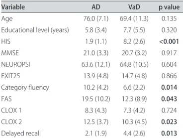
Demographic, clinical and neuropsychological data from AD and VaD groups are depicted in Table 2.
Arq Neuropsiquiatr 2010;68(2)
187
Alzheimer’s disease and vascular dementia: brief cognitive tests differention Matioli and Caramelli
the AD group presented worse performance on delayed recall. ROC curves were calculated for these tests, dis-playing low sensitivity or speciicity values (Table 3).
DISCUSSION
AD and VaD (mostly SVaD) patients evaluated in this study performed signiicantly diferent in four cognitive tests: AD patients performed worse on the delayed recall test, while VaD patients performed worse on semantic
and phonemic verbal luency and in the subtest “CLOX 2” (on copy ) of the Executive Clock Drawing Task. How-ever, ROC curve analysis revealed that these tests had low accuracy for the diferential diagnosis between the two conditions.
Greater impairment in delayed recall tests in AD in comparison to VaD is well recognized17. Delayed recall im-pairment is caused by deicits in storage of new information due to neuroibrilllary pathology of medial temporal areas, such as hippocampus, entorhinal cortex and amygdala17.
In our study, VaD patients performed significantly worse in a category luency task (animals) than the AD group. However, there is no consensus in the literature about the performance in category luency as being better or worse in VaD than in AD, with some authors reporting no diferences18 or even a reverse pattern, i.e., worse per-formance on this task in AD when compared to VaD19.
Phonemic verbal luency is a good test to assess exec-utive functions and the integrity of prefrontal cortex. In the present study performance in this task was found to be signiicantly more impaired in VaD than in AD. his inding is consistent with an early report by Canning et al. in which VaD subjects produced signiicantly less words with letter F than their AD counterparts20.
Many authors have described executive dysfunction as an important cognitive feature of VaD, especially SVaD2,17. he impaired performance in this task can be explained by damage to the dorsolateral prefrontal system, which is interconnected with the basal ganglia and thalamus in a
Table 1. Neuropsychological data from control group, AD and VaD patients.
Variable Controls AD VaD P value
MMSE 28.5 (1.6) 21.0 (3.3) 20.8 (3.2) p<0.001
NEUROPSI 102.3 (13.2) 63.6 (12.1) 64.8 (10.5) p<0.001
EXIT25 5.4 (2.9) 13.9 (4.8) 14.7 (4.8) p<0.001
Category luency 16.1 (4.4) 10.2 (4.2) 6.6 (2.2) p<0.001
FAS 30.7 (11.3) 19.5 (10.2) 12.3 (8.9) p<0.001
CLOX 1 13.8 (2.4) 8.3 (4.3) 7.3 (4.2) p<0.001
CLOX 2 14.7(1.3) 12.5 (3.7) 10.3 (4.5) p<0.001
Delayed recall 9.9 (0.3) 2.1 (1.9) 4.4 (2.6) p<0.001
AD: Alzheimer’s disease; VaD: vascular dementia; MMSE: Mini-Mental State Examination; FAS: phonemic verbal luency. Values are mean and standard deviation (in parenthesis).
Table 2. Demographic, clinical and neuropsychological data from AD and VaD groups.
Variable AD VaD p value
Age 76.0 (7.1) 69.4 (11.3) 0.135
Educational level (years) 5.8 (3.4) 7.7 (5.5) 0.320
HIS 1.9 (1.1) 8.2 (2.6) <0.001
MMSE 21.0 (3.3) 20.7 (3.2) 0.917
NEUROPSI 63.6 (12.1) 64.8 (10.5) 0.604
EXIT25 13.9 (4.8) 14.7 (4.8) 0.866
Category luency 10.2 (4.2) 6.6 (2.2) 0.014
FAS 19.5 (10.2) 12.3 (8.9) 0.043
CLOX 1 8.3 (4.3) 7.3 (4.2) 0.724
CLOX 2 12.5 (3.7) 10.3 (4.5) 0.023
Delayed recall 2.1 (1.9) 4.4 (2.6) 0.013
AD: Alzheimer’s disease; VaD: vascular dementia; HIS: Hachinski Ischemic Scale; MMSE: Mini-Mental State Examination; FAS: phonemic verbal luency. Values are mean and standard deviation (in parenthesis). Statistically signiicant p values are displayed in bold.
Table 3. Results of ROC curve analysis of cognitive tests in diferentiating VaD from AD.
Cognitive test AUC- ROC Cut-of Sensitivity Speciicity
Category luency 0.762 < 9* 86.7% 66.7%
FAS 0.716 <13* 60.0% 60.0%
CLOX 2 0.742 < 14* 93.3% 60.0%
Delayed recall 0.764 < 4# 86.7% 66.7%
Arq Neuropsiquiatr 2010;68(2)
188
Alzheimer’s disease and vascular dementia: brief cognitive tests differention Matioli and Caramelli
frontal subcortical loop, including the dorsolateral cau-date nucleus, lateral dorsomedial globus pallidus internus, and anterior and dorsomedial nucleus of the thalamus. Phonemic luency impairment can indicate that this cir-cuit is disrupted at one or more of these subcortical loci or in the white matter tracts that interconnect them with the dorsolateral prefrontal lobe21.
Royall et al.16 described that CLOX 1 places high de-mands on executive control functioning, since patients are required to perform in a novel context, whereas CLOX 2 represents a purer measure of visoconstructional abili-ty. However, their study included only patients with AD, together with a control group, but not patients with sub-cortical dementia.By contrast, we found that CLOX 1 did not discriminate AD from VaD, although VaD pa-tients performed worse in the CLOX 2 task. his ind-ing can be explained by greater executive dysfunction and visuo-constructive impairment in VaD compared to AD, as already observed by other investigators22,23. Li-bon et al.23 showed that only AD patients’ performance in CLOX 1 improved in relation to CLOX 2 when com-pared to VaD associated with SVaD. hese authors con-cluded that the copy condition may be sensitive to execu-tive dysfunction and the impaired performance of AD pa-tients in CLOX 1 might be explained by deicient seman-tic knowledge. Cosentino et al.24 administered the clock drawing (command and copy) to AD, VaD and Parkin-son dementia groups. hey considered that clock draw-ing to command demands activation of large-scale neu-ronal networks, including semantic knowledge and ex-ecutive control. hey observed that CLOX 1 was unable to distinguish dementia subtypes. hese indings support CLOX 2 as a measure of executive deicits and highlight the important role of adding a copy task when adminis-trating the clock drawing test.
he EXIT25 is an executive functions’ test described by Royall et al.15. It was selected for our study in order to identify possible diferences in executive functioning between AD and VaD. In contrast to our irst expecta-tion, we found no statistical diference between the per-formance of the two groups, either for the total score and its subtests, although it diferentiated demented patients from controls. A possible explanation for this inding is that AD and VaD groups were composed exclusively by mildly demented subjects.
ROC curve analysis showed that the statistical difer-ences described above were not suicient to attain good diagnostic accuracy for the discrimination between VaD and AD. Although considering that the evaluation of a larger sample of patients could result in a better discrim-inatory value of the current approach, we may conclude that brief cognitive tests, such as those used in this study, do not diferentiate VaD (especially SVaD) from AD and
that additional neuropsychological testing, together with neuroimaging and clinical information, are necessary for the diferential diagnosis of these two common dement-ing conditions.
REFERENCES
1. Herrera Júnior E, Silveira ACP, Nitrini R. Epidemiologic survey of dementia in a community-dwelling Brazilian population. Alzheimer Dis Assoc Disord 2002;16:103-108.
2. Róman GC, Erkinjuntti T, Wallin A, Pantoni L, Chui HC. Subcortical ischaemic vascular dementia. Lancet Neurol 2002;1:426-436.
3. Yuspeh RL, Vanderploeg RD, Crowell TA, Mullan M. Diferences in executive functioning between Alzheimer’s disease and subcortical ischemic vacular dementia. J Clin Exp Neuropsychol 2002;24:745-754.
4. Lindeboom J, Weinstein H. Neuropsychology of cognitive ageing, minimal cognitive impairment, Alzheimer’s disease, and vascular cognitive impair-ment. Eur J Pharmacol 2004;490:83-86.
5. American Psychiatric Association. Diagnostic and statistical manual of mental disorders (DSM-IV), 4th Ed. Washington, DC: American Psychiatric Association, 1994.
6. Mckhann G, Drachman D, Folstein M, Katzman R, Price D, Stadlan EM. Clinical diagnosis of Alzheimer’s disease: report of the NINCDS-ADRDA work group under the auspices of the Department of Health & Human Services Task Force on Alzheimer’s disease. Neurology 1984;34:939-944.
7. Nitrini R, Caramelli P, Bottino CM, Damasceno BP, Brucki SMD, Anghinah R. Diagnosis of Alzheimer’s disease in Brazil: diagnostic criteria and auxiliary tests. Recommendations of the Scientiic Department of Cognitive Neurol-ogy and Aging of the Brazilian Academy of NeurolNeurol-ogy. Arq Neuropsiquiatr 2005; 63:713-719.
8. Stoppe Júnior A, Jacob Filho W, Louzã Neto MR. Avaliação de depressão em idosos através da Escala de Depressão em Geriatria: resultados preliminares. Rev ABP-APAL1994;16:149-153.
9. Carthery-Goulart MT, Areza-Fegyveres R, Schultz RR, et al. Brazilian version of the Cornell depression scale in dementia. Arq Neuropsiquiatr 2007;65: 912-915. 10. Jeste DV, Finkel SI. Psychosis of Alzheimer’s disease and related dementias: di-agnostic criteria for a distinct syndrome. Am J Geriatr Psychiatry 2000;8:29-34. 11. Brucki SMD, Nitrini R, Caramelli P, Bertolucci PHF, Okamoto IH. Suggestions for utilization of the mini-mental state examination in Brazil. Arq Neuropsiquiatr 2003;61:771-781.
12. Abrisqueta-Gomez J, Ostrosky-Sollis F, Bertolucci PH, Bueno OF. Applicability of the abbreviated neuropsychologic battery (NEUROPSI) in Alzheimer dis-ease patients. Alzheimer Dis Assoc Disord 2008;22:72-78.
13. Hachinski VC, Lanssen NA, Marshall J. Multi-infarct dementia: a cause of men-tal deterioration in the elderly. Lancet 1974;2:207-210.
14. Nitrini R, Caramelli P, Herrera Júnior E, et al. Performance of illiterate and lit-erate nondemented elderly subjects in two tests of long-term-memory. J Int Neuropsychol Soc 2004; 10:634-638.
15. Royall DR, Mahurin RK, Gray KF. Bedside assessment of executive cognitive im-pairment: the Executive Interview (EXIT). J Am Geriatr Soc 1992;40:1221-1226. 16. Royall DR, Cordes JA, Polk M. Clox: an executive clock drawing task. J Neurol
Neurosurg Psychiatry 1998;64:588-594.
17. Graham NL, Emery T, Hodges JR. Distinctive cognitive proiles in Alzheimer’s disease and subcortical vascular dementia. J Neurol Neurosurg Psychiatry 2004;75:61-71.
18. Crossley M, D’Arcy C, Rawson NS. Letter and category luency in communi-ty-dwelling Canadian seniors: a comparison of normal participants to those with dementia of the Alzheimer or vascular type. J Clin Exp Neuropsychol 1997;19:52-62.
19. Jones S, Laukka EJ, Bäckman L. Diferential verbal luency deicits in the preclinical stages of Alzheimer’s disease and vascular dementia. Cortex 2006;42: 347-355. 20. Canning SJ, Leach L, Stuss D, Ngo L, Black SE. Diagnostic utility of abbreviat-ed luency measures in Alzheimer disease and vascular dementia. Neurology 2004;62:556-562.
21. Chui H, Willis L. Vascular diseases of the frontal lobes. In: Miller B, Cummings J (Eds). The human frontal lobes. New York: Guilford Press, 1999:370-401. 22. Libon DJ, Bogdanof B, Bonavita J, et al. Dementia associated with
periven-tricular and deep white matter alterations: a subtype of subcortical demen-tia. Arch Clin Neuropsychol 1997;12:239-250.
23. Libon DJ, Swenson RA, Barnoski EJ, Sands LP. Clock drawing as an assessment tool for dementia. Arch Clin Neuropsychol 1993;8:405-415.
