Chlamydia pneumoniae
AND STROKE
Is there a direct relationship?
Rubens J. Gagliardi
1, Helio H. Caiaffa-Filho
2Abstract – Objective: To investigate the possible relationship between atherothrombotic stroke and Chlamydia pneumoniae. Method: 150 patients with carotid atherothrombosis were enrolled. The casuistic was divided in three groups: ischemic stroke (IS): 65 patients; transient ischemic attack (TIA): 26 patients; and control: 59. The IS or TIA onset was up to 30 days from the beginning of the study. Carotid atheromatoses was diagnosed by Doppler-ultrasonography. Patients with cardioembolic risk or non-atherothrombotic origin were excluded. Comparisons were done between the three groups, and within each group according to the different age sub-groups, to the main arteries affected, and to the atherogenic risk factors. Bacteria detection was done using polimerase chain reaction. Results: Only one patient tested positive for C. pneumoniae belonging to the control group. Conclusion: These results do not suggest that C. pneumoniae participated in the onset of IS or TIA or that it has a role in carotid plaque destabilization.
Key WOrDS: stroke, chlamydia, atherosclerosis, risk factors, cerebral infarct.
Chlamydia pneumoniae e acidente vascular cerebral aterotrombótico: existe relação direta?
Resumo – Objetivo: Investigar a possível relação entre Chlamydia pneumoniae e acidente vascular cerebral aterotrombótico (AVC). Método: 150 pacientes com aterotrombose carotídea foram estudados. A casuística foi dividida em 3 grupos: AVC: 65 pacientes; ataque isquêmico transitório (AIT): 26 pacientes e controles: 59. O início do AVC ou AIT era até 30 dias da inclusão no estudo. A ateromatose carotídea foi diagnosticada por ultrassonografia com Doppler. Os pacientes com risco cárdio-embólico ou sem evidência de aterotrombose foram excluídos. Foram estabelecidas comparações entre os 3 grupos e dentro de cada grupo, formado sub-grupos de acordo com diferentes idades, território arterial comprometido e fatores de risco. A detecção da bactéria foi feita por reação de polimerização em cadeia. Resultados: Somente um paciente, pertencente ao grupo controle, teve resultado positivo. Conclusão: estes achados não sugerem que a C. pneumoniae participe no desencadeamento do AVC ou AIT ou que tenha papel na desestabilização da placa.
PAlAVrAS-ChAVe: acidente cerebral vascular, aterosclerose, fatores de risco, infarto cerebral, chlamydia.
Santa Casa de São Paulo, Faculty of Medical Sciences, São Paulo SP, Brazil: 1Full Professor, Neurology Discipline, Chairman Cerebrovascular Diseases
Division; 2Assistant Professor, Microbiology Discipline of Santa Casa and laboratory Medical Investigation lIM-03, FMUSP. Support: Support Center
for Scientiic Publications of Santa Casa de São Paulo – Faculty of Medical Sciences and FAP: research Support Foundation of “Santa Casa de São Paulo – Faculty of Medical Sciences”.
received 5 April 2009, received in inal form 4 June 2009. Accepted 11 June 2009.
Dr. Rubens José Gagliardi – Avenida Angélica 916 / 3o andar / Conj. 305 - 01228-000 São Paulo SP - Brasil. E-mail: [email protected]
Several studies have reported infections as a risk fac-tor for stroke (AVC)1,2 which could act in the unleashing,
the progression or the destabilization of the atheroscle-rotic plaque1. The conirmation of such hypothesis would
open an important space, both for an eventual speciic therapeutic approach in the acute phase of the stroke and for its prevention. There is an increasing volume of evi-dences suggesting the association between inlammation, infection, and atherosclerosis3,4, although that relation is
still not well established5. There are authors that defend
that the inlammation is the key for the development and the progression of atherothrombosis3,6. The most frequent
There is a lot of speculation regarding the possible role of C. pneumoniae in atherosclerosis, which could act in the unleashing, the evolution, worsening or destabiliza-tion of the plaque, making it vulnerable to thromboembo-lic compthromboembo-lications. The infection caused by C. pneumoniae
has been mentioned as an independent risk factor for vas-cular diseases, including cerebrovasvas-cular ones5-7;
howev-er that point of view is still much questionable. Opinions vary from one extreme to the other6, from the ones that
defend the microorganism would be the causal agent of atherosclerosis7,8 to the ones that believe it would merely
be an innocent bystander9. Despite the strong association
found through several epidemiologic and experimental studies, the causality of the facts cannot be simply estab-lished. There are still no safe data on the fact that pneu-moniae would be either related or not to atherosclerosis, and in case it is positive, how that relation would be9. It
could act in the beginning of the process (unleashing the histological proile), during its course, either aggravating it or in its inal phases, destabilizing the plaque. Most of the early studies used immunoluorescence assays, dosing serum levels of IgA, IgG, or IgM, as a diagnostic method for pneumoniae. The results, however, are conlicting, with some positive10 and other negative indings8,11-13, with the
methodology employed being questioned. More recent-ly, pneumoniae DNA detection by the polymerase chain reaction (PCr) has been shown to be a valid diagnostic method, and apparently more reliable than other tech-niques11. The presence of circulating DNA of
pneumoni-ae, diagnosed through the PCr in humans, may represent a persistent systemic infection by the bacteria.
The factors that leave the transformation of the as-ymptomatic plaque to sas-ymptomatic have been the tar-get of many studies and is very important in the devel-opment of ischemic stroke. This study was performed in order to analyze the possible role of pneumonia in this stage of atherosclerotic plaque.
METHOD
This is a prospective study with patients admitted to the Santa Casa de São Paulo hospital, in the period between March 2004 to August 2006; 150 patients (age range: 40–85 years), with carotid atherothrombosis, were enrolled. The patients were di-vided into three groups: ischemic stroke (IS): 65 patients (40 men); transient ischemic attack (TIA): 26 patients (11 men) and control: 59 patients (22 men). The IS or TIA onset was established up to 30 days from the beginning of the study. Stroke and TIA were diagnosed by the symptoms presented with conirmation obtained by computed tomography (CT-scan) or magnetic res-onance imaging (MrI). Carotid atheromatous plaque was diag-nosed by Doppler ultrasonography; IS and TIA by clinical and tomographic evaluation. Patients that presented cardioembolic risk (atrial ibrillation, recent myocardial infarction, intramural
thrombus, arrhythmias), lacunar infarction, cerebral hemorrhage, or other with non-atherothrombotic origin were excluded. Age, gender, and risk factor for stroke were not considered for pa-tient exclusion. The control group was formed by papa-tients that presented atherosclerotic plaques in the carotid arteries and that had not undergone a ischemic event, thus being consid-ered to be a asymptomatic plaque, different from the others with symptomatic plaque. Comparisons were done between the three groups, and within each group for the different age groups, the main arteries affected, and the atherogenic risk fac-tors were detected.
Thirth patients were between 40-55 years old (18 with IS, 4 TIA, 8 controls), 49 between 56–70 years old (21, 11, 17 respec-tively) and 71 between 71–85 years old (27, 11, 33 respecrespec-tively). The artery with the highest stenosis degree was considered: 87- internal carotid artery (37, 16, 34); 37- common carotid artery (16, 6 ,15); and 26- carotid bulb (12, 4, 10). regarding risk factors, 87 had blood hypertension (43, 17, 27); 33 were diabetic (16, 8, 9); 45 had hypercholesteremia (21, 5, 19); 22 were smokers (13, 3, 6), and 44 (13, 5, 26) did not have these classic risk factors.
Bacteria detection was performed by PCr in the venous blood.
To each 1.5 ml eppendorf tube containing blood, 1000 µ/l of the lySeS SOlUTION was added, vortexed vigorously for 15 sec., centrifuged for 2 min. at 13,000 g, and the supernatant was discarded by aspiration with a Pasteur pipette. Then, to the pel-let, 500 µ/l of TeN, 5 µ/l of SDS (10%), and 3 µ/l of protei-nase K (125 mg/ml) was added, mixed and incubated overnight in a 56oC block heater. After that, 150 µ/l of 5M NaCl was added
and the tubes were vigorously agitated for 15 sec. After centrif-ugation at 13,000 rpm for 5 minutes, the supernatant was trans-ferred to a new 1.5 ml eppendorf tube and 650 µ/l of cold iso-propanol was added. DNA was precipitated in a freezer at –20oC
overnight. After that period, the supernatant was discarded and the pellet was washed twice with cold 70% ethanol and air-dried for 30 min. The DNA pellet was dissolved in 200 µ/l of Te and stored at –20oC until use. Five microliters (5 ul) of this DNA was
used as template for PCr ampliication and 1 µ/l per tube in our “in house” Internal Control PCr (human β-Globin gene) for each sample studied.
The C. pneumoniae PCr was performed as follows: Single PCr was done in a inal volume of 20 µ/l containing 1U Taq polimerase, 1X polymerase buffer [containing 50 mMKCl, 20 mM Tris-hCl (ph8.4)], 1.5 mM MgCl2 , 200 uM dNTPs mixture,
0.5 uM of each primer using a Perkin elmer 2400 (Applied Biosys-tems). The PCr conditions were: pre-denaturation step at 94oC
for 5 minutes, followed by 40 cycles of 95oC for 45 sec, 62oC for
45 sec, and 72oC for 1 minute. A inal extension step was done at
72oC for 7 minutes. The C. pneumoniae primers used were hl-1
5’GTT GTT CAT GAA GGC CTA CT 3’ as the forward and hr-1 5’ TGC ATA ACC TAC GGT GTG TT 3’ as the reverse according to Campbell14. For each sample, a quality control PCr detecting the
mix with the primers PCO-3 5’ACA CAA CTG TGT TCA CTA GC 3’ and PCO-4 5’CAA CTT CAT CCA CGT TCA CC 3’, according to Saiki et al.15. Cycling conditions were: 5 min. at 94oC as
pre-de-naturation step, followed by 35 cycles of 1 min at 94oC, 1 min 30
sec at 62oC, and 2 min at 72oC. A inal extension step was done
at 72oC for 7 minutes.
Ten microliters of the amplified products were electro-phoresed in 1.5% agarose gel and visualized under a UV light after ethidium bromide staining. The size of the speciic C. pneumo-niae ampliied product (437 bp) and the internal control beta-globin gene (110 bp) were assessed by comparison with a com-mercial 100 bp marker.
This study was approved by the ethical Committee of Santa Casa de São Paulo (project 045/00, approved in 26.06.00).
RESULTS
Only one patient tested positive for C. pneumoniae; that patient belonged to the control group. The remain-ing results were negative.
The results by different sub-groups, are presented in the Tables (Tables 1–4).
DISCUSSION
The factors that lead to the transformation of one as-ymptomatic plaque into a sas-ymptomatic one, including the infectious and inlammatory agents, are still controversial. Why does a sick person in a speciic part of his/her life becomes symptomatic, and why do sick people with the same degree of stenosis, either symptomatic or asymp-tomatic, heave really different risk degrees for a stroke16,
are questions that really matter and that still have nor ap-propriate answer. The investigation herein seeks to help in this task, by analyzing a possible relation of with the oc-currence of a new stroke and a probable destabilization of the atherosclerotic plaque. Considering that the inclusion criteria for the study herein have been developed in order to only allow for the admission of patients with athero-thrombotic manifestations in the study group, with a good
Table 1. Distribution by age.
Age (y) Sex
IS TIA Control Total
N +CP N +CP N +CP N +CP
40–55 Men
Women
10 8
0 0
2 2
0 0
3 5
0
0 30 0
56–70 Men
Women
14 7
0 0
5 6
0 0
7 10
0
0 49 0
>71 Men
Women
15 12
0 0
6 7
0 0
14 17
1
0 71 1
y: years; IS: ischemic stroke; TIA: ischemic transient attack; N: number of patients; +CP: positivity for Chlamydia pneumoniae.
Table 2. Distribution by main affected artery.
Artery
IS TIA Control Total
N +CP N +CP N +CP N +CP
ICA 37 0 16 0 34 0 87 1
CCA 16 0 6 0 15 0 37 0
CB 12 0 4 0 10 0 26 0
ICA: internal carotid artery; CCA: common carotid artery; CB: carotid bulb; IS: ischemic stroke; TIA: ischemic transient attack N: number of patients;
+CP: positivity for Chlamydia pneumoniae.
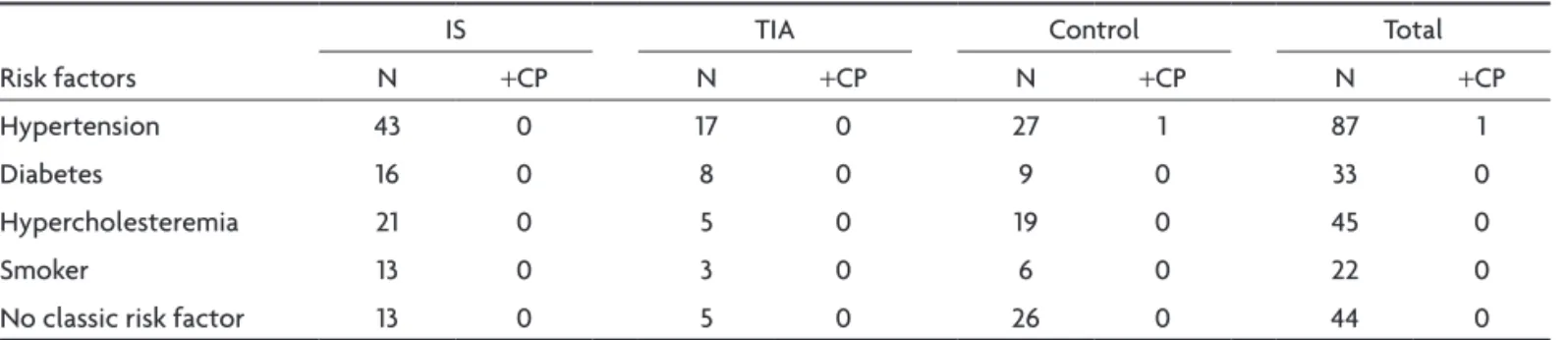
Table 3. Distribution by main risk factors.
risk factors
IS TIA Control Total
N +CP N +CP N +CP N +CP
hypertension 43 0 17 0 27 1 87 1
Diabetes 16 0 8 0 9 0 33 0
hypercholesteremia 21 0 5 0 19 0 45 0
Smoker 13 0 3 0 6 0 22 0
No classic risk factor 13 0 5 0 26 0 44 0
safety margin we could say that the elements in this study that had a stroke or a TIA had a symptomatic plaque.
In the current study we used PCr for detection of
pneumoniae for a diagnostic research; this laboratorial method allows the active infection diagnosis, being neg-ative for the patients that were previously contaminated and who are already healed. Most part of the studies are being performed using diagnosis by measuring IgG, IgA ou IgM antibodies, and that deserves some considerations. Seroepidemiological studies have important limitations, and several of them might be taken into consideration when interpreting the results obtained with that kind of study17,18. The presence of IgG antibodies simply translates
a primary infection and does not allow for the distinction between a chronic case, a persisting case, a reinfection or an old isolated infection17. The deinition of seropositivity
has been varying a lot from study to study, and there is no straight deinition for the subject. The positivity criterion varies according to different authors, with titles of IgG as
≥1:8; ≥1:16, ≥ 1:64, ≥1:128, ≥1:256, which makes comparisons and conclusions dificult. Despite the fact that the IgM an-tibody is an important marker for acute infection, it can provide a crossed reaction, with a false positive for the rheumatoid factor17,18 or other infections18. A few studies
have investigated a possible relation between the stages of atherosclerosis and the antibody classiication. labo-ratorial variation is another important aspect that many times makes it very dificult to interpret the results.
Our results (Tables 1–3) do not suggest that pneumo-niae participated in the destabilization of the plaque, for there was no positivity in patient groups with recent stroke or TIA. If that bacteria contributed for the desta-bilization of the plaque, that would make it have a great-er symptomatic probability, and we would expect to ind a larger amount of positive diagnosis in the stroke or TIA group when compared to the control group, which did not happen. The work of Ong and associates19
corrobo-rates that point of view, where by studying a largely preva-lent population for pneumoniae and atherosclerosis, they found high indexes of IgG and negativity when searching for PCr, in carotid atheroma. Such authors suggest that if there is any relation between atherogenesis and infec-tion by pneumoniae, it must be indirect and a non-essen-tial variable for the development of thrombosis19.
Tondel-la et al.20, did not ind any positivity for that research
af-ter evaluating 30 carotid araf-teries atherosclerotic plaques
using the PCr technique. Also searching for carotid ar-teries plaques obtained by endarterectomy,laBiche et al.21 have compared 37 pieces extracted from
symptom-atic patients to 57 from asymptomsymptom-atic patients, and when the diagnosis was established due to PCr, IgM and IgG, they did not ind a signiicant difference between the two groups. however, they could observe there was a relation between the symptoms that occurred and the infection, when the research was performed by IgA, with the pa-tients that have showed very high levels (>1:128)21. Such
authors remember that the single positivity for pneumo-niae is not enough to explain the event of the cerebrovas-cular process, and conclude that the bacteria has an insig-niicant role in converting a atherosclerotic plaque into the symptomatic state21. In an analysis performed with
the serum level of IgA and IgG in the carotid plaque with-drawn by endarterectomy, Vainas et al.22 conclude, in a
similar way to our casuistic, that pneumoniae is not re-lated to plaque destabilization. Such authors22 state that
this infectious agent would be related to atherosclerot-ic phenomena, probably in the irst stages of its develop-ment, but would not have any role in plaque destabiliza-tion. In a recent study, by means of the analysis of carot-id plaques obtained through endarterectomy in patients with atherothrombotic stroke, no relation to pneumoni-ae was detected12. Konya et al.13 did not ind any
correla-tion between the presence of pneumoniae in the carotid plaque and the severity of the stroke. A negativity inding in the PCr diagnosis, as in the essay herein, suggests that
pneumoniae does not play any role in complications nor in the destabilization of the plaque.
In relation to age and gender (Table 1) no difference was detected between the groups. Such data corroborate the previous reports as “The Northern Manhattan Stroke Study”8, which did not ind any difference between young
people and the elderly, men and women.
The association of pneumoniae to the classic risk fac-tors (rF) for vascular diseases is another aspect that de-served special attention. Considering the atherogenic rFs might be metabolic related and that there are reports on the interrelation of pneumoniae to the classic rFs2, that
possible issue was also a target in the investigation. We tried to identify if there would be the inluence of any rF or of an interrelation between a particular rF to the in-fection caused by the bacteria and the evolution of the medical proile. Several authors2,7 have been highlighting
that possibility and developed speciic researches on the subject, a fact that justiies the separate analysis in this casuistic. Arterial hypertension (Table 4) did not show any relation between the active infection by pneumoni-ae and the occurrence of symptoms, that could translate into an instability of the atherosclerotic plaque. Similar results were described in literature7,23, that support the
Table 4. Distribution according to the carotid stenoses degree.
Stenoses degree 1–40% 41–59% 60–99% 100%
C. pneumonia positivity 0 1 0 0
idea that arterial hypertension has no inluence over a possible atherogenic role related to the infection caused by that agent. Smoking has been frequently mentioned in literature as a related rF to pneumoniae2; though in our
casuistic (Table 4) the results did not corroborate that hy-pothesis, for there was no larger positivity among smok-ers related to the diagnosis for pneumoniae. We should point out that most reports of that positive association were established by setting the antibodies level, which could show an old infection. hypercholesterolemia is an-other atherogenic rF widely investigated, and there are several reports of a possible role linking the infection by
pneumoniae and/or contributing for the severity of the medical proile, when associated to the infection. In the present study there was no correlation found between hypercholesterolemia, infection by pneumonia,and clin-ical signs of destabilization of the atherosclerotic plaque. That inding is similar to other reports7,23. Diabetes
melli-tus was also separately analyzed; the results (Table 4), did not show any relation between the agents analyzed, sim-ilarly to the other analyses of rFs. That inding is also cor-roborated by other authors7,23.
The possibility of pneumoniae acting selectively or preferably among the main brain arteries was also stud-ied, and was not corroborated. (Table 2).
A possible relation between the stenosis level and the active infection by pneumoniae was also checked. That analysis was only carried out in the control group, in or-der to allow for an evaluation on the evolution of the stenosis in the stable plate. With that model we sought to evaluate a possible severing role before the ischemic event occurred, with a consequent plaque destabilization. The results presented in Table 4 have once again not de-tected any relation. That fact, which by our point of view is not being suficiently discussed in literature, reinforces the idea that the active infection by pneumoniae is not a risk factor for plaque destabilization.
In conclusion, our casuistic does not suggest that
pneumoniae participates in the onset of IS or TIA, and does not suggest that this bacteria plays a role in plaque destabilization.
REFERENCES
1. Elkind MS. Chlamydia pneumoniae: in your heart and on your mind. Stroke 2001;32:1976-1978.
2. Kiechl S, Werner P, Egger G, et al. Active and passive smoking, chron-ic infections, and the risk of carotid atherosclerosis: prospective results from the Bruneck Study. Stroke 2002;33:2170-2176.
3. Ross R. Atherosclerosis: an inlammatory disease. N Engl J Med 1999;
340:115-126.
4. Apfalter P, Hammerschlag MR, Boman J. Reliability of nested PCR for
the detection of Chlamydia pneumoniae in carotid artery atheroscle-rosis. Stroke 2003;34:e73-e75.
5. Rosenfeld ME, Blessing E, Lin TM, Moazed TC, Campbell LA, Kuo C. Chlamydia, inlammation, and atherogenesis. J Infect Dis 2000;181
(Suppl 3): S492-S497.
6. Hansson GK. Inlammation, atherosclerosis, and coronary artery dis
-ease. N Engl J Med 2005;352:1685-1695.
7. Fagerberg B, Gnarpe J, Gnarpe H, Agewall S, Wikstrand J. Chlamydia pneumoniae but not cytomegalovirus antibodies are associated with
future risk of stroke and cardiovascular disease: a prospective study in
middle-aged to elderly men with treated hypertension. Stroke 1999;30:
299-305.
8. Elkind MS, Lin IF, Grayston TJ, Sacco RL. Chlamydia pneumoniae and the risk of irst ischemic stroke: The Northern Manhattan Stroke Study.
Stroke 2000;31:1521-1525.
9. Cochrane M, Pospischil A, Walker P, Gibbs H, Timms P. Distribution of Chlamydia pneumoniae DNA in atherosclerotic carotid arteries: signif
-icance for sampling procedures. J Clin Microbiol 2003;41:1454-1457. 10. Shor A, Phillips JI. Chlamydia pneumoniae and atherosclerosis. JAMA
1999;282:2071-2073.
11. Glader CA, Stegmayr B, Boman J, et al. Chlamydia pneumoniae anti
-bodies and high lipoprotein(a) levels do not predict ischemic cerebral infarctions. Results from a nested case-control study in Northern Swe -den. Stroke 1999;30:2013-2018.
12. Gagliardi RJ, Silveira DR, Caffaro RA, Santos VP, Caiaffa-Filho HH.
Chlamydia pneumoniae and symptomatic carotid atherosclerotic
plaque: a prospective study. Arq Neuropsiquiatr 2007;65:385-389. 13. Konya J, Molnar S, Magyar MT, Szekeres CC, Kerenyi L, Csiba L. Se
-verity of carotid atherosclerosis unrelated to Chlamydia pneumoniae infection in acute ischemic stroke patients: a clinicopathological study.
Cerebrovasc Dis 2008;25:170-175.
14. Campbell LA. PCR detection of Chlamydia pneumoniae. In: Persing DH, Smith TF, Tenover FC, White TJ (eds): Diagnostic molecular micro
-biology: principles and applications. Washington, DC.: Amercian Soci
-ety for Microbiology, 1993: 247-252.
15. Saiki RK, Gelfand DH, Stofel S, et al. Primer-directed enzymatic ampli
-ication of DNA with a thermostable DNA polymerase. Science 1988,
239:487-491.
16. Toole JF. ACAS recommendations for carotid endarterectomy. ACAS
Executive Committee. Lancet 1996;347:121.
17. Siscovick DS, Schwarts SM, Caps M, Wang SP, Grayston JT. Chlamyd -ia pneumon-iae and atherosclerotic risk in populations: the role of
se-roepidemiology. J Infect Dis 2000;181 (Suppl 3): S417-S420.
18. Dowell SF, Peeling RW, Boman J, et al. Standardizing Chlamydia pneu
-moniae assays: recommendations from the Centers for Disease Control and Prevention (USA) and the Laboratory Centre for Disease Control (Canada). Clin Infect Dis 2001;33:492-503.
19. Ong GM, Coyle PV, D’Sa AABB, et al. Non-detection of Chlamydia spe
-cies in carotid atheroma using generic primers by nested PCR in a pop
-ulation with a high prevalence of Chlamydia pneumoniae antibody. BMC Infect Dis 2001;1:12-20.
20. Tondella MLC, Elkind MS, Guarner J, et al. Failure to detect Chlamydia
pneu-moniae in carotid atheromatous plaques. 102nd General Meeting of
Amer-ican Society for Microbiology, Salt Lake City, UT, USA., May 19-23, 2002. 21. LaBiche R, Koziol D, Quinn TC, et al. Presence of Chlamydia pneumo -niae in human symptomatic and asymptomatic carotid atherosclerotic plaque. Stroke 2001;32:855-860.
22. Vainas T, Kurvers HAJM, Mess WH, et al. Chlamydia pneumoniae serol
-ogy is associated with thrombosis-related but not with plaque-related mi
-croembolization during carotid endarterectomy. Stroke 2002;33:1249-1254.
23. Wang SS, Tondella MLC, Bajpai A, et al. Circulating Chlamydia
pneu-moniae DNA and advanced coronary artery disease. Internat J Cardiol