Increase in Vascular Injury of Sodium
Overloaded Mice May be Related to Vascular
Angiotensin Modulation
Cintia Taniguti Lima1, Juliane Cristina de Souza Silva1, Katia Aparecida da Silva Viegas1, Thais Cristina de Souza Oliveira1, Rariane Silva de Lima1, Leandro Ezequiel de Souza2, Danielle Aragão3, Dulce Elena Casarini3, Maria Claudia Irigoyen2, Silvia Lacchini1*
1Department of Anatomy, Institute of Biomedical Sciences, University of São Paulo, São Paulo, São Paulo, Brazil,2Hypertension Unit, Heart Institute, University of São Paulo Medical School, São Paulo, São Paulo, Brazil,3Nephrology Division, Department of Medicine, Federal University of São Paulo, São Paulo, São Paulo, Brazil
Abstract
This study aimed to analyzing the effect of chronic sodium overload upon carotid and femo-ral injury, and its relation to vascular angiotensin modulation. Male C57Bl6 mice were divid-ed in: control (cont), receiving 1% NaCl solution for 2 weeks (salt-2) or 12 weeks (salt-12). Two-weeks before the end of the study, a 2mm catheter was implanted around the left fem-oral and carotid arteries to induce injury. Blood pressure (BP) and heart rate (HR) were measured at the end of the study by tail plethysmography. Arteries were collected and pre-pared for histological analysis to determine arterial thickening and perivascular collagen de-position. Angiotensin II and Ang(1-7) were quantified in fresh arteries using the HPLC method. There were no differences in body weight, BP and HR. Intima/media ratio had a similar increase in both injured arteries of cont and salt-2 mice, but a more pronounced in-crease was observed in salt-12 mice (31.1±6%). On the other hand, sodium overload modi-fied perivascular collagen deposition, increasing thick fibers (cont: 0.5%; 2: 3.4%; salt-12: 0.6%) and decreasing thin fibers (cont: 7.4%; salt-2: 0.5%; salt-salt-12: 6.8%) in non-injured arteries. Injured arteries presented similar collagen fiber distribution. Angiotensin quantifica-tion showed increased Ang(1-7) in salt treated mice (salt-2: +72%; salt-12: +45%) with a concomitant decrease in Ang II (salt-2: -54%; salt-12: -60%). Vascular injury increased sig-nificantly Ang(1-7) in salt-12 mice (+80%), maintaining Ang II reduction similar to that of a non-injured artery. The lack of changes in BP and HR suggests that the structural changes observed may be due to non-hemodynamic mechanisms such as local renin-angiotensin system. Collagen evaluation suggests that sodium overload induces time-related changes in vascular remodeling. The increase of artery injury with concomitant increase in Ang(1-7) in 12-week treated mice shows a direct association between the duration of salt treatment and the magnitude of vascular injury.
OPEN ACCESS
Citation:Lima CT, Silva JCdS, Viegas KAdS, Oliveira TCdS, Lima RSd, Souza LEd, et al. (2015) Increase in Vascular Injury of Sodium Overloaded Mice May be Related to Vascular Angiotensin Modulation. PLoS ONE 10(6): e0128141. doi:10.1371/journal.pone.0128141
Academic Editor:Masanori Aikawa, Brigham and Women's Hospital, Harvard Medical School, UNITED STATES
Received:October 31, 2014
Accepted:April 22, 2015
Published:June 1, 2015
Copyright:© 2015 Lima et al. This is an open access article distributed under the terms of the Creative Commons Attribution License, which permits unrestricted use, distribution, and reproduction in any medium, provided the original author and source are credited.
Data Availability Statement:All relevant data are within the paper and its Supporting Information files.
Introduction
At an epidemiological level, it is still debatable whether a high sodium intake causes an eleva-tion in blood pressure or increases the rate of progression into a hypertensive level [1]. Some studies in rats have reported an age-related increase in blood pressure [2] while others have not found any significant changes [3]. More recently, we have shown that high sodium intake in rats changes autonomic regulation of HR, increasing tachycardic response to arterial pressure (AP) reductions [4]. Other experimental studies in rats have found an association of salt intake with target-organ effects, such as cerebral, renal [5], and cardiac [6] arteries and respective or-gans. Until the last decade, it has been generally assumed that adverse cardiovascular and renal effects of increased salt intake were mediated via increase in blood pressure. Moreover, several newer experimental and clinical studies have clearly demonstrated that in addition to these pressure-independent outcomes, salt overload also exerts direct harmful effects [7]. The find-ings related to the effect of salt intake upon cardiovascular system have pointed hypertension as the mechanism underlying these effects.
In this context, some studies using hypertensive high salt diet- treated rats have reported ar-terial stiffness [8], carotid hypertrophy [9–10], and intimal lesions [11]. However, it should be emphasized that any hemodynamic modification will interfere with vascular wall. Thus, the comprehension of vascular adaptations to salt intake, regardless of blood pressure, requires a clear understanding of the role that each factor plays regardless of hemodynamic factors.
Renin-angiotensin system (RAS) is directly modulated by sodium intake, and it is closely re-lated to RAS activity in hypertension and cardiovascular injury [12], inflammatory response [13], and to the structure of retinal arterioles [14]. On the other hand, components of RAS have been implicated in the pathogenesis of atherosclerosis in a variety of manners and as such they should be seen as important therapeutic targets. A large body of data indicates that angio-tensin II (Ang II) is associated with the production of reactive oxygen species, inflammatory cytokines, and the activation of adhesion molecules [15–16]. This is also consistent with the fact that angiotensin converting enzyme (ACE) is activated in the inflammatory cells present in vascular lesions; it is also consistent with the activation of RAS components during differentia-tion of monocytes to macrophages [17]. ACE activity is also increased in the neointima associ-ated with vascular injury in rats [18–19], in human carotid artery plaques [1], and in the stent-induced inflammatory mechanism [20].
There are many factors involved in the development of cardiovascular diseases; some stud-ies have pointed the vascular structure and function derangement as a feature underlying car-diovascular diseases, and it has been associated with inflammation and endothelial activation [3–21]. Furthermore, the development of atherosclerotic lesions has as characteristic the inti-mal thickening which is promoted in an early essential stage of disturb. Many experimental models have been created to study this pathologic condition and to evaluate the efficacy of therapeutic strategies. The cuff-induced neointimal thickening is closely related to a well de-scribed inflammatory reaction [22–23]. In this context, we have previously shown that vascular ACE activity is directly related to neointima formation in induced vascular injury in mice [24].
Despite the relevance of potential associations described above, few reports to date have as-sociated vascular adaptations to remodeling under sodium overload, and stated their relevance to vascular neointima formation after injury, particularly in normotension. The purpose of this study was then to evaluate whether high sodium intake can induce vascular remodeling under normotensive conditions and whether this condition may lead to vascular susceptibility to injury.
Materials and Methods
Animals
Experiments were performed with isogenic male C57Bl/6 mice obtained from the animal care unit of the Department of Anatomy of the Institute of Biomedical Sciences at University of São Paulo. The mice received standard laboratory chow and waterad libitum, and were distributed
five per cage in a temperature-controlled room (22°C) with a 12-h dark-light cycle. All proto-cols used were in accordance with the Guidelines for Ethical Care of Experimental Animals from the International Animal Care and Use Committee. This study was approved by the Uni-versity of Sao Paulo Medical School Ethical Committee (044/11). The mice were randomly as-signed to one of three groups: control, receiving filtered water (Cont); 1% NaCl solution to drink for two weeks (salt-2); 1% NaCl solution to drink for twelve weeks (salt-12).
The treatments started on the 4-week old mice, and were continued until they reached 16 weeks of age. All mice have undergone surgical implantation of perivascular cuff two weeks be-fore the end of treatments (14-weeks of age), in order to induce vascular injury and neointima formation.
Blood Pressure Measurements
In the last week of treatment, pulse blood pressure (BP) was indirectly measured by tail plethys-mography. During the week prior to the measurement, the animals were adapted to the system. BP and heart rate (HR) were measured 10 times for two consecutive days. For this, peak systolic BP were captured by the BP-2000 series II system, which consists of an electromagnetic trans-ducer located in the tail and connected to an amplifier and to an analog-to-digital converter. To measure tail plethysmography, the mouse was placed in restraining box previously heated to promote vasodilatation in the tail thereof. The sphygmomanometer adapted to mice was placed at the tail and inflated to total obstruction of blood flow to the artery flow. The flow obstruction was decreased slowly in order to capture the first peak of systolic blood pressure.
Femoral cuff placement
Two-weeks before the end of the study, it was performed a surgical procedure to induce femoral artery injury. To this procedure, it was used the femoral cuff placement method as described in detail previously [24]. The animals were anesthetized with an injection of Ketamine (90 mg/kg) and Xylazine (10 mg/kg) ip. Body temperature was controlled by placing the animal on operating table with heating plate to maintain the body temperature at 38°C. After isolating the left femoral artery from the surrounding tissues, a 2.0-mm polyethylene cuff (inner diameter 0.56 mm, outer diameter 0.965 mm; Becton Dickinson) was inserted around the artery, and then closed with a 4–0 cotton suture. To ensure that the blood flow was not obstructed, the cuff used was larger than the vessel. The right femoral artery did not receive the perivascular cuff, and was used as a control.
Carotid cuff placement
used was larger than the vessel. Also, the right carotid artery was used as a control. After recov-ery from anesthesia, the animals were given a standard diet and water ad libitum.
Tissue harvesting and histological staining
Fourteen days after implantation, mice were euthanized with an intraperitoneal injection of Ketamine (180 mg/kg) and Xylazine (20 mg/kg), received sodium heparin (50 U), and were subsequently perfused with 0.9% NaCl solution at constant pressure (80–90 mmHg) followed by a buffered 4% formalin solution. Tissues were maintained in formalin for 24–48 h to com-plete the fixation process, while arteries were studied using the adjacent tissues to preserve their integrity. After processing the tissues, it was embedded in paraplast for further histologi-cal analysis. It was performed different types of staining depending on the evaluation needed. To assess cellular morphology was performed Hematoxylin-Eosin staining, while to identify elastic lamina was used Verhoeff-Van Gieson staining; Picrosirius staining was used to identify collagen fiber deposition.
Morphometry. Morphometric analyses were performed on Verhoeff-Van Gieson, Picros-sirius, and Weigert-stained tissues. Histomorphometric analyses were performed blinded to the identity of experimental groups. Five-micrometer sections were obtained every 50μm, to-taling 10 sections along the 500μm of vessel length, corresponding to almost 25% of the total cuff-induced vascular lesion. Femoral and carotid cross sections images were digitized by com-puter image analysis (AxioVision 4.8 System) coupled to a Zeiss Axio Scope II microscope. For each section, the areas of the intima and media were calculated. The intimal area was calculated from the difference between the area of the inner elastic lamina and the luminal area; the area of the media layer was calculated by the difference between the area of the outer elastic lamina and the area of the inner elastic lamina. For collagen fiber deposition, slides were stained with Picrosirius and viewed by polarized light. For adequate staining slides were stained with 0.1% Sirius Red solution and counterstained with Harris's hematoxylin. Picrosirius-stained sections were evaluated by ordinary polychromatic and polarized light microscopies. Percent area of mature (red and yellow fibers) was compared to immature (green) fibers under polarized light. Elastic system fibers were studied using Weigert's resorcin-fuchsin staining. Elastic laminae were counted in 5 slices for each artery.
Vascular ACE immunohistochemistry. Angiotensin I converting enzyme (ACE) was measured by immunohistochemistry performed in slices of each artery stained with IgG anti-ACE antibody (Santa Cruz Biotechnology, Santa Cruz, CA). In brief, slices were deparaffinized, rehydrated, and the endogenous peroxidase activity was blocked with hydrogen peroxide (3% in water) for 20 minutes. Following rehydration, the slices were rinsed with phosphate-buffered saline (PBS). Bovine serum albumin 2% (BSA in PBS) was used to block the nonspecific sites for 60 minutes at room temperature. The primary antibody was diluted to 1:500 in TBS-TC, and applied to the sections for 16–18 hours at 4°C. Subsequently, the samples were washed and incubated with the biotinylated secondary antibody (Zymed Laboratories, South San Francisco, CA) for 60 minutes at room temperature, followed by incubation with streptavidin-peroxidase complex (1: 1000) for 60 minutes at room temperature. Finally, a 3,3’—diaminobenzidine solu-tion (DAB, Vector Labs.) was applied. The slices were coded and then assessed by two indepen-dent blinded observers using an optical microscope (Zeiss Axio Scope II) equipped with a 40x objective and coupled to an image analyzer (Axio Vision 4.8 System).
Angiotensin quantification
fragments of femoral arteries were collected. The mice were subsequently euthanatized by an overdose of the same anesthetic and the carotid arteries were removed. To harvest control ves-sels (without injury) we collected approximately 2mm segments from the region corresponding to that of vascular injury (in the contralateral vessel), while for the arteries with injury, we col-lected a 2mm fragment surrounded by cuff. Tissues were removed, immediately frozen and kept at -80°C until measurement. These femoral and carotid arteries were used for quantifica-tion of angiotensin I, II and 1–7 [25]. Angiotensin extraction was performed using in Sep-Pak-C18 columns (Waters, Milford, MA), and then, peptides were measured by HPLC, as described by Almeida et al., 2006 [26]. Peptides were identified according to retention time. The identity of eluted ANG I, ANG II, and ANG 1–7 was confirmed by direct sequencing (protein sequenc-er PPSQ-23; Shimadzu, Tokyo, Japan). For concentration detsequenc-ermination, commsequenc-ercially avail-able peptides were employed to develop a standard curve. Peptide levels were expressed as pictograms per artery, as described in a previous study [24]. These were not normalized ac-cording to total protein concentration since total protein was at very low concentrations.
Statistical analysis
All values are expressed as means ± SD. Morphometric evaluations were first tested by two-way ANOVA, while angiotensins were tested by one-two-way ANOVA. When ANOVA demon-strated significant differences, Tukey's post hoc analysis was used to compare groups. For all statistical analyses,P0.05 was considered statistically significant.
Results
Body weight and hemodynamic measurements
Body weight at the start and at the end of protocol was similar between the three groups. Indi-rect measurements showed that blood pressure (Fig 1A) and heart rate levels (Fig 1B) remained essentially the same in all groups regardless of the high salt treatments (S1 Table).
Neointima thickening in femoral and carotid arteries
Upon cuff-induced injury, all mice developed vascular neointima thickening regardless of ei-ther fresh water or salt intake.Fig 2Ashows the relative area of neointima, comparing non-in-jured and innon-in-jured carotid arteries in control and in two and twelve-week treated mice. The neointima-to-media ratio showed a significant increase in cont and salt-2 groups, and a greater increase in salt-12 mice.Fig 2Balso shows that in femoral arteries the neointima-to-media ratio cont and salt-2 groups, with a larger increase in the salt-12 group (S2 Table). The area of the media was not different among the four groups regardless of neointima formation (data not shown), thus indicating an injury process without vascular remodeling.
Elastic lamellae analysis
Fig 3presents the counting of elastic lamellae in carotid and femoral arteries. As can be ob-served, there were no changes in this elastic component associated with salt intake. Also, vascu-lar injury did not change lamellae distribution in femoral and carotid arteries.
Collagen fibers deposition
fiber (in green color), was found, together with a relative low deposition of mature (red) and in-termediary (yellow) fibers (Fig 4). Interestingly, 2-weeks of salt intake changed this pattern, with an important reduction of young fibers, and increase in intermediary and mature collagen fibers. This response was found for both non-injured carotid and femoral arteries and for in-jured carotid and femoral arteries. On the other hand, a 12-week salt intake showed a return to a pattern similar to that of control mice. However, after 12-weeks of salt intake the injured ca-rotid presented an intense deposition of all mature, intermediary and young collagen fibers.
ACE immunohistochemistry
ACE was evaluated by the staining score in histological prepared vessels.Fig 5presents a semi-quantitative analysis of this immunohistochemistry in carotid (5A) and femoral (5B) arteries.
The results were similar to those obtained for intima/media ratio, when all injured vessels Fig 1. Hemodynamic measurements.Systolic blood pressure (1A) and heart rate (1B) assessed by tail plethysmography. As can be observed, there was no difference between the groups.
Fig 2. Intima/media ratio in femoral and carotid arteries.A: carotid artery, and B: femoral artery. Observe the increase in I/M ratio after vascular injury in all treatments. White bar: control, non-injured artery; black bar: injured artery. ¥ p<0.05 when compared to non-injured artery; # p<0.05 when compared to injured artery of cont and salt2 groups.
presented increases in antibody staining, with a larger increase found for 12-week salt treated mice.
Angiotensin evaluation
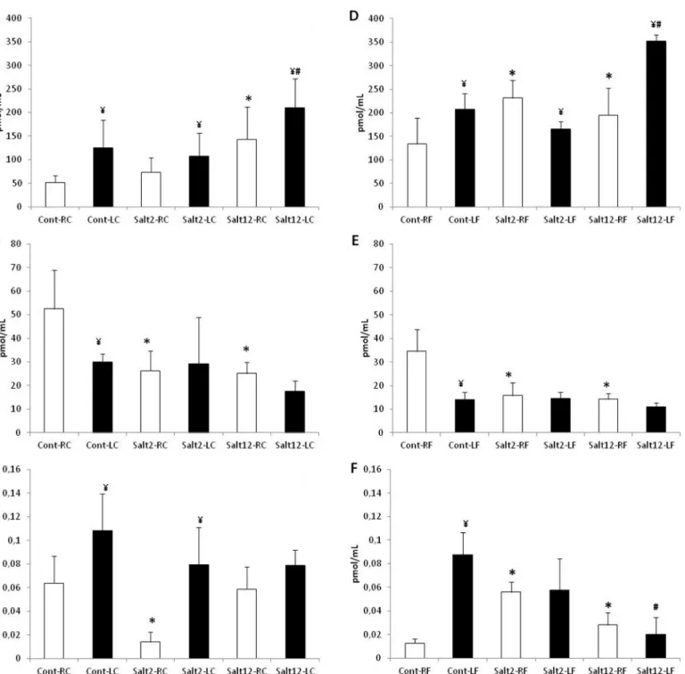
Angiotensin I, II and (1–7) were measured in 2mm of carotid and femoral arteries, and the re-sults obtained are presented inFig 6. It can be observed that in carotid artery (6A, 6Band 6C)
Fig 3. Number of elastic lamellae in femoral and carotid arteries.A: carotid artery, and B: femoral artery. White bar: control, non-injured artery; black bar: injured artery. No differences were observed between treatments.
salt intake increases Ang (1–7) and decreases Ang II, with low interference in Ang I. However, after injury, we did not observe Ang II reduction in salt-2 and salt-12 mice with a concomitant reduction of Ang I. A similar response was found for femoral arteries (6D, 6Eand 6F).
Discussion
This study assessed the effect of salt overload upon femoral and carotid injury in mice. The main findings of the present study were that high salt intake increases the magnitude of vascu-lar injury without blood pressure increments. In addition, high salt intake interferes with vas-cular collagen deposition and Ang II and Ang (1–7) synthesis, which may be associated with vascular injury.
We observed that high salt treatment did not change hemodynamic parameters, such as ar-terial pressure and heart rate. These findings are consistent with previous studies in rats [24] and mice [27], showing that there was no blood pressure increase in normotensive animals re-ceiving 1% saline to drink.
Many clinical and experimental studies have associated high salt consumption to blood pressure increase [28]. However, other studies have suggested that a high salt intake may be di-rectly associated with the risk of stroke, left ventricular hypertrophy, and other conditions without increments on blood pressure [29,30,31].
Femoral and carotid arteries morphometry
The tunica intima/ media thickness ratio has been widely used to quantify the magnitude of vascular injury, as it clearly informs the neointimal response after injury. Moreover, it is well established that implantation of a perivascular catheter is capable of inducing a local inflamma-tory reaction, which triggers neointima formation. Although we have not observed changes in blood pressure, we might speculate that salt overload indicates that some sort of adjustment Table 1. Percent values of collagen fibers composition.
Femoral artery
without injury injury
Cont Thinfibers 89.41±6.57 90.17±3.10
Thickfibers 10.59±6.57 9.83±3.10
Salt2 Thinfibers 3.77±2.71* 3.417±1.63
Thickfibers 96.23±2.71 ¥ 96.59±1.63
Salt12 Thinfibers 80.16±6.64 88.09±8.90
Thickfibers 19.84±6.64 ¥ 11.91±. 8.90**
Carotid artery
without injury injury
Cont Thinfibers 71.21±9.63 75.07±7.92
Thickfibers 28.79±9.63 24.93±7.92
Salt2 Thinfibers 8.85±6.06* 6.72±5.28
Thickfibers 91.15±6.06 ¥ 93.28±5.28
Salt12 Thinfibers 71.21±16.17 70.70±8.70
Thickfibers 28.79±16.17 29.30±8.70
Red: thickfibers; green: thinfibers; yellow: intermediaryfibers. *p0.05 compared to thinfibers of control group
¥ p0.05 compared to thickfibers of control group **p0.05 compared to non-injured artery.
Fig 4. Vascular collagen fiber deposition.Picrosirius red-stained sections assessed by bright field (A, C, E—left column) and polarized light (B, D, E—right
must have influenced the magnitude of this lesion. The difference in injury magnitude mea-sured in animals undergoing 12 weeks of salt overload suggests that there is some kind of im-balance of regulatory factors. Clinical studies have shown that salt-sensitive patients display increased inflammatory markers, such as p-selectin, e-selectin and monocyte chemotactic pro-tein type 1 (MCP-1) when compared to salt-resistant ones [32]. Moreover, experimental obser-vations in normotensive rats have found that sodium loading promotes neointimal formation, low deposition of mature (thick, red) and intermediary (yellow) collagen fibers in control group. On the other hand, salt2 group showed a significant reduction of thin fibers and increase of intermediary and thick collagen fibers. Salt12 group showed a pattern similar to the control group. (Magnification 1000x. Groups: Control, A-B; Salt2, C-D; Salt12, E-F. White arrow: thin fiber; Yellow arrow: thick fibers).
doi:10.1371/journal.pone.0128141.g004
Fig 5. Score evaluation of ACE immunohistochemistry in femoral and carotid arteries.A: carotid artery, and B: femoral artery. Observe the increase of staining after vascular injury in all treatments. White bar: control, non-injured artery; black bar: injured artery. ¥ p<0.05 compared to non-injured artery; # p<0.05 compared to injured artery of cont and salt2 groups.
even in normotensive rats, and that hypertension further stimulates this neointimal formation [11–33].
Carotid and femoral quantification of elastic lamellae by Weigert’s staining showed no change in the treated groups when compared to control, both in the carotid arteries and in the Fig 6. Vascular quantification of angiotensin in femoral and carotid arteries.A: Ang (1–7) quantification in carotid artery, B: Ang II quantification in
carotid artery, C: Ang I quantification in carotid artery, D: Ang (1–7) quantification in femoral artery, E: Ang II quantification in femoral artery, F: Ang I
quantification in femoral artery. Observe the increase in I/M ratio after vascular injury in all treatments. White bar: control, non-injured artery; black bar: injured artery.*p<0.05 compared to control group; ¥ p<0.05 compared to non-injured artery; # p<0.05 compared to injured artery of cont and salt2 groups.
femoral arteries. The fact that we did not measure the differences between treatments suggests that the stimulus offered by the treatments was not enough to determine a significant change in the elastic system. However, a more detailed assessment, which would include a closer look at elastic fibers, may find whether salt overload effectively alters the elastic system.
On the other hand, the analysis of collagen fibers in both femoral and carotid arteries shows a percent distribution with a predominance of less mature thin fibers, thus suggesting a con-stant renewal of collagen system, a pattern that would hold after vascular injury. However, salt overload treatments seem to alter the synthesis, degradation and/or deposition of collagen fi-bers, since after 2 weeks of salt overload there is a substantial reduction of thin fibers (possibly along with reduced synthesis), while intermediate fibers (more mature) are increased, probably due to a reduction in the degradation of the existing fibers. Another point worth mentioning lies in the fact that in the more chronically treated mice with saline, the distribution pattern of collagen fibers closely resembles that of the control group.
Interestingly, the injury did not modify this collagen composition in femoral and carotid ar-teries. However, since the thin-thick fiber ratio in femoral artery of 12-week treated group is similar to control group, the number of thick fibers increases, thus suggesting an arterial stiff-ening process. The percent distribution of collagen fibers in the femoral arteries showed similar results to those observed in the carotid arteries, for which there is a different distribution of col-lagen fibers in the group receiving saline overload for 2 weeks. The increase in colcol-lagen deposi-tion was previously described in rats [34]. In that study, a 1% saline solution promoted carotid collagen deposition in stroke-prone spontaneously hypertensive rats [34], as well intimal pro-liferations and necrotic lesions in SHR intrarenal arteries [11] or associated to cardiac fibrosis in SHR [35]. These findings offer a very interesting perspective, suggesting that the collagen system is sensitive to the amount of salt intake, and may indeed play a role in the mechanism of arterial stiffening. However, we should not neglect the hypertension-related findings of those previous studies. Since hypertension increases inflammatory markers which may con-tribute to hypertension“per se”, to collagen deposition and vascular injury, the present study reinforces the argument that some processes may take place in the vascular wall, regardless of increases in blood pressure.
Effect on renin-angiotensin system
It should be noted that the intensity of ACE staining by immunohistochemistry corresponds to the intensity of vascular injury observed in both carotid and femoral arteries. In a previous study, we described the relationship between the increase of Ace gene copies, ACE vascular ac-tivity and vascular injury in genetic modified mice [24]. Also, we found that vascular injury is related to an increase in the ACE staining in all groups; however, mice receiving saline overload for 12 weeks showed a more intense staining than the other groups. On the other hand, the quantification of vascular angiotensin by HPLC lends support to the results obtained in the immunohistochemistry study. We found that both femoral and carotid arteries showed a change in the Ang II—Ang (1–7) ratio in animals receiving saline overload. In these groups, we found a significant reduction of Ang II and progressive increase in Ang (1–7), and this may work as a protective mechanism in vascular physiological conditions, possibly by altering the activity of enzymes forming Ang II (reduced ACE) and Ang (1–7) (increased ACE2).
addition, the evaluation of the carotid and femoral arteries undergoing vascular injury showed that the increase in Ang (1–7) observed in animals which received salt overload persisted for 12 weeks.
Recently, Mas receptor of Ang (1–7) [35], and plasma renin activity [36] have been associat-ed with salt-sensitive hypertension. Previous studies have shown a rassociat-eduction of cardiac Ang (1–7) associated to incapacity to control hypertension in spontaneously hypertensive rats (SHR) receiving high sodium intake [37]. However, similar to our results, Roks and coworkers (2004), also found a modulation of Ang (1–7) function through increased sodium intake in normotensive rats [38]. In this sense, a high sodium intake increases the contribution of Ang (1–7) for the maintenance of blood pressure in normotensive models. Considering these re-sults, we may suggest that Ang (1–7) production along with Mas receptor synthesis serves as an endogenous feedback mechanism against Ang II, and would be related to a vascular adapta-tion to high salt intake.
On the other hands, it should be emphasized that further reductions of Ang II in injured ar-teries of mice with saline overload were not detected and that these vessels might have in-creased ACE activity, as suggested by both reduced Ang I and inin-creased positive staining for ACE in immunohistochemistry. In this context, this Ang II amount should be directly related to the increased of vascular injury. It is well established that Ang II leads to vascular smooth muscle cells migration, extracellular matrix deposition and vascular remodeling [39], and the AT1 receptor blockade prevents vascular lesion [40]. The results observed suggest that this non-reduced Ang II in cuff injured arteries leads to the observed increase in intima/media ratio, characterizing the increased vascular lesion.
Collectively, our data suggests that 1% saline intake increases neointima formation after in-jury and vascular collagen deposition. This increase in neointima formation is probably related to local interaction between angiotensin II and angiotensin (1–7).
Supporting Information
S1 Table. Hemodynamic values obtained in Cont, Salt2 and Salt12 groups.Systolic blood Pressure (SBP) and heart hate were determined in control (cont), Salt2 and Salt12. Data are means ± SDM.
(PDF)
S2 Table. Morphometric and biochemical results.Intima-to-media ratio, elastic lamellae and biochemical analysis of ACE, Ang I, Ang II and Ang-(1–7) were determined in control (cont), Salt2 and Salt12. LC: left carotid artery; RC: right carotid artery; LF: left femoral artery; RF: right femoral artery. Data are means ± SDM.
(PDF)
Author Contributions
Conceived and designed the experiments: SL. Performed the experiments: CTL JCSS LES DA DEC MCI. Analyzed the data: CTL JCSS KASV TCSO. Contributed reagents/materials/analy-sis tools: CTL JCSS DEC MCI SL. Wrote the paper: CTL JCSS RSL SL.
References
1. Fukuhara M, Geary RL, Diz DI, Gallagher PE, Wilson JA, Glazier SS, et al. Angiotensin-converting en-zyme expression in human carotid artery atherosclerosis. Hypertension. 2000; 35: 353–359. PMID:
10642324
3. Lusis AJ. Atherosclerosis. Nature. 2000; 407: 233–241. PMID:11001066
4. Lacchini S, Ferlin EL, Moraes RS, Ribeiro JP, Irigoyen MC. Contribution of nitric oxide to arterial pres-sure and heart rate variability in rats submitted to high sodium intake. Hypertension. 2001; 38: 326–31.
PMID:11566899
5. Safar ME and Benetos A. Factors Influencing Arterial Stiffness in Systolic Hypertension in the Elderly: Role of Sodium and the Renin-Angiotensin System. AJH. 2003; 16: 249–258. PMID:12620707
6. Ahn J, Varagic J, Slama M, Susic D, Frohlich ED. Cardiac structural and functional responses to salt loading in SHR. Am J Physiol Heart Circ Physiol. 2004; 287: H767–H772. PMID:15059772
7. Susic D and Frohlich ED. Salt consumption and cardiovascular, renal, and hypertensive diseases: clini-cal and mechanistic aspects. Curr Opin Lipidol. 2012; 23: 11–16. doi:10.1097/MOL.
0b013e32834d9c52PMID:22123673
8. Mercier N, Labat C, Louis H, Cattan V, Benetos A, Safar ME, Lacolley P. Sodium, arterial stiffness, and cardiovascular mortality in hypertensive rats. American J Hypertens. 2007; 20: 319–325.
9. Labat C, Lacolley P, Lajemi M, de Gaspero M, Safar ME, Benetos A. Effetc of valsartan on mechanical proterties of the carotid artery in spontaneously hypertensive rats under high-salt diet. Hypertension. 2001; 38:439–443. PMID:11566919
10. Partovian C, Benetos A, Pommiès JP, Mischler W, Safar ME. Effects of a chronic high-salt diet on large artery structure: role of endogenous bradykinin. Americam J Physiol. 1998; 274: H1423–H1428. PMID:
9612345
11. Limas C, Westrum B, Limas CJ, Cohn JN. Effect of salt on the vascular lesions of spontaneously hyper-tensive rats. Hypertension. 1980; 2 (4): 477–489. PMID:7399629
12. Resende MM and Mill JG. Effect of high salt intake on local rennin-angiotensin system and ventricular dysfunction following myocardical infarction in rats. Clinical and Experimental Pharmacology and Phys-iology. 2007; 34: 274–279. PMID:17324137
13. Yilmaz R, Akoglu H, Altun B, YildirimT, Arici M and Erdem Y. Dietary salt intake is related to inflamma-tion and albuminuria in primary hypertensive patients. European Journal of Clinical Nutriinflamma-tion. 2012; 66 (11):1214–1218. doi:10.1038/ejcn.2012.110PMID:22909578
14. Raff U, Harazny JA, Titze SI, Schmidt BM, Michelson G, Schmieder RE. Salt intake determines retinal arteriolar structure in treatment resistant hypertension independent of blood pressure. Atherosclerosis. 2012; 222: 235–240. doi:10.1016/j.atherosclerosis.2012.02.006PMID:22386068
15. Ruiz-Ortega M, Lorenzo O, Rupérez M, Esteban V, Suzuki Y, Mezzano S, et al. Role of the renin-angio-tensin system in vascular diseases—expanding the field. Hypertension.2001;38: 1382–1387.PMID:
11751722
16. Tedgui A, Mallat Z. Cytokines in atherosclerosis:pathogenic and regulatory pathways. Physiol Rev. 2006; 86: 515–581. PMID:16601268
17. Suzuki Y, Ruiz-Ortega M, Lorenzo O, Ruperez M, Esteban V, Egido J. Inflammation and angiotensin II. Int J Biochem Cell Biol. 2003; 35: 881–900. PMID:12676174
18. Fernandez-Alfonso MS, Martorana PA, Licka I, van Even P, Trobisch D, Scholkens BA, et al. Early in-duction of angiotensin I-converting enzyme in rat carotid artery after balloon injury. Hypertension. 1997; 30: 272–277. PMID:9260992
19. Rakugi H, Kim DK, Krieger JE, Wang DS, Dzau VJ, Pratt RE. Induction of angiotensin converting en-zyme in the neointima after vascular injury. Possible role in restenosis. J Clin Invest. 1994; 93: 339–
346. PMID:8282805
20. Ribichini F, Pugno F, Ferrero V, Bussolati G, Melissano G, Chiesa R, et al. Angiotensin-converting en-zyme tissue activity in the diffuse in-stent restenotic plaque. Circulation. 2000; 101: E33–E35. PMID:
10637219
21. Ross R. Atherosclerosis: an inflammatory disease. N Engl J Med. 1999; 340: 115–126. PMID:
9887164
22. Booth RF, Martin JF, Honey AC, Hassall DG, Beesley JE, Moncada S.Rapid development of athero-sclerotic lesions in the rabbit carotid artery induced by perivascular manipulation. Atherosclerosis. 1989; 76:257–268. PMID:2659008
23. Kockx MM, De Meyer GR, Andries LJ, Bult H, Jacob WA, Herman AG. The endothelium during cuff-in-duced neointima formation in the rabbit carotid artery. Arterioscler Thromb. 1993; 13: 1874–1884.
PMID:8241110
25. Prieto MC, González-Villalobos RA, Botros FT, Martin VL, Pagán J, Satou R, et al. Duct renin in Gold-blatt hypertensive rats ACE2/ANG 1–7 are associated with enhanced collecting. Am J Physiol Renal
Physiol. 2011; 300: F749–F755. doi:10.1152/ajprenal.00383.2009PMID:21209009
26. Almeida WS, Maciel TT, Di Marco GS, Casarini DE, Campos AH, Schor N.Escherichia coli lipopolysac-charid inhibits renin activity in human mesangial cells. Kidney International. 2006; 69: 974–980. PMID:
16528246
27. Johansson ME, Bernberg E, Andersson IJ, Bie P, Skøtt O, Gan L, et al. High-salt diet combined with el-evated angiotensin II accelerates atherosclerosis in apolipoprotein E-deficient mice. Journal of Hyper-tension. 2009; 27: 41–47. PMID:19145766
28. Frisoli TM, Schmieder RE, Grodzicki T, Messerli FH. Salt and Hypertension: Is Salt Dietary Reduction Worth the Effort? The American Journal of Medicine. 2012; 125: 433–439. doi:10.1016/j.amjmed.
2011.10.023PMID:22482843
29. Ferreira DN, Katayama IA, Oliveira IB, Rosa KT, Furukawa LNS, Coelho MS, et al. Salt-Induced Cardi-ac Hypertrophy and Interstitial Fibrosis Are Due to a Blood Pressure-Independent Mechanism in Win-star Rats. J. Nutr. 2010; 140: 1742–1751. doi:10.3945/jn.109.117473PMID:20724490
30. Nagata C, Takatsuka N, Shimizu N, Shimizu H. Sodium Intake and Risk of Death From Stroke in Japa-nese Men and Women. Stoke. 2004; 35: 1543–1547. PMID:15143292
31. Takeda Y, Yoneda T, Demura M, Miyamori I, Mabuchi H. Sodium-Induced Cardiac Aldosterone Syn-thesis Causes Cardiac Hypertrophy. Endocrinology. 2000; 141: 1901–1904. PMID:10803602
32. Larrousse M, Bragulat E, Segarra M, Sierra C, Coca A, de La Sierra A. Increased Levels of Atheroscle-rosis Markers in Salt-Sensitive Hypertension. Am J Hypertens. 2006; 19: 87–93. PMID:16461197
33. Takeda R, Suzuki E, Takahashi M, Oba S, Nishimatsu H, Kimura K, et al. Calcineurin is critical for sodi-um-induced neointimal formation in normotensive and hypertensive rats. Am J Physiol Heart Circ Phy-siol. 2008; 294: H2871–H2878. doi:10.1152/ajpheart.00031.2008PMID:18456738
34. Levy BI, Poitevin P, Duriez M, Guez DC, Schiavi PD, Safar ME. Sodium, survival, and the mechanical properties of the carotid artery in stroke-prone hypertensive rats. J Hypertens. 1997; 15 (3): 251–258.
PMID:9468452
35. Varagic J, Frohlich ED, Susic D, Ahn J, Matavelli L, Lopez B, et al. AT1 receptor antagonism attenuates target organ effects of salt excess in SHRs without affecting pressure. Am J Physiol Heart Circ Physiol. 2008; 294: H853–H858. PMID:18055516
36. Chandramohan G, Bai Y, Norris K, Rodriguez-Iturbe B, Vaziri ND. Effects of Dietary Salt on Intrarenal Angiotensin System, NAD(P)H Oxidase, COX-2, MCP-1 and PAI-1 Expressions and NF- kB Activity in Salt-Sensitive and-Resistant Rat Kidneys. Am J Nephrol. 2008; 28:158–167. PMID:17951998
37. Varagic J, Ahmad S, Brosnihan KB, Groban L, Chappell MC, Tallant EA, et al. Decreased cardiac Ang-(1–7) is associated with salt-induced cardiac remodeling and dysfunction. Ther Adv Cardiovasc Dis.
2010; 4 (1): 17–25. doi:10.1177/1753944709353337PMID:19946038
38. Roks AJM, Nijholt J, van Buiten A, van Gilst WH, de Zeeuw D, Henning RH. Low sodium diet inhibits the local counter-regulator effect of angiotensin-(1–7) on angiotensin II. Journal of Hypertension. 2004;
22: 2355–2361. PMID:15614030
39. Touyz RM. Intracellular mechanisms involved in vascular remodeling of resistence arteries in hyperten-sion: role of angiotensin II. Exp Physiol. 2005; 90.4: 449–455. PMID:15890798