Universidade Nova de Lisboa
Instituto de Higiene e Medicina Tropical
Improved nucleic acid testing strategies to
detect and discriminate veterinary relevant
Mycobacterium tuberculosis
complex
members
Pedro Miguel Nisa Costa
DISSERTATION PRESENTED TO OBTAIN THE Ph.D. DEGREE IN BIOMEDICAL SCIENCES, SPECIALIZATION MICROBIOLOGY
Universidade Nova de Lisboa
Instituto de Higiene e Medicina Tropical
Improved nucleic acid testing strategies to detect and
discriminate veterinary relevant
Mycobacterium
tuberculosis
complex members
Author: Pedro Miguel Nisa Costa
Supervisor: João José Inácio Silva
Co-supervisor: Miguel Viveiros Bettencourt
Tutorial Commission:
Doutor João José Inácio Silva
Professor Doutor Miguel Viveiros Bettencourt Professora Doutora Isabel Leitão Couto
Dissertation presented in fullfilment of the necessary requirements to obtain the Ph.D. degree in Biomedical Sciences, specialization Microbiology.
with the work presented in this dissertation:
• Costa P., Ferreira A. S., Amaro A., Albuquerque T., Botelho A., Couto I., Cunha M.
V., Viveiros M. and Inácio J. 2013. Enhanced detection of tuberculous mycobacteria in animal tissues using a seminested probe-based real-time PCR. PLoS One 8(11): e81337 (DOI:10.1371/journal.pone.0081337).
• Costa P., Couto I., Viveiros M., Inácio J. 2014. Nested and multiplex real-time PCR
using dual-labeled probes: detecting and discriminating Mycobacterium tuberculosis
complex members in cultures and animal tissues. In Cunha MV, Inácio J. (Eds). Molecular Diagnostics and High-Throughput Strategies in Veterinary Infection Biology. Springer Science + Business Media, LLC, NY, USA (accepted).
• Costa P., Viveiros M., Inácio J. 2014. Improved nucleic acid testing strategies to
detect and discriminate veterinary relevant Mycobacterium tuberculosis complex
members. Frontiers in Molecular Diagnostics (Review by invitation; accepted).
• Costa P., Ferreira A. S., Amaro A., Machado D., Albuquerque T., Couto I., Botelho
A., Viveiros M. and Inácio J. 2014. Rapid identification of veterinary-relevant
Mycobacterium tuberculosis complex species using 16S rDNA, IS6110 and Regions
of Difference-targeted dual-labelled hydrolysis probes. Journal of Microbiological Methods (accepted).
Papers in peer-reviewed international scientific journals directly related with the work presented in this dissertation (submitted/in preparation):
• Costa P., Ferreira A. S., Amaro A., Albuquerque T., Couto I., Botelho A., Viveiros
M. and Inácio J. 2014. Identification of Mycobacterium tuberculosis and M. bovis
•
for identification of mycobacteria from the Mycobacterium tuberculosis complex.
Clinical Microbiology and Infection 16: 1464–1469 (DOI: 10.1111/j.1469-0691.2010.03120.x).
• Silva LB., Veigas B., Doria G., Costa P., Inácio J., Martins R., Fortunato E.,
Baptista PV. 2011. Portable optoelectronic biosensing platform for identification of mycobacteria from the Mycobacterium tuberculosis complex. Biosensors &
Bioelectronics 26: 2012–2017 (DOI: 10.1016/j.bios.2010.08.078).
• Santos M., Soares R., Costa P., Amaro A., Inácio J., Gomes J.. 2012. Revisiting the
Tams1-encoding gene as a species-specific target for the molecular detection of
Theileria annulata in bovine blood samples. Ticks and Tick-borne Diseases 4: 72-77
(DOI:10.1016/j.ttbdis.2012.07.006).
The work described in this dissertation was presented in three oral presentations and nine posters at national and international conferences:
Oral presentations
• Costa, P., Monteiro, M., Mendonça, P., Carvalho, P., Neves, M., Albuquerque, T.,
Botelho, A., Inácio, J., Basto, M. P., Rosalino, L. M., Bandeira, V., Fonseca, C., Cunha, M. V. 2014. New evidences on the circulation of Mycobacterium tuberculosis complex in wild carnivores from mainland Portugal. VI Congresso
Sociedade Portuguesa de Ciências Veterinárias, 3rd-5th April, Lisbon, Portugal.
• Costa P., Amaro A., Couto I., Viveiros M., Inácio J. 2013. Avaliação da presença de
micobactérias do complexo Mycobacterium tuberculosis em tecidos animais por
PCR em tempo real. 2º Congresso Nacional de Medicina Tropical, 22nd and 23rd April, Lisbon, Portugal.
• Costa P., Viveiros M., Inácio J. 2012. Detecção e identificação de Mycobacterium
bovis e de outros membros do complexo M. tuberculosis por PCR em tempo real.
pertencentes ao complexo Mycobacterium tuberculosis em tecidos animais por PCR
em tempo real em formato nested. IV Jornadas Científicas do IHMT, 13rd December,
Lisbon, Portugal.
• Costa P., Ferreira A., Amaro A., Albuquerque T., Botelho A., Couto I., Cunha M.
V., Viveiros M., Inácio J. 2013. Direct detection in animal tissues of mycobacteria belonging to the Mycobacterium tuberculosis complex using a semi-nested
probe-based real-time PCR. Microbiotec13, 6th-8th December, Aveiro, Portugal.
• Costa P., Ferreira A., Amaro A., Couto I., Viveiros M., Inácio I. 2013. Evaluation of
the presence of mycobacteria belonging to the Mycobacterium tuberculosis complex
in animal tissues by real-time PCR. 34th Annual Congress of the European Society of Mycobacteriology, 30th June to 3rd July, Florence, Italy.
• Costa P., Pereira C., Soares R., Ferreira A., Ferronha H., Gomes J., Inácio J., Amaro
A. 2011. Evaluation of IS6110-targeted conventional and real-time PCR assays for
detecting Mycobacterium tuberculosis Complex in paraffin embedded bovine tissues.
Microbiotec11, 1st-3rd December, Braga, Portugal.
• Costa P., Ferreira A., Amaro A., Botelho A., Inácio J. 2011. Rapid detection and
identification of Mycobacterium bovis and other M. tuberculosis complex members
using a TaqMan-based real-time PCR approach. V Congresso Sociedade Portuguesa de Ciências Veterinárias, 13rd-15th October, Vale Santarém, Portugal.
• Costa P., Ferreira A., Amaro A., Botelho A., Viveiros M., Inácio J. 2011. Detection
and identification of Mycobacterium bovis and other M. tuberculosis complex
members using Regions of Difference targeted TaqMan probes. 4th Congress of the Federation of European Microbiologists, 26th-30th June, Geneva, Switzerland.
• Costa P., Amaro A., Botelho A., Inácio J. 2011. Identification of Mycobacterium
tuberculosis complex members using Regions of Difference targeted real-time PCR
potential of these species in Portugal. Jornada Científica do IHMT, 13th December, Lisbon, Portugal.
• Costa P., Baptista P., Amaro A., Botelho A., Inácio J. 2010. Molecular strategies for
differentiation of members of the Mycobacterium tuberculosis complex: Regions of
“Nowhere in these ancient communities of the Eurasian land mass, where it is so common and feared, is there a record of its beginning. Throughout history, it had always been there, a familiar evil, yet forever changing, formless, unknowable. Where other epidemics might last weeks or months, where even the bubonic plague would be marked forever afterwards by the year it reigned, the epidemics of tuberculosis would last whole centuries and even multiples of centuries. Tuberculosis rose slowly, silently, seeping into homes of millions, like an ageless miasma. And once arrived, it never went away again. Year after year, century after century, it tightened its relentless hold, worsening whenever war or famine reduced the peoples' resistance, infecting virtually everybody, inexplicably sparing some while destroying others, bringing the young down onto their sickbeds, where the flesh slowly fell from their bones and they were consumed in the yearslong fever, their minds brilliantly alert until, in apocalyptic numbers, they died, like the fallen leaves of a dreadful and premature autumn.”
i
Acknowledgments
It is with great pleasure that I express here my genuine thanks to all those who made possible the realization of the work presented in this dissertation. That wouldn't be possible without the help and support of several people. I would like to thank the ones that directly or indirectly have contributed for this work:
João Inácio, my supervisor, thank you for giving me the opportunity to learn so much about each one of the contents of this dissertation and for teaching me how to become a scientist. Thank you for the orientation, incentive, dedication and friendship of the last five years.
Miguel Viveiros, my co-supervisor, for guiding the present investigation, the commitment and availability shown since the beginning of this project, and the excellence of the scientific orientation, essential factors for the realization of this work. Isabel Couto for her support and "brainstorm" suggestions that have greatly improved this work and careful revisions of our papers and posters.
Pedro Baptista, for the commitment, support and availability shown since the beginning of my research career, and also by the interest and contribution to the development of this work.
Ana Amaro for her friendship, constant support and interest in my work and careful revisions.
Ana Botelho for her valuable contribution to our studies and all the support.
Mónica Cunha for her dedication to the study of tuberculosis and her incentive to younger scientists, as myself, to pursue research in this area.
Helena Ferronha and Ivone Correia for their interesting contributions, comments and suggestions provided for the development of this project and support shown throughout the work.
Unidade de Micobactérias of Instituto de Higiene e Medicina Tropical for the warm
ii My colleagues of Unidade de Micobacterias, Sofia Costa, Diana Machado, Jorge
Ramos, Cláudia Palma, Carlos Serra, Vânia Silva; of Instituto Nacional de Investigação Agrária e Veterinária, Ana Sofia, Célia Leão, Inês Guinote, Neuza Reis, Cláudia
Pereira, Ricardo Soares, Filipa Dias, Marcos Santos, Sofia Santos, Susana Serrano; of 315 lab, João Rosa, Jorge Dias, Miguel Larguinho, Gonçalo Dória, André Pinheiro, Bruno Veigas, João Conde, thank you all for your friendship, companionship and support. A special thank to Ana Sofia, with whom I had the pleasure to work with, for their precious help that strongly contributed to the progress of this work, and of course, to ours friendship.
To all my friends who accompanied me during this process. For supporting me, the conviviality and patience during all the ups and downs of this process.
My family, especially my parents, for their love and patience and for always believing in me.
Last but not least, to you, for the unconditional support, motivation and understanding. I have no words to describe the gratitude that I have for you. Many thanks for all this and so much more. Thanks for doing me a better man.
Without them it would not have been possible, nor would give me so much satisfaction to be writing these lines.
They are those whom I wish to thank particularly among many, I hope that they know what it means the support that they have given me.
iii
Resumo
Improved nucleic acid testing strategies to detect and discriminate veterinary
relevant Mycobacterium tuberculosis complex members
Palavras-chave: complexo Mycobacterium tuberculosis; Mycobacterium bovis;
tuberculose bovina; testes de DNA; diagnóstico molecular.
Os membros do complexo Mycobacterium tuberculosis (MTC) são agentes causadores
de tuberculose em humanos e animais. A tuberculose bovina tem sido sujeita nas últimas décadas a programas de erradicação bastante dispendiosos, na maioria dos países desenvolvidos, envolvendo a análise laboratorial de tecidos de animais suspeitos para a detecção dos membros do MTC, nomeadamente Mycobacterium bovis. O
diagnóstico definitivo é obtido através da cultura bacteriológica, o que pode levar 6-12 semanas, período durante o qual a carcaça do animal suspeito e a exploração de origem permanecem sob embargo sanitário. Neste trabalho, descreve-se um protocolo de extracção de DNA de fácil utilização adaptado aos tecidos, o qual é acoplado a um
semi-nested PCR em tempo real, utilizando como alvo a IS6110, por forma a melhorar a
detecção directa de bactérias pertencentes ao MTC em animais, abreviando o período necessário ao diagnóstico. O ensaio foi avaliado num grupo de 128 amostras de tecido provenientes de bovinos, javalis, veados e raposas. O desempenho global do teste corresponde a uma sensibilidade e especificidade de diagnóstico de 98,2% e 88,7%, respectivamente. Foi observado um coeficiente kappa de 0,859 entre o ensaio de semi-nested PCR e a cultura bacteriológica. Este ensaio permite a detecção rápida de
micobactérias tuberculosas em amostras de animais com alta sensibilidade e especificidade, sendo acessível e de baixo custo para uma utilização num laboratório de diagnóstico veterinário.
As espécies do MTC são geneticamente muito semelhantes, mas podem divergir na sua epidemiologia, nomeadamente na distribuição geográfica e preferência pelo hospedeiro, factores de virulência e padrões de susceptibilidade antimicrobiana. No entanto, o diagnóstico laboratorial convencional não diferencia rotineiramente as espécies do MTC. Foi desenvolvido um algoritmo de identificação rápido e robusto, baseado em PCR em tempo real, dirigido para cinco alvos genómicos para a identificação das espécies do MTC vulgarmente associadas à tuberculose nos bovinos e outros animais. O primeiro passo permite a confirmação dos membros do MTC nas culturas, através da detecção da IS6110, ou como uma espécie micobacteriana, pela presença do 16S rDNA.
Se uma espécie do MTC for identificada, o segundo passo do algoritmo permite avaliar a presença ou ausência das regiões genómicas RD1, RD4 e RD9. O padrão correspondente permite inferir a espécie do isolado como M. tuberculosis (se todas as
RDs estiverem presentes), M. caprae (se apenas a RD1 e RD4 estiverem presentes) ou M. bovis (se apenas a RD1 estiver presente). O algoritmo de identificação desenvolvido
demonstrou um coeficiente kappa de 0,970 com o resultado da análise bacteriológica. O
iv Tem-se registado uma procura crescente por métodos de diagnóstico de doenças infecciosas rápidos, de fácil utilização e acessíveis, passíveis de serem utilizados em pontos-de-decisão. A detecção dos membros do MTC é geralmente realizada por diversos métodos convencionais baseados na cultura, que normalmente necessitam de oito semanas. Foram também desenvolvidas estratégias de diagnóstico molecular, mas a maioria requer operadores qualificados e equipamentos e infra-estruturas sofisticadas. Recentemente, a técnica de Loop-Mediated Isothermal Amplification (LAMP)
mostrou-se promissora para o demostrou-senvolvimento de testes rápidos, de baixo custo, mostrou-sensíveis e específicos para a detecção de agentes patogénicos. Neste trabalho, foram optimizados dois sistemas LAMP em formato duplex (dLAMP) para a identificação do MTC e Mycobacterium tuberculosis, e do MTC e M. bovis, apresentando valores de
sensibilidade e especificidade comparáveis a outras abordagens em que se utiliza o PCR convencional. Os resultados das amplificações são avaliados colorimetricamente utilizando dispositivos de fluxo lateral, simples e comercialmente disponíveis, para a detecção de ácidos nucleicos (NALF).
v
Abstract
Improved nucleic acid testing strategies to detect and discriminate veterinary
relevant Mycobacterium tuberculosis complex members
Keywords: Mycobacterium tuberculosis complex; Mycobacterium bovis; bovine
tuberculosis; nucleic acid testing; molecular diagnosis
Members of Mycobacterium tuberculosis complex (MTC) are causative agents of
tuberculosis in both humans and animals. Bovine tuberculosis has been tackled for decades by costly eradication programs in most developed countries, involving the laboratorial testing of tissue samples from allegedly infected animals for detection of MTC members, namely Mycobacterium bovis. Definitive diagnosis is usually achieved
by bacteriological culture, which may take up to 6-12 weeks, during which the suspect animal carcass and herd are under sanitary arrest. In this work we describe a user-friendly DNA extraction protocol adapted for tissues and coupled with an IS6110
-targeted semi-nested real-time PCR assay to enhance the direct detection of MTC bacteria in animal specimens, reducing the time to achieve a diagnosis. The assay was evaluated with a group of 128 fresh tissue specimens collected from bovines, wild boars, deer and foxes. Overall, the full test performance corresponds to a diagnostic sensitivity and specificity of 98.2% and 88.7%, respectively. An observed kappa coefficient was estimated in 0.859 for the overall agreement between the semi-nested PCR assay and the bacteriological culture. This novel IS6110-targeted assay allows the
fast detection of tuberculous mycobacteria in animal specimens with very high sensitivity and specificity, being amenable and cost effective for use in the routine veterinary diagnostic laboratory with further automation possibilities.
MTC species are genetically very similar but may differ in their epidemiology, namely geographic distribution and host preferences, virulence traits and antimicrobial susceptibility patterns. However, the conventional laboratory diagnosis does not routinely differentiate between the species of the MTC. We developed a rapid and robust two-step five-target probe-based real-time PCR identification algorithm, based on genomic deletion analysis, to identify the MTC species most commonly associated with TB in livestock and other animals. The first step allows the confirmation of the cultures as MTC members, by targeting their IS6110 element, or as a mycobacterial
species, if only a 16S rDNA product is detected in the duplex amplification reaction. If a MTC member is identified, the second amplification step allows to assess the presence or absence of the RD1, RD4 and RD9 genomic regions. The correspondent pattern allows to infer the species of the isolate as M. tuberculosis (if all RDs are present), M. caprae (if only RD1 and RD4 are present) and M. bovis (if only RD1 is present). The
vi Rapid, user-friendly and affordable diagnostic tests for use in the point-of-decision or point-of-care settings are in high demand globally. Detection of MTC members is generally performed by cumbersome conventional culture-based methods, which usually takes up to eight weeks. Molecular diagnosis strategies were also developed but most of these requires skilled operators and sophisticated equipments and facilities. More recently, the Loop-Mediated Isothermal Amplification (LAMP) technique showed promise for the development of rapid, low-cost, sensitive and specific tests for detecting pathogens. In this work we have optimized duplex LAMP (dLAMP) assays for the identification of MTC and Mycobacterium tuberculosis, and MTC and M. bovis,
presenting similar sensitivities and specificities when compared to standard PCR approaches. The amplification results are assessed colorimetrically by using simple and commercially available nucleic acid lateral flow (NALF) strips.
vii
Table of Contents
Acknowledgments .……… i
Resumo ……….. iii
Abstract ……….. v
Index of Figures ………...……….. x
Index of Tables ..……… xii
List of Abbreviations ………..………... xiii
List of Units ...……… xv
CHAPTER 1. General Introduction 1.1. Tuberculous mycobacteria and tuberculosis ..………..…….….. 1
1.1.1. Classification and general characteristics of mycobacteria ………... 2
1.1.2. The Mycobacterium tuberculosis complex …...………... 4
Phylogenetic relationships and evolutionary scenario ..………..……... 7
Clonal complexes of Mycobacterium bovis………...…. 9
1.2. Bovine tuberculosis …….……….... 12
1.2.1. Implications for public health ………...…. 14
1.2.2. Epidemiological features ……….………...… 15
Transmission of Mycobacterium bovis……….………..……... 15
Global epidemiological situation ……….………...…. 16
Tuberculosis in free-ranging wildlife populations ..………...…. 17
1.3. Diagnosis of bovine tuberculosis ………. 20
1.3.1. Conventional methodologies ……….…. 21
Immunologically-based approaches ……… 21
Anatomopathological diagnosis ………...………... 22
Bacteriological diagnosis ………...……. 23
1.3.2. Rationale of the molecular-based strategies for the assessment of the MTC ……… 25
viii
DNA amplification-based assays ………. 25
Isothermal DNA amplification ………. 30
Immunochromatographic assays ……….. 31
Nanotechnology and nanodiagnosis ………. 33
Characterization and genotyping …...……….. 36
Spoligotyping ………... 36
MIRU-VNTR ………... 39
1.4. Dissertation objectives and outline ………..… 41
1.5. References ………... 43
CHAPTER 2. Enhanced detection of tuberculous mycobacteria in animal tissues using a semi-nested probe-based real-time PCR 2.1. Summary ………...……... 68
2.2. Introduction ………... 69
2.3. Materials and Methods ……….... 70
2.4. Results ………. 79
2.5. Discussion ……….……... 82
2.6. References ………...…… 85
CHAPTER 3. Rapid identification of veterinary-relevant Mycobacterium tuberculosis complex species using 16S rDNA, IS6110 and Regions of Difference-targeted dual-labelled hydrolysis probes 3.1. Summary ………...……... 90
3.2. Introduction ………... 90
3.3. Materials and Methods ………...…. 93
3.4. Results ………. 104
3.5. Discussion ………... 107
ix
CHAPTER 4. Identification of Mycobacterium tuberculosis and M. bovis
using duplex Loop-Mediated Isothermal DNA Amplification (dLAMP) and colorimetric lateral flow devices
4.1. Summary ………...……... 120
4.2. Introduction ………...………….. 120
4.3. Materials and Methods ………...……. 122
4.4. Results ………. 132
4.5. Discussion ………... 137
4.6. References ………... 139
CHAPTER 5. Final Conclusions and Perspectives 5.1. Final Conclusions ……….……... 142
5.2. Perspectives ………...….. 148
x
Index of Figures
Figure 1.1. Colonies of Mycobacterium tuberculosis, strain H37Ra, grown on
Middlebrook 7H10 solid medium………...……... 3
Figure 1.2. Phylogenetic tree of mycobacteria based on 16S rRNA genes
sequences………..……….. 4
Figure 1.3. Phylogeny of the Mycobacterium tuberculosis complex based on the
analysis of Regions of Difference and Single Nucleotide
Polymorphisms ...……… 8
Figure 1.4. Evolution of members of Mycobacterium tuberculosis complex
lacking the RD9 ………….……….………... 9
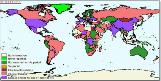
Figure 1.5. Worldwide distribution of Bovine tuberculosis .………..…… 16
Figure 1.6. Schematic representation of TaqManchemistry ………...……... 28
Figure 1.7. Schematic representation of Loop-mediated Isothermal
Amplification assay ……… 31
Figure 1.8. Schematic representation of immunochromatographic dipstick assay. 33
Figure 1.9. Gold nanoprobe assay ………... 35
Figure 1.10. Schematic representation of the structure of the DR locus in the
mycobacterial genome …………...………. 37
Figure 1.11. Illustration of a spoligotype profile (M. caprae) and its
correspondent binary code ………….…………...……….. 38
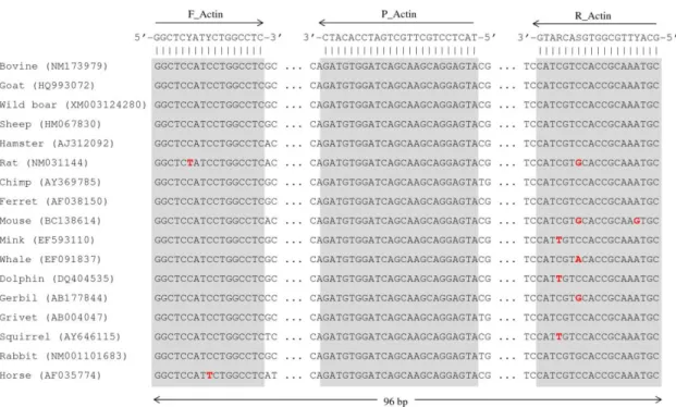
Figure 2.1. Complementary targets of the mammals β-actin gene targeted probe
and flanking primers ……….. 76
Figure 2.2. Complementary targets of the MTC-specific IS6110-targeted probe
and flanking primers ……….. 76
Figure 2.3. IS6110-targeted real-time PCR amplification curves obtained in
specificity tests ………... 80
Figure 2.4. Testing of spiked samples with the semi-nested duplex real-time
PCR assay ……….…….… 81
xi
Figure 3.2. Complementary targets of the Rv3875-esat6 (RD1) targeted probe
and flanking primers ……….…….
98
Figure 3.3. Complementary targets of the Rv2073c (RD9) targeted probe and
flanking primers ………... 99
Figure 3.4. Illustration of the real-time PCR amplification curves obtained ….… 106
Figure 4.1. Location of the complementary regions of LAMP primers and
FITC-labelled probes ………... 131
Figure 4.2. Specificity tests of the dLAMP-LFD assays ……...……….…… 134
Figure 4.3. Detection limit of the dLAMP-LFD assys ………….……….………. 135
Figure 4.4. Examples of the dLAMP-LFD amplification results obtained with
xii
Index of Tables
Table 1.1. Mycobacteria species frequently found in Medicine, according to the
risk of infection ……….. 1
Table 1.2. Species belonging to the Mycobacterium tuberculosis complex and
respective hosts …..……… 6
Table 1.3.Prevalence of bovine tuberculosis in Portugal ....………….…………... 13
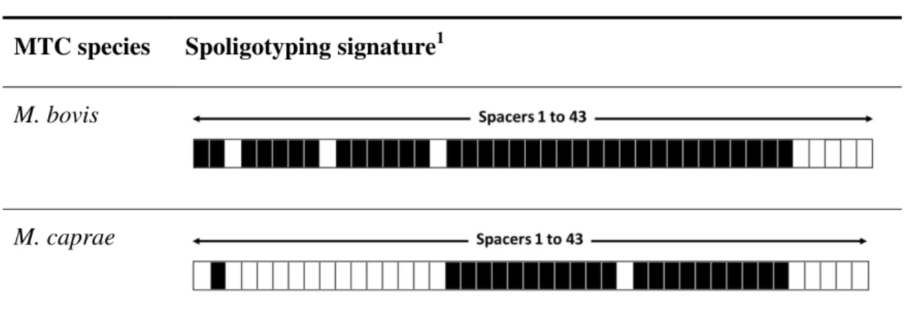
Table 1.4. Spoligotyping profiles characteristic of M. bovis and M. caprae ……... 38
Table 2.1. Bacterial reference strains and clinical isolates whose cultures were
used in the present study for the evaluation of specificity of the amplification assays and respective results ..……….. 71
Table 2.2. Typology of tissue samples used in this study and respective results of
the histological, bacteriological and semi-nested duplex real-time PCR
analyses ……….. 73
Table 2.3. Sequences of primers and probes used in this study ………... 75
Table 3.1. Reference and clinical strains used in the present study and respective
results for the duplex and triplex genomic targets amplification … 93
Table 3.2. Sequences of primers and probes used in this study ………... 96
Table 3.3. Results of the application of the duplex and triplex real-time PCR
amplification assays to DNA templates extracted from BACTEC liquid cultures of TB-suspected animal tissues ……….. 102
Table 4.1. Reference and clinical bacterial strains used to assess the specificity of
the dLAMP-LFD assays and respective amplification results .……….. 123
Table 4.2. Results of the dLAMP-LFD #A assay tested with DNA templates
extracted from human isolates .……….. 125
Table 4.3. Results of the dLAMP-LFD #B assay tested with DNA templates
extracted from isolates from TB-suspected animal tissues .…………... 127
xiii
List of abbreviations
Af1 - African 1 Complex Af2 - African 2 Complex AFB - Acid Fast Bacilli Ag - Silver
Au - Gold
AuNPs - Gold Nanoparticles
BAAR - Acid-Alcohol Resistant Bacilli BCG - Bacillus Calmette-Guérin
Bst - Bacillus stearothermophilus
bTB - Bovine Tuberculosis
CPC - Hexadecyl Pyridine Chloride Ct– Threshold cycle
DGAV - Direcção Geral de Alimentação e Veterinária
DGS - Direcção Geral de Saúde
DIG – Digoxigenin
DNA - Deoxyribonucleic Acid DR - Direct Repeat
dsDNA - Double Stranded DNA DVR - Direct Variable Repeat
EDTA - Ethylenediamine tetraacetic acid
ELISA - Enzyme-Linked Immunosorbent Assay ESAT-6 - 6 kDa early secretory antigenic target Eu1 - European 1 Complex
Eu2 - European 2 Complex
FITC - Fluorescein Isothiocyanate
FRET - Fluorescence Resonance Energy Transfer GC - Guanine and Cytosine
gyrB - Coding gene for the B subunit of DNA gyrase
HIV - Human Immunodeficiency Virus
xiv
List of abbreviations (Cont.)
IFN-γ - Interferon-Gamma
IHMT/UNL - Instituto de Higiene e Medicina Tropical/Universidade Nova de Lisboa
INIAV - Instituto Nacional de Investigação Agrária e Veterinária
IS - Insertion Sequence IVD - In Vitro Diagnostics
LAMP - Loop-Mediated Isothermal Amplification LFD – Lateral Flow Dipstick
LNIV - Laboratório Nacional de Investigação Veterinária
LSPs - Large Sequence Polymorphisms
M. - Mycobacterium
MIRU - Mycobacterial Interspersed Repetitive Units MTC - Mycobacterium tuberculosis complex
NAT - Nucleic Acid Testing NPs - Nanoparticles
NPV - Negative Predictive Value
OIE - World Organisation for Animal Health OMS –Organização Mundial de Saúde
PBS - Phosphate Buffered Saline PCR - Polymerase Chain Reaction POC - Point-of-Care
PPD - Purified Protein Derivative PPV - Positive Predictive Value Pt - Platinum
RD - Region of Difference
RFLP - Restriction Fragment Length Polymorphism RNA – Ribonucleic Acid
rRNA - Ribosomal Ribonucleic Acid SDS - Sodium Dodecyl Sulfate
xv
List of abbreviations (Cont.)
Spoligotyping - Spacer Oligonucleotide Typing SPR - Surface Plasmon Resonance
Taq - Thermus aquaticus
TB - Tuberculosis
TCH - Thiophene-2-carboxylic acid hydrazide VLA - Veterinary Laboratories Agency VNTR - Variable Number Tandem Repeats WHO - World Health Organization
List of Units
bp – pase pair ºC – degrees Celsius rpm – rotation per minute
msec – millisecond; sec – second; min – minute nm – nanometer
µl – microliter; ml – milliliter
fg – fentogram; pg – picogram; ng – nanogram; µg – microgram pmol – picomole
µM – micromolar
CHAPTER 1
1
1.1. Tuberculous mycobacteria and tuberculosis
Tuberculosis (TB) is an epidemic and serious infectious disease of global proportions, responsible for the death of approximately two million people per year, being estimated that one third of the world's population is latently infected [1]. Although curable, tuberculosis is still the leading cause of death in humans in many countries, and increasing numbers of cases of disease are reported in many regions of the world. The disease also affects animals, particularly livestock, with tremendous economic impacts. This disease is thus still a serious public health problem [1]. Despite being one of the most documented infectious diseases since the first records of the mankind [228], the main etiological agent of human tuberculosis, the Koch bacillus, was discovered by Robert Koch only in 1882 [2]. Today, it is known that tuberculosis is caused by several members of the Mycobacterium tuberculosis complex (MTC), a group of
phylogenetically-related species including: M. tuberculosis, the principal agent of
human tuberculosis; and M. bovis and M. caprae, most associated with tuberculosis in
cattle and goats, respectively [3]. In the genus Mycobacterium, besides the MTC
members, there are more than 120 recognized species, of which at least 20 to 30 may cause disease in humans [4] (Table 1.1).
Table 1.1. Mycobacteria species frequently found in Medicine, according to the risk of
infection (adapted from [6]).
Classification Species
Strict pathogens M. tuberculosis; M. bovis; M. africanum; M. leprae; M. ulcerans; M. szulgai; M. marinum
Potential or
opportunistic pathogens
M. avium; M. intracellulare; M. scrofulaceum; M. kansasii; M. xenopi; M. haemophilum; M. genavense; M. simiae; M. malmoense.
Rare pathogens, commensal or saprophytic
M. fortuitum; M. peregrinum; M. chelonae; M. abscessus; M. thermoresistibile; M. gordonae; M. triviale; M. gastri; M. terrae; M. flavescens
2
1.1.1. Classification and general characteristics of mycobacteria
The genus Mycobacterium is one of the oldest bacterial genus described. The formal
classification of mycobacteria started in 1896 when Lehmann and Neumann proposed for the first time the creation of the genus Mycobacterium, encompassing a number of
microorganisms whose growth, forming branching filamentous forms on the surface of the liquid culture media, was similar to fungi (the Greek prefix "myco" means "fungus") (Figure 1.1).
Mycobacteria are dispersed in nature, either as saprophytic or as pathogenic species, having the last ones a wide range of hosts, ranging from plants to humans (Table 1.1). The genus Mycobacterium belongs to the family Mycobacteriaceae of the order Actinomycetales and, as mentioned above, includes more than 120 validly described
species [4, 5]. Some of these species are strict or opportunistic (or potential) pathogens that affect both humans and animals. Among the strict pathogens, the principal species affecting humans include Mycobacterium tuberculosis, the causative agent of
tuberculosis, M. leprae, which causes leprosy, and M. bovis that causes tuberculosis in
animals but can also cause disease in humans. Opportunistic pathogens comprise a variety of mycobacterial species, including M. avium and M. kansasii, among others [6]. Mycobacteria are Gram-positive, rod-shaped, aerobic, non-motile, non-sporulating, and
3
Figure 1.1. Colonies of Mycobacterium tuberculosis, strain H37Ra, grown on
Middlebrook 7H10 solid medium (original from Pedro Costa).
Among other features, the mycobacterial cell wall gives the property of acid-alcohol resistance and mainly contributes to the hydrophobic nature of the cells surface, which leads to the formation of lumps when grown in liquid medium. The differences in the number of carbon atoms, and of their chemical bonds, in mycolic acids are important for the classification of mycobacteria. Other microorganisms such as Corynebacterium, Nocardia and Rhodococcus also produce mycolic acids, although with different
molecular structures and lower molecular weight [6].
The different growth rates in specific culture media have led to the traditional division of mycobacteria into two groups: the rapidly growing and the slow growing mycobacteria (Figure 1.2). This traditional division scheme distinguishes mycobacteria that grows in less than seven days, without additional growth factors, from those whose growth occur after seven days and requiring additional growth factors added to the media. The first group includes for example saprophytic species such as M. smegmatis
and M. fortuitum. The second slow-growing group encompasses pathogenic species,
including M. tuberculosis, M. bovis, M. avium subsp. avium and M. avium subsp. paratuberculosis. The identification of mycobacteria is conventionally performed on the
basis of their phenotypic characteristics, including morphological, cultural and biochemical features [6].
The pathogenic mycobacteria were initially separated into two groups, the
4 mycobacteria species, being their discrimination performed according to phenotypic identification criteria in the majority of clinical diagnostic laboratories [129].
The evolution of genomic studies have permitted to establish relationships between the genetic and phenotypic characteristics of mycobacteria, e.g., leading to the genetic differentiation between the slow and fast growing species and to the description of new species and reclassification of the existing ones [8] (Figure 1.2). Overall, the members of the genus Mycobacterium have a high content of guanine and cytosine in genomic
DNA, in the order of 62% to 70% (except for M. leprae, which is about 58%), and
share, at least, 94.3% of similarity in the sequences of the 16S ribosomal gene [6].
Figure 1.2. Phylogenetic tree of mycobacteria based on the 16S rRNA gene sequences
(Adapted from [9]).
1.1.2. The Mycobacterium tuberculosis complex
The species of Mycobacterium that are agents of tuberculosis (tuberculous
mycobacteria) are gathered in the so-called Mycobacterium tuberculosis complex
(MTC). These species share about 99.95% of sequence homology, with a greatly reduced genetic diversity at the nucleotide level, and present identical sequences in the 16S ribosomal gene, [10, 11]. The genome of the MTC members is longer than in other
intracellular microorganism’s genomes, with approximately 4.4 million base pairs,
5 parasites, and also contains a higher GC content of about 65%. The exchange of genetic material, such as horizontal gene transference, is virtually non-existent in these species which results mainly in a clonal evolution, relying on extensive deletions of the nucleotide sequences (LSPs, Large Sequence Polymorphisms), rearrangements and chromosome point mutations [3, 12].
The MTC is currently constituted by various species and subspecies with human and veterinary clinical importance: M. canettii, M. africanum, M. pinnipedii, M. microti, M. caprae, M. bovis, M. tuberculosis, M.mungi, M. orygis and the Dassie bacillus [13-17].
MTC mycobacteria are strict pathogens, depending on the host for its survival. Despite of the high degree of conservation of their genomes, the MTC species demonstrate important phenotypic differences, adaptations to different hosts and show different degrees of virulence [18]. For example, M. tuberculosis, the main agent of human
tuberculosis, is almost always associated with the human. Otherwise, M. bovis is found
primarily associated with cattle, M. caprae to goats and M. pinnipedii to marine
mammals [18]. However, the adaptation of these species to each host is not necessarily strictly as it is for other pathogenic bacteria. All members of the MTC can cause disease in humans and other mammals that were not initially considered as primary hosts (Table 1.2) [4]. It is noteworthy that M. bovis and M. caprae represents a significant potential
of zoonotic transmission to Human [19, 20]. There is evidence pointing to the possibility of person-to-person transmission of the zoonotic Mycobacterium species
[21], but the main routes of transmission are the contact with infected animals and intake of dairy improperly pasteurized or unpasteurized [1, 22]. Mycobacterium bovis
6
Table 1.2. Species belonging to the Mycobacterium tuberculosis complex and
respective hosts.
Species Primary
Host
Other Hosts Characteristics
M. tuberculosis Human Other mammals Species almost exclusively found in humans.
However, there are records of infections in other mammals [24-28].
M. bovis Cattle Sheep, goats,
dogs, cats and humans
Species with the highest number of possible hosts [6, 29-32].
M. caprae Goats Human Formerly considered a subspecies of M. bovis,
but genetic differences have led to it being considered a distinct species [33].
M. africanum Human - Species phenotypically intermediate between M. tuberculosis and M. bovis and whose area
of focus is almost exclusively restricted to the African continent. Two varieties were described [34].
M. microti Rodents Human Pathogenicity level lower than the remaining
species, being a potential substitute for BCG vaccine strains [35-37].
M. canettii Human - Species most divergent from MTC and
allegedly nearest the ancestral species precursor of the complex [3, 38, 39].
M. pinnipedii Seals Humans and other
mammals
Species first isolated in seals in South America with lesions of tuberculosis [40] and has also been associated with cases of tuberculosis in mammals in other zoos [41].
M. mungi Banded
mongooses
- The causative TB agent among banded
mongooses, first isolated in Botswana. Host spectrum and transmission dynamics remain unknown [13].
Dassie bacillus Hyrax or Dassie
- An infrequent variant of the M. tuberculosis
complex characterized as being most similar to
M. microti, is the causative agent of
tuberculosis in the dassie (Procavia capensis)
[42].
M. orygis ? Mammals Oryx bacilli have been isolated from members
of the Bovidae family, i.e., oryxes, gazelles,
7
Phylogenetic relationships and evolutionary scenario
As mentioned above, the MTC members share 99.95% of genetic homology, with a greatly reduced genetic diversity at the nucleotide level. Although genetically very similar, the members of the complex can be distinguished from each other by stable molecular differences, such as deletions or single nucleotide polymorphisms (SNPs), which have been the basis for evolutionary studies of these mycobacteria [3, 43]. The irreversible deletions of chromosomal regions, called Regions of Difference (RDs), were recognized in a much larger number in the genome of M. bovis compared to other
members of the complex [3]. The analysis of these deletions allowed Brosch and collaborators [3] to outline a new phylogenetic tree for the MTC, later on confirmed by another independent study [11], in which M. bovis and M. bovis BCG appear as the last
descendants of the complex (Figure 1.3). The vaccine strains of M. bovis BCG still
exhibit an additional deletion in RD1, exclusive of these strains, and whose absence is associated with lower virulence [44]. The various members of the MTC that infect animals have in common the absence of RD9, which is present in the other members, like M. tuberculosis [3]. Other genomic data, including that resulting from the
whole-genome sequencing of M. bovis [45] and M. tuberculosis [46], corroborate the
evolutionary scenario proposed by Brosch et al. (2002) for the MTC, in which M. bovis
was the last descendant in the phylogeny (Figure 1.3). This new hypothesis came to oppose the previous one, based on epidemiological evidence, that M. bovis was the
precursor species due to its wider hosts preferences range, and have served as a basis for the evolution of M. tuberculosis, by adaptation phenomena and specialization to Human
8
Figure 1.3. Phylogeny of the Mycobacterium tuberculosis complex based on the
analysis of Regions of Difference and Single Nucleotide Polymorphisms. Each orange box represents a deletion of one or more specific RDs in a given species [4]. Blue boxes indicate the SNPs; superscripts mark the position of the mutation at either the nucleotide (n) or the codon (c) of the respective genes. Adapted from [3], including information from [4, 14, 17, 50, 158].
Other phylogenies were produced for the MTC using additional molecular markers, such as deletions of spacer sequences of the Direct Repeat (DR) region and the identification of Single Nucleotide Polymorphisms (SNPs) [48-52], which are consistent with the proposal presented by Brosch et al. (2002). In 2009, in an overall
phylogenetic analysis of the MTC bringing together all these molecular markers, this group of microorganisms appears as a set of ecotypes adapted to different hosts, corresponding the different affinities of the host to different niches (Figure 1.4) [4, 53]. The MTC members harbouring animals as their main hosts share the absence of RD9, which is present in M. tuberculosis, and also other common features (e.g. deletion of
9
Figure 1.4. Evolution of members of Mycobacterium tuberculosis complex lacking the
RD9. The predecessor species are numbered from Anc1 to Anc6 (Adapted from [4, 53]).
Clonal complexes of Mycobacterium bovis
Bovines are the main hosts of M. bovis, which determines the disease called bovine
tuberculosis (bTB). This term is also often used to describe the infection by M. bovis in
other species, including wild animals and humans, to demonstrate the cattle as a source of infection. Although bovine tuberculosis has been reported on every continent where there are breeding of cattle, it was assumed to have originated in a particular geographic location, and since then has spread throughout the globe [54]. Historical data indicate that bovine tuberculosis has its origins in Europe and, especially during the colonial period, was distributed from that continent to the rest of the world. Myers and Steele (1969) suggested that M. bovis reached the northern Italy and from there moved to
10 transmission of M. bovis, especially in the absence of appropriate control and
eradication measures [56].
Modern methods of typing, based on the analysis of genetic characteristics, have determined progress in the knowledge of the geographical distribution of M. bovis and
allowed the identification of dominant clonal complexes within broader geographic areas [57]. The cattle trade between neighbouring regions and countries leads to the dispersion of M. bovis and the dominance of clonal complexes in large areas [58]. These
clonal complexes were mainly defined by the presence of specific patterns of deletions, namely in the DR region, by a reverse-hybridization typing technique named spoligotyping. Phylogeographic analysis have confirmed the existence of four major M. bovis clonal complexes: African 1 (Af1), African 2 (Af2), European 1 (Eu1) and
European 2 (Eu2) [54, 58-60]. Two of these complexes are geographically located in Africa (Af1 and Af2), a third complex exhibit a global distribution (Eu1), and the fourth complex has a strong geographical predominance in the Iberian Peninsula (Eu2). The Af1 clonal complex (dominant in Cameroon, Nigeria, Mali and Chad) and the Af2 complex (common in East Africa) were identified by Berg et al. (2011) and Muller et al. (2009), the clonal complex Eu1 (common in Ireland, UK and overseas countries)
was identified by Smith et al. (2011), and the clonal complex Eu2 (predominantly in the
Iberian Peninsula) was described by Rodriguez-Campos et al. (2012).
11 most other strains that have only one or fewer copies. The deletion RDAf2 were intact in Af1 clonal complex strains, as well as RDAf1 deletion showed intact for the Af2 strains, allowing to conclude that the strains of clonal complexes Af1 and Af2 are mutually exclusive and do not share any phylogenetic history with the latest common ancestor of each clonal complex, suggesting that the mixing between the cattle populations in that countries is uncommon [54, 59].
The clonal complex Eu1 is characterized by the deletion of the chromosomal region RDEu1 and was identified by the analysis of spoligotyping patterns available in the database of M. bovis (www.mbovis.org). Over 1000 strains from over 30 countries were
analysed. This complex is frequently found in the Republic of Ireland and UK, represent less than 14% of the isolates in France, Spain and Portugal and proved rare in other European Community countries and Iran. However, Eu1 complex was found with high frequency in countries that had cattle trade relations with the UK (USA, South Africa, New Zealand, Australia and Canada). In the American continent, with the exception of Brazil, the Eu1 was also found with high frequency in Argentina, Chile, Ecuador and Mexico. Isolates from Korea and Kazakhstan were also associated with Eu1 clonal complex. The simplest explanation for the worldwide distribution of Eu1 is that it was spread by infected cattle from the UK, such as the Hereford cattle breed created in the 18th century, to old trading partners, although there is no evidence of secondary spread since then. This was the first identification of a clonal complex of M. bovis globally
dispersed, which tries to explain much of the current global distribution of this major causative agent of bTB [54]. The Eu2 clonal complex, unlike the other clonal complexes mentioned above, is characterized by the absence of the spacer 21 and by a SNP in the guaA gene [61]. The Eu2 isolates of M. bovis are from the Iberian Peninsula,
where they are found with high frequency. Eu2 is also found in France, Italy and the British Isles but with a very low frequency. Previous studies have revealed that about 70% of the strains of M. bovis in Portugal [62] and Spain [63] were characterized by the
absence of spacer 21.
12 scenario for the MTC, based on regions of difference [3]. SNPs have also been used, however with a more targeted application for phylogenetic studies [39, 53]. The analysis of the clonal complexes of M. bovis currently represents a new tool for
molecular epidemiologists, allowing the creation of hypotheses to investigate the demographic distribution and the pathogenicity of these important zoonotic agents of global significance [53].
1.2. Bovine tuberculosis
As mentioned above, M. bovis is the major etiological agent of bovine tuberculosis, a
most important chronic and debilitating animal disease. Besides cattle, M. bovis can also
cause tuberculosis in other mammalian species, including humans. Like human tuberculosis, bovine tuberculosis has a worldwide distribution and importance. It is a zoonosis with a high socio-economic impact, due to the low productivity of cattle and imposed restrictions on animal trade and products thereof, as well as costs associated with the implementation of programs to control and eradicate the disease, consisting also a major risk to public health [65, 66]. The disease has important implications in public health, especially in developing countries [1, 67].
Bovine tuberculosis has a predominantly chronic clinical evolution, with the development of typical granulomatous lesions, mainly in the lungs and lymph nodes, although any organ can be affected. The main routes of infection of bovine tuberculosis in animals are the direct infection by inhalation, ingestion and, less frequently, by congenital route. The mode of transmission more common is by aerosols inhalation, also due to the high survival rate of the agent in aerosols [68]. The infection by indirect route can also occur since M. bovis is resistant to adverse environmental conditions,
surviving for more than 74 days in fomites and from two to six months in faeces, depending on the temperature and humidity conditions [69]. The anatomical location of the lesions suggests the most likely route of infection: hepatic lesions indicate congenital infection, intestinal lesions the infection by the gastrointestinal tract and lesions in the respiratory route the infection by aerogenous dissemination.
13 developing countries [1, 22, 70]. In Portugal, every year, the costs are high, resulting from the compensation to producers, slaughter of infected animals and initiatives to control and eradicate the disease. Despite the efforts made by the veterinary authorities, the eradication of bovine tuberculosis has not yet been achieved in Portugal, although the prevalence is low in the European context. A decrease of the incidence of bovine tuberculosis has been recorded since 1997 [71, 72]. In 2011, the reported prevalence in cattle was 0.17% against 0.07% and 0.09% in 2001 and 2002, respectively. As we can see in Table 1.3, there has been an apparent upsurge in the prevalence of the disease in Portugal, also potentially related with the maintenance of M. bovis in wildlife [104,
229].
Table 1.3. Prevalence of bovine tuberculosis in Portugal (Adapted from [72]).
Year Total number of animals tested
Number of positive animals
% Positive animals (Animal prevalence)
2001 783.680 546 0.07
2002 776.231 716 0.09
2003 958.306 1.221 0.13
2004 984.527 856 0.09
2005 976.532 647 0.07
2006 976.893 425 0.04
2007 1.006.908 414 0.04
2008 1.032.586 264 0.03
2009 1.060.831 885 0.08
2010 1.036.310 2.702 0.26
14
1.2.1. Implications for public health
Since ancient times that M. bovis infects man [73], although M. tuberculosis is the main
causative agent of human tuberculosis. In the early twentieth century, cases of tuberculosis caused by M. bovis in humans showed up as extrapulmonary forms, from
consumption of unpasteurized milk and affecting mainly children [74]. The prevalence of human tuberculosis caused by M. bovis decreased dramatically in countries where
pasteurization of milk and the campaigns of control and eradication of tuberculosis in cattle were implemented [75]. Contact with infected animals and ingestion of unpasteurized milk and dairy products, or incorrectly pasteurized, are the main routes of transmission of these zoonotic species [23, 75]. As mentioned above, in industrialized countries, human TB cases attributed to M. bovis were 0.5 to 7.2% in the last twenty
years [23]. However, it is estimated that over 94% of the world´s population live in countries where there are no strategies for the control of infections caused by M. bovis
[76]. In these countries, many of which belonging to the African continent, bovine tuberculosis poses a significant threat to public health [1, 67, 70], where it is estimated to be responsible for 10 to 15% of new cases of human tuberculosis [23].
In Portugal, according to the World Health Organization (WHO), the last recorded cases of human tuberculosis caused by M. bovis occurred in 2003 and 2004 (Euro TB
Reports, 1999-2006), but usually, only the Mycobacterium tuberculosis complex is
identified in clinical laboratories. It was suggested that 3.4% of tuberculosis cases detected in Hospital Amadora-Sintra, between 1999 and 2002, were caused by strains
of M. bovis [77], but subsequent studies have not confirmed the classification of these
isolates [78, 79].
Tuberculosis is especially important in individuals with immunodeficiency, particularly in patients infected by human immunodeficiency virus (HIV), which are particularly susceptible to infection by mycobacteria [67, 70, 75]. The emergence of multi-resistant strains of M. bovis in humans [80], as is the case with M. tuberculosis, and the fact that
transmission between humans can occur [21, 81], reveals worrying.
Human tuberculosis caused by M. bovis, from the clinical and pathological point, is
indistinguishable of the tuberculosis caused by M. tuberculosis. Discrimination between
15 and molecular methods. However, in the laboratorial diagnosis of human tuberculosis culture media are generally not suitable for isolation of M. bovis (needs to be
supplemented with pyruvate – see 1.3.1) and, on the other hand, is carried out only the identification of the MTC, raising up the suspicion that M. bovis can be an undervalued
cause of morbidity and mortality [67, 82]. This finding is particularly relevant in developing countries where, due to limited resources, the diagnosis is based only on viewing acid-fast bacilli (AFB) in smears [67].
1.2.2. Epidemiological features
Transmission of Mycobacterium bovis
The aerial route is the main and most important route of transmission of bovine tuberculosis [76], being facilitated by the high survival rate of M. bovis in aerosols [68].
The direct contagion by inhalation thus constitutes the most common route of infection. Another important way of transmitting the infection is by ingestion, which may occur through contaminated water or pastures, or even through infected replacer milk administered to calves [9]. The congenital infection and vertical transmission, as well as genital transmission, are rare, especially in regions where eradication plans operate [83, 84]. It is also uncommon the transcutaneous transmission, which may occur in situations of bitten by infected animals [85]. There is reference to the arthropods as vectors of infection, since viable M. bovis were detected in ticks, but this transmission
path is very unlikely [69].
The infection by indirect way can occur since M. bovis have some resistance to the
environmental conditions, being its survival dependant of factors such as temperature, humidity and sunlight [69, 86]. These mycobacteria can survive in the environment for a limited period (3 to 14 days) when exposed to sunlight, but can survive for a longer period (six weeks) under conditions of darkness and moisture. Eventually, mycobacteria can resist in fomites and faeces for two to six months [69, 86]. The survival of M. bovis
in protozoa suggests that these organisms may provide protection against adverse environmental conditions [87], thus representing an environmental reservoir of M. bovis
16
Global epidemiological situation
Bovine tuberculosis is a disease of worldwide geographical distribution (Figure 1.5), with high incidence in many developing countries, where it has great importance [1, 70]. Thanks to the implementation of eradication programs, several EU countries, including Austria, Denmark, Slovakia, Finland, Holland, Luxembourg, Czech Republic and Sweden, have managed to eradicate bovine tuberculosis, while other countries, including Portugal, only reduced the incidence of the disease (OIE, 2013).
Figure 1.5. Worldwide distribution of Bovine tuberculosis (OIE, 2013).
In many countries, the eradication of bovine tuberculosis has been hampered by the presence of several wild animals which act as reservoirs in the maintenance of M. bovis,
among which are the badger (Meles meles), the opossum (Trichosurus vulpecula), bison
(Bison bison), the African buffalo (Syncerus caffer) and the white-tailed deer
(Odocoileus virginianus) [89, 104, 229].
17 potential risk of transmission it is important to use all the resources for the early detection of infected animals and herds, including serological and molecular ante and post-mortem laboratory diagnostic tests [143, 230]. The rapid detection of M. bovis is
pivotal in contributing to the control and eradication of bovine tuberculosis.
Tuberculosis in free-ranging wildlife populations
Mycobacterium bovis and other MTC species have been recognized as a global threat at
the wildlife-livestock-human interface. Several wildlife species have been identified as maintenance hosts. Spill over of infection from these species to livestock or other wildlife species may have economic and conservation implications, and potential infection of humans poses public health concerns. The existence of natural reservoirs (maintenance hosts) of the disease, capable of reintroducing it into farms free of tuberculosis, has complicated the control and eradication of bovine tuberculosis in many countries [76, 91].
18 Although tuberculosis has been detected in various sylvatic species, does not mean that all these populations constitute a natural reservoir of the disease. Most of these species are accidental hosts, i.e., can acquire the disease, but if removing the source of infection, there is a reduction in the prevalence of tuberculosis, which is not maintained in the population [95]. On the other hand, there are species that are considered maintenance hosts, because the infection may persist in the population only by horizontal transmission. This ability to act as a reservoir and transmit the disease to other susceptible species depends on the prevalence of the disease in the population of the maintenance host, their behaviour and their relationships in the ecosystem [93, 96]. There are several examples of these sylvatic reservoirs of tuberculosis, responsible for the transmission of infection to domestic cattle, hampering the efforts to eradicate the disease in certain countries. Some of the most studied are the European badger (Meles meles) in the UK and Ireland and the possum (Trichosurus vulpecula) in New Zealand,
countries where there was an observed decrease in the prevalence of the disease in domestic cattle after campaigns based on the elimination of infected populations of these sylvatic animals [96, 97].
In the state of Michigan, USA, the white-tailed deer (Odocoileus virginianus), also
known as the Virginia deer or simply as the whitetail, is considered the responsible for the re-emergence of the disease in cattle, after bad options of management of this cinegetic species. Consequently, due to the practice of supplementary feeding in the winter, their number grew beyond the capacity of the ecosystem, encouraging behaviours that increase the risk of intra- and interspecific transmission. This maintenance host is still the source of outbreaks in several species of carnivores, such as the coyote (Canis latrans), raccoon (Procyon lotor), the black bear (Ursus americanus)
or red fox (Vulpes vulpes), and a potential danger to the hunters [92]. The water buffalo
or domestic Asian water buffalo (Bubalus bubalis) in Australia is an example of a
sylvatic maintenance host associated with a well succeed eradication campaign of tuberculosis. This required the total slaughter of chronically infected herds and restocking with animals originating from a disease-free population [96, 97]. In Africa, the most studied natural reservoir of the disease is the African buffalo or Cape buffalo (Syncerus caffer), which has proved a source of spreading of the disease to the
19 increasing concerns in the preservation of endangered species, including the larger felid species, such as lions (Panthera leo), cheetah (Acinonyx jubatus) and leopard (Panthera pardus) [98].
The first studies on tuberculosis in sylvatic species in the Iberian Peninsula were performed in Spain. Several studies have identified the wild boar (Sus scrofa) and the
red deer (Cervus elaphus) as important reservoirs of the disease, reaching dispersed
geographically populations and presenting a high prevalence of tuberculosis [95, 99, 100]. Although in certain European countries the wild boar is considered an accidental host, various molecular, anatomopathological and epidemiological studies have strengthened the role of the wild boar as a reservoir in Mediterranean ecosystems [101-103].
The population density of wild boar and deer has increased significantly, either by the lack of natural predators, or by the practices of cinegetic management of these species, such as the improvement of the hunting reserves fences (not relevant in the case of wild boar) and the food supplementation in the summer when the pastures are scarce [100]. In addition to the features mentioned above, also the behaviour of these animals and the evidence that they share M. bovis isolates with the same spoligotype profile as domestic
cattle [95], make the wild boars and deer a risk factor in the reintroduction of the disease in disease-free farms. Also other domestic animals can be infected, including goats and Iberian pigs, in farms of extensive-type regime. These natural reservoirs of disease constitute a potential threat to public health (especially to the hunters and handlers of carcasses) and have also been implicated in transmission to endangered species such as the Iberian lynx (Lynx pardinus), endangering their preservation [100].
20 slaughter, but this is seldom adopted by economic, logistical, social and nature protection reasons [98].
In Portugal, the recognition of the presence of tuberculosis in wild boar and deer is longstanding [104, 229]. However, there is no national plan of surveillance of this disease in sylvatic species, however, exists only a few actions/studies in certain fields. Thus, it is not known the infection status of these populations. Currently, this information is summarized in some recent works about tuberculosis in wild boar [102, 104] and about the genotyping of strains responsible for tuberculosis in deer and wild boar [62, 105]. In Portugal, the prevalence of tuberculosis in cattle and the occurrence of the disease in wild boars are significantly associated, suggesting a relationship between the disease in these species [102]. Furthermore, according to this author, the presence of tuberculosis in wild boar is also significantly associated with the density and diversity of sylvatic ungulates. Therefore, the wild boar appears to be the maintenance host of M. bovis in Portugal, and satisfies the theoretical conditions for
being regarded as the reservoir of tuberculosis in our country [102]. Nevertheless, the great diversity of patterns found in isolates of deer from the same geographical area suggests that these animals are accidental hosts and not reservoirs of the disease. However, there is strong evidences of transmission of M. bovis between cattle and deer
or wild boar, although not fully understood the meaning of it [62].
1.3. Diagnosis of bovine tuberculosis
In cattle, symptoms of tuberculosis often manifest later, namely by the presence of lesions in tissues, so the clinical diagnosis is rare, especially in the course of control and eradication plans [23]. In this context, the screening of tuberculosis in live animals (in vivo or ante-mortem diagnosis) depends on immunological diagnostic tests, such as the
single intradermal cervical tuberculin test (SICCT) and the interferon-gamma (IFN-γ)
test, which are based on the detection of cell-mediated immune responses. The use of these tests represents an important step for the eradication and monitoring of the disease. The confirmation of the presence of infection and consequently the definitive diagnosis of bovine tuberculosis, is made post-mortem usually based on
![Figure 1.2. Phylogenetic tree of mycobacteria based on the 16S rRNA gene sequences (Adapted from [9])](https://thumb-eu.123doks.com/thumbv2/123dok_br/15766813.640603/33.892.147.785.432.757/figure-phylogenetic-tree-mycobacteria-based-rrna-sequences-adapted.webp)
![Table 1.3. Prevalence of bovine tuberculosis in Portugal (Adapted from [72]).](https://thumb-eu.123doks.com/thumbv2/123dok_br/15766813.640603/42.892.161.779.529.964/table-prevalence-bovine-tuberculosis-portugal-adapted.webp)