Instituto de Higiene e Medicina Tropical
Characterization of the Hepatitis Delta Virus Small
Antigen: Intracellular Localization, Structure,
Multimerization and RNA Binding Ability
Carolina Alpalhão Mantero de Mendonça Alves
DISSERTAÇÃO PARA A OBTENÇÃO DO GRAU DE DOUTOR EM CIÊNCIAS BIOMÉDICAS, ESPECIALIDADE DE BIOLOGIA MOLECULAR E CELULAR
Instituto de Higiene e Medicina Tropical
Characterization of the Hepatitis Delta Virus Small
Antigen: Intracellular Localization, Structure,
Multimerization and RNA Binding Ability
Carolina Alpalhão Mantero de Mendonça Alves
Supervisor: Professor Celso Cunha (IHMT, UNL)
Co-supervisor: Professor Emeritus John Taylor (Fox Chase Cancer Center)
Tutorial Comittee: Professor Celso Cunha (IHMT, UNL)
Professor Emeritus John Taylor (Fox Chase Cancer Center) Professor Aida Esteves (IHMT, UNL)
Dissertation to obtain a Doctoral Degree in Biomedical Sciences, Specialization in Molecular and Cellular Biology
i
Acknowledgements
I want to gratefully acknowledge those that directly or indirectly contributed to the work presented here.
To Professor Celso Cunha for everything – for his guidance and encouragement throughout all these years, for the knowledge he shared and his support and sense of humor!
To Professor John Taylor, for his guidance and support throughout this project, and for receiving me in his lab at Fox Chase Cancer Center.
To everybody who contributed to my research at the Fox Chase Cancer Center. Particularly, to Severin Gudima for his support and shared knowledge. To Ziying Han and Ning Chai, for their help and support. And to William Mason for his constructive criticism throughout the project.
To all my colleagues and former colleagues at Instituto de Higiene e Medicina
Tropical. Particularly Ana Casaca, Marta Mendes and Cristina Branco, who “suffered”
the most!
To my parents, my brother and Eduardo for their unconditional support and especially for their infinite patience.
Last, but definitely not least, to my American family, specially my “mom”
iii
Caracterização do Antigénio Pequeno do Vírus da Hepatite Delta: Localização Intracelular, Estrutura, Multimerização e Capacidade de Ligação ao RNA
Carolina Alpalhão Mantero de Mendonça Alves
Resumo
O vírus da hepatite delta (HDV) é o agente patogénico responsável por uma das formas mais severas de hepatite viral. O genoma consiste numa molécula circular de RNA de cadeia simples de polaridade negativa e apresenta uma única proteína viral, o antigénio delta pequeno (S-HDAg). Na sequência de um mecanismo de editing outra
proteína viral é traduzida, o antigénio delta grande (L-HDAg). Apesar de partilharem grande parte da sua sequência, as duas proteínas desempenham funções distintas. O S-HDAg é essencial para a acumulação de RNAs virais enquanto o L-S-HDAg inibe a replicação viral e é necessário para o empacotamento. O HDV depende extensivamente de factores do hospedeiro para completar o seu ciclo de replicação. Pensa-se que a polymerase II (pol II) do hospedeiro é redireccionada para transcrever o RNA viral.
No presente trabalho procurou-se caracterizar o S-HDAg e clarificar o seu papel no ciclo de replicação do HDV.
Observamos que quando o S-HDAg é expresso na presença de replicação do RNA viral, o antigénio co-localiza com a pol II. Contudo, a co-localização com pol II verifica-se mesmo na presença de RNA viral incapaz de ser replicado e na ocorrência de inibição da replicação viral, sugerindo que o S-HDAg não participa directamente na transcrição do RNA viral. Assim, propomos que o S-HDAg é essencial para acumulação de RNAs virais protegendo ou estabilizando os RNAs. Observamos ainda que na ausência de RNA viral o S-HDAg co-localiza com a nucleolina nos nucléolos. Contudo, na presença de RNA incapaz de ser replicado, o antigénio desloca-se para o nucleoplasma mantendo-se a nucleolina nos nucléolos, sugerindo que o S-HDAg não interage directamente com a nucleolina.
Ao estudarmos as características estruturais do S-HDAg verificámos, utilizando um preditor de desordem intrínseca, que apresenta um elevado grau de desordem. A previsão foi confirmada in vitro por dicroísmo circular observando-se que apenas 30%
dos amino ácidos adoptam uma conformação de hélice . A ausência de uma estrutura rígida pode conferir ao antigénio flexibilidade para se adaptar a diferentes parceiros e participar em vários passos do ciclo de replicação viral.
A multimerização do S-HDAg foi analisada por dispersão de luz dinâmica. Os resultados indicam que o antigénio recombinante purificado é capaz de formar multímeros de 12 moléculas. Adicionalmente, foram observados multímeros de seis a oito moléculas em gel de poliacrilamida desnaturante, após cross-linking. Os mesmos
multímeros foram observados para S-HDAg presente em partículas virais sugerindo que a multimerização do antigénio ocorre in vivo.
iv
com RNA e DNA. A falta de especificidade observada pode dever-se apenas a interacções electrostáticas entre o S-HDAg de carga positiva (+12) e ácidos nucleicos de carga negativa. Propomos que, in vivo, a fosforilação extensiva do S-HDAg reduza a
carga positiva contribuindo para que a interacção seja específica para os RNAs virais.
v
Characterization of the Hepatitis Delta Virus Small Antigen: Intracellular Localization, Structure, Multimerization and RNA Binding Ability
Carolina Alpalhão Mantero de Mendonça Alves
Abstract
Hepatitis delta virus (HDV) is the causative agent of one of the most severe forms of viral hepatitis. It has a small single-stranded circular RNA genome of negative polarity and only one viral protein, the small delta antigen (S-HDAg). Following site-specific RNA editing, a second longer protein is translated, the large delta antigen (L-HDAg). Although these viral proteins share most of their sequence they play distinct roles. S-HDAg is essential for the accumulation of HDV RNAs whereas L-HDAg inhibits HDV replication and is necessary for viral assembly. With such a limited coding capacity HDV must rely extensively on host cell components to complete its replication cycle. The host DNA-directed RNA polymerase II (pol II) is thought to be re-directed to transcribe HDV RNAs.
The objective of this study was to further characterize S-HDAg and clarify its role(s) during the HDV replication cycle.
We observed that when S-HDAg was expressed in vivo along with replicating
HDV RNA it co-located with host pol II. However, such co-localization was also observed in the presence of non-replicating HDV RNAs or when replication was inhibited by specific doses of -amanitin. Thus, we propose that S-HDAg is essential for HDV RNA accumulation by stabilizing or protecting the viral RNAs rather than acting as a direct player in HDV RNA transcription. Additionally, we observed that S-HDAg located in nucleolus when expressed in the absence of HDV RNA, and co-located with host nucleolin. However, in the presence of non-replicating HDV RNAs, S-HDAg moved to the nucleoplasm whereas nucleolin was unchanged. This suggests that S-HDAg is not interacting directly with nucleolin.
In our examination of S-HDAg‟s structural features we applied a meta-predictor of intrinsic disorder, PONDR-FIT. It predicted that full-length S-HDAg has extensive intrinsic disorder. This result was confirmed in vitro by circular dichroism
measurements that indicated no more than 30% of S-HDAg amino acids adopted an -helical structure. Such a lack of a well-defined rigid structure is expected to grant flexibility to the antigen allowing it to interact with several partners and perform distinct roles during the HDV replication cycle.
vi
Finally we examined the ability of S-HDAg to bind nucleic acids in vitro. Both
multimers and monomers bound to conformations of both RNA and DNA. Such a lack of specificity was probably due to electrostatic interactions between the positively-charged S-HDAg (+12) and negatively-positively-charged nucleic acids. We propose that in vivo,
extensive post-translational phosphorylation of S-HDAg reduces the positive charge, thereby contributing to interactions more specific for HDV RNAs and possibly dependent upon protein multimerization.
Despite our observations presented here, some issues relating to our aims remain unresolved.
vii
Publications
Han, Z., Alves, C., Gudima, S. and Taylor, J. (2009) Intracellular localization of hepatitis delta virus proteins in the presence and absence of viral RNA accumulation. J. Virol., 83, 6457-6463.
Alves, C. Cheng, H., Roder, H. and Taylor, J. 2010. Intrinsic disorder and oligomerization of the hepatitis delta virus antigen. Virology, 407, 333-340
ix
Table of contents
Acknowledgements ... i
Resumo ... iii
Abstract ... v
Publications ... vii
Abbreviations ... xi
1. Introduction ... 1
1.1. Hepatitis Delta Virus ... 2
1.1.1. Epidemiology ... 2
1.1.2. Clinical Expression ... 4
1.1.3. Diagnosis and Treatment ... 6
1.2. HDV Biology ... 11
1.2.1. HDV Virions and Putative Host Cell Receptors ... 11
1.2.2. HDV RNAs ... 12
1.2.3. HDV RNA Replication ... 14
1.2.4. HDV Origin ... 17
1.3. Delta Antigens ... 19
1.3.1. Small Delta Antigen ... 22
1.3.2. Host Interactions ... 26
1.4. Intrinsically Disordered Proteins ... 28
1.4.1. Intrinsic Disorder Prediction ... 29
1.4.2. ID Prevalence ... 30
1.4.3. Biophysical Characterization of Disorder ... 31
1.4.4. Viral IDPs ... 32
1.5. Final Remarks ... 35
2. Specific Aims ... 37
x
4. Intrinsic Disorder and Oligomerization of the Hepatitis Delta Virus Antigen47 5. Multimerization of the Delta Antigen is not Essential for In Vitro Nucleic Acid
Binding ... 61
6. Discussion and Future Perspectives... 87
7. References ... 92
xi
Abbreviations
3-D 3-dimensional
ADAR1 Adenosine deaminase acting on RNA
Ala Alanine
Arg Arginine
Asn Asparagine
CCD Coiled-coil domain
CD Circular dichroism
CPEB3 Cytoplasmic polyadenylation element-binding protein 3
Cys Cysteine
DIPA Delta interacting protein A DNA Deoxyribonucleic acid
ERK1/2 Extracellular signal-related kinases 1 and 2
Gln Glutamine
Glu Glutamic acid
Gly Glycine
HBV Hepatitis B virus
HBsAgs Hepatitis B virus surface antigens HDAg Hepatitis delta antigen
HDV Hepatitis delta virus
IC Internal control
IDP Intrinsically disordered protein IDR Intrinsically disordered region IFN- Interferon α
IgG Immunoglobulin G
IgM Immunoglobulin M
xii
Leu Leucine
L-HDAg Large hepatitis delta antigen
Lys Lysine
mRNA messenger RNA
NELF-A Negative elongation factor, subunit A NES Nuclear export signal
NLS Nuclear localization signal NMR Nuclear magnetic resonance
ORF Open reading frame
PEG-IFN- Pegylated interferon α
Phe Phenylalanine
Pol II DNA-dependent RNA polymerase II Poly(A) Polyadenylated
Pro Proline
PTMs Post-translational modifications
RNA Ribonucleic acid
RNPs Ribonucleoproteins
RT-qPCR Real-time quantitative polymerase chain reaction
Ser Serine
S-HDAg Small hepatitis delta antigen
SUMO1 Small ubiquitin-related modifier isoform 1
Thr Threonine
Trp Tryptophan
Tyr Tyrosine
1
2
1.1. Hepatitis Delta Virus
In 1977, a novel antigen was found in the nucleus of hepatocytes from patients with a more severe form of hepatitis B (Rizzetto et al., 1977). It was first thought to be a previously unknown marker of hepatitis B virus (HBV; Rizzetto et al., 1979). Only later, it was found that the delta antigen was not part of HBV but of a separate defective virus that requires the presence of HBV for infection (Rizzetto et al., 1980). The newfound virus was designated hepatitis delta virus (HDV) and, by 1986, its RNA genome was cloned and sequenced (Wang et al., 1986). This peculiar virus has been classified as the only member of the genus Deltavirus due to its uniqueness (Murphy,
1996). The HDV virion is a hybrid particle, composed of the delta antigen and HDV RNA enclosed by the surface antigens of HBV (HBsAgs). HDV has the smallest RNA genome of all known animal viruses. However, it is comparable, although larger, to viroid RNAs, pathogenic agents of higher plants (Rizzetto et al., 1980).
1.1.1. Epidemiology
HDV infection is distributed worldwide, although not uniformly, and it is estimated that 5% of HBsAgs carriers are also infected with HDV, which signifies that there might be between 15 and 20 million HDV infected individuals (Rizzetto and Ciancio, 2012). This is a very rough number because it lacks data from areas where HBV is highly prevalent and HDV is poorly studied.
HDV is highly endemic in Mediterranean countries, the Middle East, Northern parts of South America, and Central Africa (Radjef et al., 2004). HDV also has high prevalence in Turkey (Bahcecioglu et al., 2011; Değertekin et al., 2008), Western Pacific populations (Dimitrakakis and Gust, 1991), Central Asia (Tsatsralt-Od et al., 2005), and the Amazonian region of Western Brazil (Paraná et al., 2006).
3 the number of HDV infected HBsAgs carriers in Europe increased to 8-12% (Gaeta et al., 2007; Wedemeyer et al., 2007a). This increase has been attributed to immigration of individuals from highly endemic regions (Wedemeyer et al., 2007a). Another report claims that the increase in HDV incidence is not only due to immigration but also to other factors associated with HDV modes of transmission (Gaeta et al., 2007). Drug addiction and other risk behaviors, such as multiple sexual partners, tattooing and piercing or uncontrolled medical procedures, have been shown to contribute to the spread of hepatitis D in Italy (Gaeta et al., 2007). In fact, in Western countries the virus is highly prevalent in intravenous drug addicts with chronic HBV infection (Gaeta et al., 2007; Wedemeyer et al., 2007a).
More recent and reliable data are needed, especially from poorly studied high endemic regions. Only in the last couple of years are we getting to know numbers from certain areas of the world. In the last monothematic conference on HDV, held in Istanbul in 2010, new HDV epidemiologic data was presented for several countries, some of which had no previous data. In countries such as Albania (Sadiku and Basho, 2010), Libya (Elhaasi et al., 2010) and Mauritania (Mansour et al., 2010) high HDV prevalence was reported, with 10-19% of HBsAgs carriers being anti-HDV antibody positive. In Cameroon, 7.9% of pregnant women HBsAgs positive were anti-HDV antibody positive (Abgueguen et al., 2010), and a study in Tyva Republic, in the Russian Federation, reported that 2.5% of healthy individuals present in a trial were anti-HDV antibody positive (Kozhanova et al., 2010). An epidemiological study in the Republic of Korea has reported that only 1 patient in 226 HBsAgs carriers was positive for anti-HDV antibody, showing very low prevalence in that part of Asia (Jung et al., 2010). In Portugal, the only epidemiological study on HDV, dating back to 1987, reported that 8.4% HBsAgs carriers had chronic HDV infection (Ramalho et al., 1987).
4
worldwide, present mainly in Europe, Middle East, North America and Northern Africa. It is associated with both severe and mild forms of the disease (Su et al., 2006). Genotype 2 is more common in the Far East, being present in Japan, Taiwan and parts of Russia (Hughes et al., 2011). Genotype 2 is associated with a milder disease course (Su et al., 2006). HDV genotype 3 is exclusively found in the Amazon Basin (Paraná et al., 2006) and genotype 4 is present in Japan and Taiwan (Wu et al., 1998). Genotypes 5 to 8 were found in African patients that had migrated to Northern Europe (Le Gal et al., 2006; Radjef et al., 2004). Phylogenetic reconstructions based on the delta antigen coding sequence have shown a probable ancient radiation of African lineages (Radjef et al., 2004).
1.1.2. Clinical Expression
Hepatitis delta virus usually induces a severe form of hepatitis but, as will be discussed in this Section, the range of clinical manifestations is very wide going from asymptomatic cases to fulminant hepatitis.
Regarding HDV transmission, like its helper virus HBV, it is parenterally transmitted through exposure to infected blood or body fluids. Intrafamilial spread is naturally common in highly endemic regions. Tests made in chimpanzees have shown that very small inocula are sufficient to transmit infection (Ponzetto et al., 1987). Hence, transmission rates are high amongst intravenous drug users. Also, people with high-risk sexual activity have an increased risk of infection (Gaeta et al., 2007). However, blood transfusion recipients or patients subject to haemodialysis are no longer at risk of infection in developed countries because prior screening of blood products is performed.
HDV requires the presence of HBsAgs to form new infectious virions and propagate HDV infection. Thus, hepatitis D only occurs in individuals infected with
HBV. Consequently, there are two major patterns of infection: “co-infection” with HBV and HDV or “super-infection” of patients already infected with HBV. A rare third
pattern has been reported; it can occur after liver transplantation for an HDV-infected
5 apparent help from HBV. Such an infection remains asymptomatic unless reactivated by HBV appearance (Ottobrelli et al., 1991).
For an HBV and HDV acute co-infection the most common outcome (95%) is viral clearance (Hughes et al., 2011). However, such a co-infection can be more severe than an acute HBV mono-infection, resulting in some cases in acute liver failure (Govindarajan et al., 1984). Acute hepatitis strikes after an incubation period of 3-7 weeks, beginning with a period of non-specific symptoms such as fatigue, lethargy or nausea (Farci and Niro, 2012). This pre-icteric phase is also characterized by elevated serum alanine aminotransferase. The subsequent icteric phase is not always observed but when it occurs it is characterized by high levels of serum bilirubin (Farci and Niro, 2012).
HDV super-infection of chronic HBV patients also causes severe acute hepatitis but in this case, for up to 80% of patients, it progresses to chronicity (Smedile et al., 1982). The processes, which determine whether a patient clears HDV spontaneously or becomes chronically infected, remain unclear. When chronic HDV infection is established, the pre-existing liver disease caused by HBV is usually aggravated (Smedile et al., 1981). It has been claimed that during the acute phase of HDV infection, HBV replication is suppressed to very low levels and that this suppression can persist once a chronic HDV infection is established (Farci et al., 1988). Patients with HDV super-infection suffer a more rapid progression to cirrhosis (Fattovich et al., 1987; Saracco et al., 1987), increased liver decompensation and eventually death (Fattovich et al., 2000; Romeo et al., 2009), when compared with patients with HBV mono-infection. Despite the higher rates of progression to cirrhosis not all published studies refer to an increased rate of hepatocellular carcinoma (Cross et al., 2008). One explanation of this may be the abovementioned suppression of HBV replication by HDV, since other studies assert that higher HBV DNA serum levels correlates with a greater risk of carcinoma (Chen et al., 2006).
6
multiorgan failure, so that a patient can go from a healthy status to near death within just 2 to 10 days (Farci and Niro, 2012).
1.1.3. Diagnosis and Treatment
There are three serological markers specific for an HDV infection: HDV RNA, delta antigen (HDAg), and anti-HDV antibodies. The presence of HDAg in serum marks an acute infection, while anti-HDV IgG antibody reflects a past or chronic infection, and anti-HDV IgM antibody is characteristic of the period between the appearance of HDAg and the development of IgG anti-HDV in chronic infections (Shattock and Morris, 1991).
Since HDV is a satellite virus of HBV, every HBsAgs positive patient should be screened for co-infection with HDV; that is, patients should be tested, at least once, for anti-HDV antibodies. A negative result does not justify testing for HDV RNA as, so far, it seems that every individual infected with HDV develops anti-HDV antibodies (Wedemeyer and Manns, 2010). In contrast, a positive result for anti-HDV antibodies requires confirmation of continued HDV infection, through detection of HDV RNA in serum. Anti-HDV antibodies may be present even after HDV RNA has disappeared during recovery from the infection (Wedemeyer and Manns, 2010).
Currently, there is no need for quantification of the HDV RNA levels in serum during the diagnosis step. There is no evidence that a correlation exists between the stage of liver disease and the levels of HDV RNA (Zachou et al., 2006). Thus, a liver biopsy is still the major tool for evaluating the stage of delta hepatitis in patients (Wedemeyer and Manns, 2010). However, a quantitative assay of HDV RNA is useful during the therapy stage to monitor the treatment response of patients undergoing therapy. Unfortunately, very few data are available on the levels of HDV RNA during the different stages of the disease. Thus, there is no accepted threshold level at which one might recommend treatment.
7 specialized laboratories using in-house protocols, which unfortunately become irrelevant outside the laboratory of origin. Such assays typically lacked an internal control (IC) and were limited to only one genotype. Furthermore, there was no international reference standard to make results from different laboratories comparable (Chudy, 2010; Pawlotsky, 2010). As proposed elsewhere, an HDV RNA reference preparation should be defined by the World Health Organization to be used as an international standard (Olivero and Smedile, 2012).
In 2012, two standard protocols were proposed to detect and quantify HDV RNA from clinical samples (Ferns et al., 2012; Scholtes et al., 2012). The method proposed by Caroline Scholtes et al. is described as able to be automated to accurately
quantify the major HDV genotypes present in Europe (genotype 1 and the migrant African strains 5-8; Scholtes et al., 2012). It uses a commercial kit to extract nucleic acids from samples, and includes an internal control. The IC is added to the samples before extraction to enable monitoring of the overall performances of the assay. The one-step RT-qPCR makes use of another commercial kit. To detect HDV nucleic acids two forward primers and one reverse primer were designed to bind conserved parts of the delta antigen coding sequence. In addition, in vitro-transcribed HDV RNAs were
consecutively diluted and used as standards.
The other standardized test, as proposed by R. B. Ferns and colleagues, uses a Brome Mosaic virus RNA as an internal control and also requires a one-step RT-qPCR, using a commercial kit (Ferns et al., 2012). They describe the protocol as being able to detect and quantify all HDV genotypes (Ferns et al., 2012).
Application of standardized procedures is crucial to improve our understanding of HDV RNA kinetics during the course of disease. It will improve patient management, as data can be gathered that will help in the decision to start treatment, as well as monitoring the response to therapy in chronic patients. Also it will contribute to the screening of HDV infections in the endemic areas, providing more reliable epidemiological data. Overall, acceptance of standardization will help clarify the pathophysiology of HDV infections.
8
the liver become persistently undetectable and a complete resolution is achieved when HBsAgs clearance is also obtained.
However, at this time there is no efficient therapy. Prolonged treatment with recombinant interferons is the only therapy that has shown antiviral activity against HDV. Such therapies, which last up to 2 years, have been reported as only 20-40% efficient (Wedemeyer et al., 2011).
In general, when searching for a treatment for viral disorders the first and preferred targets analyzed are the viral components, such as enzymes involved in the virus replication cycle. But HDV lacks any specific enzymatic function to target. Since the only known enzymatic activity the virus possesses is a ribozyme, the virus relies on the host cell to provide for all other enzymatic activities needed for its life cycle. This represents a serious challenge in finding an HDV-specific therapeutic target.
Puzzlingly, the nucleoside and nucleotide analogues used for treatment of HBV infection are inefficient against HDV. Although they block HBV DNA synthesis in chronic patients, they have little impact on HDV and do not even enhance interferon treatments (Wedemeyer et al., 2011). Famciclovir, lamivudine and adefovir, all used in HBV treatment, have been shown to lack any significant antiviral activity against HDV (Niro et al., 2005; Wedemeyer et al., 2007b; Yurdaydin et al., 2002). Ribavirin, a nucleotide analogue, which inhibits HDV replication in cell culture, when administered alone or in combination with interferon also failed to increase rates of HDV RNA clearance (Garripoli et al., 1994; Niro et al., 2006).
9 a 10 to 20% chance of HDV clearance and in a 2-years treatment trial, 20% of patients were cleared (Farci et al., 2007). The rate of response is proportional to the dose of IFN-; patients treated with doses of 9 million units responded better than those treated with only 3 million units, and relapse was common when the IFN- dose was reduced (Niro et al., 2005). Unfortunately, a prolonged treatment with high doses of IFN- is tolerated by only a minority of patients (Wedemeyer and Manns, 2010). IFN- side effects include flu-like symptoms, fatigue and weight loss as well as severe psychiatric disturbances. Patients have a tendency to become deeply depressed; suicides and attempted suicides have been reported (Niro et al., 2005). The severity of reactions tends to be proportional to IFN- dose and intermittent use of IFN-, observed in drug abusers, increased incidence and severity of side effects (Niro et al., 2005).
By 2006, IFN- was largely replaced by longer-lasting pegylated IFN- (PEG-IFN-; Castelnau et al., 2006; Erhardt et al., 2006; Niro et al., 2006). Clearance of HDV RNA was obtained for 6 out of 14 patients in a 1-year treatment plan (Castelnau et al., 2006). However, in a similar study only 2 patients in 12 were cured (Erhardt et al., 2006). In a third study, 8 patients out of 38 became HDV RNA-negative after 72 weeks of treatment (Niro et al., 2006). Ribavirin was also used in this trial but without any apparent beneficial effect (Niro et al., 2006).
The Hep-Net International hepatitis D intervention trial, which included 90 patients from Germany, Greece and Turkey, tested PEG-IFN-2a alone or with adefovir and adefovir alone (Wedemeyer et al., 2007b). HDV RNA clearance was only observed in patients who had received treatment including PEG-IFN-2a, showing an antiviral efficacy in more than 40% of patients, and 25% became HDV RNA negative (Wedemeyer et al., 2007b). Adefovir showed little efficacy in reducing HDV RNA levels but a PEG-IFN-2a plus adefovir therapy was superior in reducing HBsAgs serum levels (Wedemeyer et al., 2007b).
10
11
1.2. HDV Biology
1.2.1. HDV Virions and Putative Host Cell Receptors
An infectious HDV virion is an enveloped, roughly spherical particle, of around 36 nm in diameter (He et al., 1989). The outer coat of the virion contains host lipids and the HBsAgs. As illustrated in Figure 1, there are three size classes of HBsAg, referred to as large (L-), medium (M-) and small (S-). The envelope surrounds an inner nucleocapsid consisting of viral ribonucleoproteins (RNPs) with the genomic RNA and about 200 molecules of HDAg per genome (Gudima et al., 2002).
Since HDV and HBV share the same envelope proteins it is often assumed that attachment and cell entry occur via similar mechanisms. Attachment of HBV to cultured cells has been reported to require glycosaminoglycans but it is not known if this is also true for HDV or for hepatocyte attachment (Leistner et al., 2008; Schulze et al., 2007).
Figure 1: HBV surface antigens. Schematic representation of the three HBsAgs: S-HBsAg, M-HBsAg,
and L-HBsAg. The C-terminus region, common to the three antigens is represented as S. preS2 represents the domain unique to M-HBsAg and L-HBsAg; preS1 represents the domain exclusive to L-HBsAg. The average size of each region is referred to by the number of amino acids (aa).
12
Many studies have aimed to discover the host receptors for HBV (and maybe HDV). Many candidates have been proposed but not confirmed (Glebe and Urban, 2007).
As a recent example, it has been suggested that functional purinergic receptors are required for HDV entry as compounds that block the activation of such receptors inhibited HDV and HBV infection of primary human hepatocytes (Taylor and Han, 2010).
In contrast to all previous studies an important new report by Yan and colleagues demonstrates that a necessary and sufficient receptor for HBV and HDV is the sodium taurocholate co-transporting polypeptide (Yan et al., 2012). This protein is a multiple transmembrane transporter expressed in the liver. Silencing expression of this protein in primary hepatocytes using small interfering RNAs inhibited HBV and HDV infection. Expression of this protein in human liver cell lines rendered them susceptible to infection by HBV and HDV. Therefore, it is now possible for the first time to study the infection processes for these viruses in vitro, using established human liver cell
lines, which are much more convenient and reproducible than primary hepatocyte cultures.
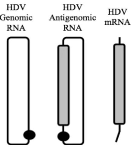
1.2.2. HDV RNAs
Since its discovery as an HBV satellite virus, HDV has puzzled scientists. HDV has a small circular RNA genome with only 1700 nucleotides; this sequence length varies by no more than 30 nucleotides among HDV isolates (Dény, 2006). In native conditions the RNA folds into an unbranched rod-like structure due to intramolecular base pairing involving around 74% of its nucleotides (Kuo et al., 1988).
13 HDV contains one functional open reading frame (ORF), encoding the delta antigen (Chen et al., 1986). This ORF is not encoded by the genomic RNA but by another RNA species that arises during replication, the HDV antigenome, an exact complement of the genome.
The delta antigen is transcribed from a third RNA species, a linear 0.8 Kb
messenger RNA (mRNA) of antigenomic polarity and a 5‟-cap and 3‟-polyadenylated tail (Hsieh and Taylor, 1991). The different HDV RNA species are represented in Figure 2. In an infected cell, the three HDV RNA species accumulate in very different amounts, although genomic RNA is the only species assembled into HDV virions. HDV genomic RNA is the most abundant, around 300,000 copies accumulate in an infected cell whereas 100,000 copies of the antigenome are present (Chen et al., 1986). The HDV mRNA is considerably less abundant with approximately 500 copies per cell (Gudima et al., 2000).
Figure 2: Schematic representation of three HDV RNA species. The HDV genomic RNA is a
single-stranded circular RNA with 1700 nucleotides. It forms an unbranched rod-like structure due to intramolecular base pairing. The HDV antigenomic RNA is the exact complement of the genomic RNA, both RNAs have site-specific ribozymes indicated by the black circle. The HDV antigenomic RNA contains the open reading frame for the delta antigen, represented in grey, but the antigen is translated from another RNA species, the mRNA. The mRNA is 800 nucleotides long with a 5‟-cap and a 3‟ -polyadenylated tail.
14
ribozyme RNAs have been crystallized and an atomic structure solved (Ferré-D'Amaré et al., 1998). They enhance HDV RNA self-cleavage by a 106- to 107-fold when compared with uncatalyzed cleavage (Been, 2006; Ferré-D'Amaré et al., 1998). Although ribozymes are characteristic of viroids, their structures are different from HDV ribozymes, which are actually more related to the cytoplasmic polyadenylation element-binding protein 3 (CPEB3) ribozyme, a conserved mammalian sequence within an intron of the CPEB3 gene (Salehi-Ashtiani, 2006). In fact, numerous HDV-like ribozymes have since been found in several eukaryotic species (Webb et al., 2009).
1.2.3. HDV RNA Replication
HDV RNAs are transcribed in the nucleus of infected cells, but the details of this process remain poorly defined. As mentioned before, during HDV replication three RNA species accumulate in infected cells: the genome, antigenome and mRNA (Figure 2). Each of these is the product of post-transcriptional processing.
The precursors, from which these species arise, are thought to be transcribed by a double-rolling circle mechanism, exemplified in Figure 3. In this model the circular genome RNA is used as a template to produce multimeric species of opposite polarity (Taylor, 1990). These greater than unit-length RNAs are subsequently self-cleaved by the HDV ribozymes and re-ligated, producing unit-length circular antigenomic RNAs. The re-ligation step is thought to involve a host ligase (Reid and Lazinski, 2000) although it has been shown that the HDV ribozyme can self-ligate in vitro (Sharmeen et
al., 1989). Through a similar mechanism the unit-length circular antigenomic RNA acts as a template for the transcription of multimeric species, which are processed to produce genomic RNA. The genomic RNA also acts as a template for transcripts that are processed into mRNA.
15
Figure 3: Model of HDV replication through a rolling-circle mechanism. The HDV genomic RNA is used as a template for the precursors of HDV mRNA (Steps 1-2) and also acts as a template for multimeric RNAs of antigenomic polarity (Step 3). These multimeric RNAs contain at least two copies of the HDV ribozyme and are thus self-cleaved to produce linear unit-length HDV antigenomes (Step 4), which are then ligated to produce circular antigenomic RNA (Step 5). In turn, the new antigenomic RNA is a template for multimeric RNAs of genomic polarity (Step 6) that are similarly self-cleaved and subsequently ligated to produce new circular genomic RNA (Steps 7-8).
Most RNA viruses use a virus-encoded RNA-directed RNA polymerase for replication. This is consistent with the dogma that host cell RNA polymerases are DNA-dependent and do not accept RNA templates. An important exception are retroviruses which first synthesize a DNA intermediate using a virus-encoded reverse transcriptase, to produce a DNA intermediate, which becomes integrated into the host genome and is then transcribed into RNA, using a host RNA polymerase. However, HDV has no known DNA intermediate (Taylor, 2009), and the only HDV protein, the delta antigen, is too small to be a polymerase. This means that HDV RNA must somehow redirect host DNA-dependent RNA polymerases to use HDV RNAs as templates. How this is achieved and which host polymerase(s) is (are) involved has been extensively studied but the results remain somewhat controversial.
16
have shown that inhibition of pol II by low concentrations of the specific inhibitor α -amanitin blocks HDV RNA synthesis of both the genomic and antigenomic strands (Chang et al., 2008). One possible explanation is that the rod-like conformation of HDV RNAs may trick pol II into accepting the RNA as a double-stranded DNA template. It has been shown, through immunoprecipitation assays, that pol II binds the terminal stem-loop regions of HDV genome (Greco-Stewart et al., 2007). It has also been reported that, after binding to the stem-loop, pol II is able to elongate multimeric RNA species, carrying out transcription (Filipovska and Konarska, 2000). Such elongation was observed on a partial antigenomic RNA stem-loop and originated a chimeric molecule of newly synthesized transcript covalently bound to the 5‟-end of the template. Thus, it is not clear if such elongation is biologically relevant.
Despite being shown that pol II interacts with genomic HDV RNA it has been suggested that a different host polymerase is responsible for the synthesis of antigenomic HDV RNA (Macnaughton et al., 2002; Modahl et al., 2000). The idea that at least two different host polymerases are involved in the HDV replication cycle is based on the observation, that in transfected cells, the synthesis of new HDV
antigenomic RNA was not inhibited by concentrations of α-amanitin that would inhibit pol II activity (Modahl et al., 2000). This has led to the speculation that pol I copies genomic HDV RNA to produce new antigenomic RNA. This is contrary to the afore mentioned nuclear run-on assays, which have shown that both genomic and
antigenomic RNA synthesis are sensitive to low doses of α-amanitin, consistent with pol II (Chang et al., 2008).
An additional complication arises from in vitro studies, which indicate that
fragments of the HDV RNA genome interact not only with pol II but also with pol I and pol III (Greco-Stewart et al., 2009). However, such in vitro interactions may not have
biological relevance, especially since they do not lead to RNA-directed transcription. The HDV mRNA possesses characteristics of a pol II transcript that is processed
17 The controversy regarding the transcription process is thus limited to whether genomic RNA is transcribed by pol II or another polymerase, possibly pol I (Taylor, 2012).
In addition to the post-transcriptional processing to make the abovementioned three HDV RNAs, there is an important RNA-editing event. During the virus replication cycle some of the antigenomes are edited at a specific site by a host adenosine deaminase (ADAR1). This changes the adenosine in the amber codon to inosine. After subsequent RNA-directed RNA synthesis, it leads to the replacement of inosine with guanosine (Polson et al., 1996). That is, the UAG stop codon is changed to a UGG tryptophan codon. In this way the delta antigen ORF is extended by 19 amino acids; that is, to the next stop codon. The specificity of the editing site is in part directed by the specific folding of the HDV antigenomic RNA (Casey, 2006).
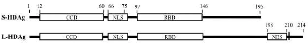
Therefore, although HDV has only one ORF, it encodes two proteins: the small delta antigen (S-HDAg) of 195 amino acids and the large delta antigen (L-HDAg) with 214 amino acids, which will be described in Section 1.3.
1.2.4. HDV Origin
The resemblance of HDV RNA to that of plant viroids could indicate a viroid ancestor. The similarities are indeed extensive: the small genome size and circular conformation, the presence of ribozyme domains and the proposed rolling-circle mechanism for HDV replication (recently reviewed by Flores et al., 2012). Viroids are nevertheless significantly smaller than HDV, ranging from 250 to 400 nucleotides, and they are also non-coding RNAs (Tsagris et al., 2008). HDV could have evolved to
encode the delta antigen and HDAg coding sequence accounts for HDV‟s longer
18
Like HDV RNA, viroid RNAs are circular molecules that display intramolecular folding achieving almost rod-like structures. Some viroids, known as avsunviroids, replicate via a symmetrical rolling-circle mechanism as the one proposed for HDV (Taylor, 2009; Tsagris et al., 2008).
Despite the similarities that support a proposed evolution from the viroid world, there are important differences that advocate a different origin. For example, although ribozymes are characteristic of viroids, the two HDV ribozymes are structurally very different from those of the viroids. In contrast, several HDV-like ribozymes have been found in eukaryotes (Webb et al., 2009). This more supports the hypothesis that HDV may be derived from the human transcriptome rather than a plant or ancestral RNA world. Also, the HDV genome is larger than viroids and its antigenome encodes a viral protein.
19
1.3. Delta Antigens
HDV has only one open reading frame, which, as mentioned in Section 1.2.3, due to some post-transcriptional site-specific RNA editing, ultimately encodes two proteins, S-HDAg and L-HDAg. The two isoforms share 195 amino acids and differ only in that the large form has 19 extra amino acids on the C-terminus. As such, S-HDAg and L-S-HDAg share several functional domains within the common amino acid sequence, as illustrated in Figure 4. The delta antigens contain a nuclear localization signal (NLS) comprised by amino acids 66 through to 75 (Alves et al., 2008); a coiled-coil domain (CCD), also referred to as dimerization domain, within amino acids 12 to 60; and an RNA binding domain within amino acids 97 and 143 (Lee et al., 1993). L-HDAg has, within its extra sequence, a nuclear export signal (NES) spanning amino acids 198 to 210 (Lee et al., 2001).
Figure 4: Functional domains of S-HDAg and L-HDAg. The delta antigens share most of their
sequence differing only in the 19 amino acids extension at the C-terminal of L-HDAg. They have, within the common sequence, as represented, a coiled-coil domain (CCD), a nuclear localization signal (NLS), and an RNA binding domain (RBD). Also indicated on L-HDAg are the nuclear export signal (NES) and the unique cysteine, residue 211, which is the target for farnesylation. The numbers indicate the position of the amino acid residues.
Both delta antigens undergo post-translational modifications (PTMs) by several host enzymes. Several groups have investigated the impact these PTMs may have on the
antigens‟ functions but the precise significance of most of these modifications remains
uncertain.
20
There are other PTM events, ones shared by both forms of the delta antigen. These involve phosphorylation, methylation, acetylation and sumoylation (Chen et al., 2008; Hong and Chen, 2010; Li et al., 2004; Mu et al., 2004; 1999; Tseng et al., 2008).
Phosphorylation has been observed at multiple sites, mostly at serine and threonine residues. Different phosphorylation patterns were observed for S-HDAg and L-HDAg, and, if relevant, the distinct patterns may in part account for their distinct biological functions (Mu et al., 1999). Several host enzymes have been reported to phosphorylate delta antigens at different sites: casein kinase II on Ser2 and Ser213 (Yeh et al., 1996); double-stranded RNA-activated protein kinase R on residues Ser177, Ser180, and Thr182 (Chen, 2002); extracellular signal-related kinases 1 and 2 (ERK1/2) on Ser177 (Chen et al., 2008); and protein kinase C on residue Ser210 (Yeh et al., 1996). It has been alleged that S-HDAg phosphorylation increases replication of genomic HDV RNA from the antigenomic strand (Chen et al., 2008). By enhancing the expression of ERK1/2 in cells transfected with plasmids expressing S-HDAg and dimeric HDV antigenomic RNA, an increase in the accumulation of HDV genomic RNA was observed, but not for antigenomic RNA (Chen et al., 2008). More recently, it has been suggested that phosphorylation of S-HDAg at Ser177 can work as a switch in HDV antigenomic RNA replication from the initiation to the elongation stage (Hong and Chen, 2010).
Acetylation of Lys72 on S-HDAg, by host p300 acetyltransferase, is thought to regulate nucleocytoplasmic shuttling of viral RNA (Huang et al., 2008b; Mu et al., 2004). Note that this amino acid is within the NLS of the HDAgs (Alves et al., 2008). Thus such a modification could be expected to have an impact on nuclear import. Acetylation of S-HDAg, has also been suggested to function as a switch in the synthesis of the different viral RNA species as this PTM was reported to be essential for HDV genome and mRNA synthesis but dispensable for antigenomic RNA synthesis (Tseng et al., 2008).
Methylation of Arg13 on S-HDAg, by protein arginine methyltransferase I, has been observed in vitro and has also been proposed to have a switching effect on HDV
21 cells, the mutant S-HDAg reduced genomic RNA synthesis and almost completely suppressed HDV mRNA synthesis (Li et al., 2004; Tseng et al., 2008).
Finally, sumoylation of multiple lysine sites, by small ubiquitin-related modifier isoform 1 (SUMO1), has been reported. Such PTM was detected on S-HDAg but not on L-HDAg (Tseng et al., 2010). And this PTM was proposed to enhance genomic RNA and mRNA synthesis based on experiments where SUMO1 was fused to S-HDAg, so as to mimic sumoylated S-HDAg (Tseng et al., 2010).
Although the two delta antigens share sequence and functional domains they play very distinct roles in the HDV replication cycle. S-HDAg is essential for HDV RNA accumulation whereas L-HDAg acts as a dominant negative inhibitor of HDV replication (Chao et al., 1990) and also is essential for the assembly, via HBsAgs, of HDV RNA into new virus particles. There is, however, a common function attributed to both antigens: it has been observed that both can downregulate HBV replication (Williams et al., 2009).
22
1.3.1. Small Delta Antigen
The objective of my research has been to increase the understanding of the structure and function of S-HDAg, the one viral protein that is essential for the accumulation of processed HDV RNA transcripts. Therefore, in this Section I will review, in more detail, prior knowledge and uncertainties relating to this protein.
Three groups have purified recombinant S-HDAg, but have been unable to go on and produce distinct crystals. Thus, at this time a detailed molecular structure for the protein is unavailable. A region comprising amino acids 12 to 60 was predicted to have alpha-helical structure and also be responsible for the protein‟s ability to form dimers (Xia and Lai, 1992; Wang and Lemon, 1993). To test this, one lab obtained large amounts of the corresponding synthetic peptide (Rozzelle et al., 1995). As a follow-up to this synthesis, it was found that the peptide readily formed crystals, leading to a successful molecular structure (Zuccola et al., 1998).
In this structure dimers were observed. More specifically, the largely alpha-helical peptides formed dimers by an anti-parallel coiled-coil interaction; hence this region has been referred to as the coiled-coil domain or CCD. Furthermore, the crystal structure indicated how such dimers might interact to form higher multimers. To test this hypothesis, the authors expressed and purified full-length recombinant S-HDAg, and showed, by prior cross-linking followed by mass spectrometry, that complexes as high as 8-mers could be detected (Zuccola et al., 1998).
Earlier studies had demonstrated that the ability to form dimers was essential for S-HDAg‟s role in promoting HDV RNA accumulation (Lazinski and Taylor, 1993). The molecular structure of the CCD was used to predict amino acids that might be important in the dimer and/or multimer interactions. Some of these predictions were tested and it was confirmed that there are amino acids, which are critical for dimer formation and, when mutated, affect S-HDAg‟s ability to support accumulation of HDV RNAs (Moraleda et al., 2000).
23 Several different approaches have been used to demonstrate how S-HDAg binds to RNA. In some of these studies the binding was considered to be specific for HDV rod-like RNA structure (Chao et al., 1991; Defenbaugh et al., 2009; Lin et al., 2010). One study showed a minimum requirement of 311 nucleotides in a rod-like folding (Defenbaugh et al., 2009). In addition, multimerization of the S-HDAg was also required (Lin et al., 2010). In these two studies, the researchers examined purified recombinant S-HDAg that was C-terminally truncated to become HDV RNA specific and did not report the same assays with full-length S-HDAg. An earlier study, performed with S-HDAg fusion proteins, also reported that the HDV RNA rod-like structure is a prerequisite for the HDV RNA to be recognized by the antigen (Chao et al., 1991). A different group observed that the removal of the dimerization domain did not prevent S-HDAg from binding to the viral RNA (Lin et al., 1990). Thus, while it is agreed that S-HDAg is an RNA binding protein there remains controversy as to whether it is an HDV RNA specific interaction or if multimerization of S-HDAg is a prerequisite for the interaction.
As summarized in Table 1, several roles have been attributed to S-HDAg during the HDV life cycle.
Table 1: Roles attributed to S-HDAg. Putative and observed functions of S-HDAg during the HDV replication cycle.
Function Observations
Achieve nuclear import of HDV RNAs S-HDAg-mediated nuclear import of HDV RNA has been observed in vivo (Chou et al., 1998; Tavanez et al., 2002)
Facilitate ribozyme cleavage (chaperone)
S-HDAg stimulates HDV RNA ribozyme cleavage in vitro (Wang et al., 2003)
Regulate HDV RNA editing S-HDAg expression in transfected cells suppresses editing of HDV antigenomic RNA (Polson et al., 1998)
Facilitate accumulation of processed
HDV RNA transcripts S-HDAg is essential for the accumulation of full-length HDV RNAs in transfected cells (Chao et al., 1990)
24
S-HDAg is present in the virions forming viral RNPs with the HDV genome. One of the first tasks it performs is the transport of the viral genome into the nucleus of infected cells, where RNA-directed RNA synthesis takes place. This transport is achieved by the presence of the previously described NLS and RBD. Nuclear import may be facilitated by karyopherin 2, since this importin interacts with S-HDAg in vitro (Chou et al., 1998).
Another role attributed to S-HDAg is the regulation of HDV RNA editing, particularly the deamination by ADAR-1. This editing seems to occur at multiple locations on HDV RNAs, but it is focused on the antigenomic RNA at the stop codon adenosine (Polson et al., 1998). S-HDAg has been found to suppress editing at this stop codon when expressed in transfected cells at levels close to those observed during HDV replication (Polson et al., 1998). This observation suggests that the antigen plays a role in limiting HDV RNA editing, as excessive editing has been shown to inhibit HDV RNA accumulation (Jayan and Casey, 2002).
It has been known for more than two decades that the small form of the delta antigen is essential for the accumulation of processed HDV RNAs (Chao et al., 1990). Several theories have been proposed for the precise role(s) it may play, as will be discussed ahead.
S-HDAg has been shown to interact with host pol II. In a pull-down assay, both S-HDAg and L-HDAg fused with a glutathione S-transferase tag were able to bind pol II from HeLa nuclear extracts (Yamaguchi et al., 2001). In the same study S-HDAg, was observed to enhance pol II elongation, presumably by displacing the subunit A of the negative elongation factor (NELF-A). S-HDAg was thus reported as an elongation enhancer of DNA-templated pol II transcription in vitro (Yamaguchi et al., 2001).
However, the observed enhancement appears to be limited to 3‟-OH end additions, rather than transcription. In a subsequent study Yamaguchi et al., reported that S-HDAg
25 between pol II and S-HDAg loosens what, from molecular structure studies, is considered to be a pol II clamp, thereby reducing transcriptional fidelity and allowing the recognition of the atypical RNA template.
Amidst all the reports that S-HDAg actively participates in HDV RNA transcription there is a contradictory result showing that the presence of S-HDAg is not required for the accumulation of processed short HDV transcripts, although full-length transcripts, genomic or antigenomic, do require S-HDAg, or even L-HDAg (Lazinski and Taylor, 1994). As an explanation it was proposed that full-length HDV RNAs are susceptible to nucleolytic degradation in the absence of S-HDAg and, due to their size, such RNAs are more prone to be degraded than smaller RNAs. In other words, S-HDAg interacts with HDV RNAs to protect them and thereby allow their accumulation in infected cells.
Another role attributed to S-HDAg is that of HDV RNA chaperone. In vitro
studies have reported that S-HDAg can stimulate HDV RNA ribozyme activity (Wang et al., 2003). From such studies it is inferred that in vivo S-HDAg may be directly
involved in post-transcriptional processing of nascent multimeric transcripts by enhancing cleavage into unit-length molecules. It should be noted however, that the abovementioned studies of Lazinski and Taylor indicate that in vivo, HDAg is not
directly needed for ribozyme cleavage and subsequent ligation (Lazinski and Taylor, 1994).
26
1.3.2. Host Interactions
HDV has a very small RNA genome, as mentioned earlier, and encodes only one viral protein, HDAg. Albeit the fact that a second isoform of the HDAg appears later in the replication cycle, S-HDAg and L-HDAg are not sufficient for HDV to complete its replication cycle. None of these two HDAg isoforms has replicase activity or any other known enzymatic activity and, despite the fact that HDAgs are essential, HDV must rely extensively on host cell factors to complete its replication cycle. Many S-HDAg-interacting proteins have been identified through different affinity approaches, as summarized in Table 2. But, as will be discussed below, the role of most of these interactions remains unclear and is still being investigated.
Table 2: Identification of S-HDAg-interacting proteins. Summary of the proteomic studies to identify host proteins that interact with S-HDAg.
Reference Experimental Approach Number of Identified Proteins
Cao et al., 2009 Immunopurification followed by mass spectrometry > 100
Casaca et al., 2011 Yeast two-hybrid screening 30
A comprehensive study using immunopurification followed by mass spectrometry identified over 100 host proteins that associated with a tagged S-HDAg (Cao et al., 2009). This set included 9 of the 12 subunits of the pol II complex, further supporting the idea that pol II is involved in HDV RNA transcription (Cao et al., 2009). In another study, a yeast two-hybrid approach identified 30 proteins encoded by a human liver cDNA library that interacted with S-HDAg (Casaca et al., 2011). Only three proteins from this study had also been identified by the previously mentioned immunopurification approach.
27
Table 3: S-HDAg-interacting proteins. Summary of S-HDAg-interacting proteins with putative or observed function (adapted from Greco-Stewart and Pelchat, 2010).
Host Protein Function Reference
Casein kinase II
Phosphorylation of Ser2 observed in vivo as inhibition of casein kinase II suppresses Ser2
phosphorylation Yeh et al., 1996
Double-stranded RNA-activated protein
kinase R
Phosphorylation of Ser177, Ser180 and Thr182
observed in vitro and in vivo Chen et al., 2002
ERK1/2
Phosphorylation of Ser177 observed in vitro, and in vivo activation of endogenous ERK1/2 increases
Ser177 phosphorylation Chen et al., 2008
Arginine
methyltransferase 1 Methylation of Arg13 observed in vitro Li et al., 2004; Tseng et al., 2008
p300 acetyltransferase Acetylation of Lys72 observed in vivo Mu et al., 2004
SUMO 1 Sumoylation of multiple lysine sites observed in vitro and in vivo by co-transfection with
S-HDAg-expressing plasmid Tseng et al., 2010
Karyopherin 2 Nuclear import of viral RNPs (interacts S-HDAg) in vitro with Chou et al., 1998
Pol II S-HDAg enhances pol II elongation might be limited to 3‟-OH end additions in vitro but Yamaguchi et al., 2001
28
1.4. Intrinsically Disordered Proteins
1The notion that a rigid 3-Dimensional (3-D) structure is a prerequisite for a protein to be functional was unquestioned for a long time. The paradigm can be dated back to the lock-and-key hypothesis, proposed in 1894 by Emil Fischer, to explain enzymatic specificity (Lemieux and Spohr, 1994). This concept was later validated as the crystal structures of proteins were beginning to be solved by X-ray diffraction (reviewed by Uversky, 2011a). So, for a long time, the conventional view was that a functional protein folds into a unique and stable 3-D structure, perfectly matching the substrate to which it should bind.
Up until a couple of decades ago, the idea that disordered proteins could also have specific functions was considered outlandish. Nevertheless, occasionally, flexible but functional proteins were discovered or re-discovered. Information on these flexible
proteins was scarce and shallow, since they didn‟t fit the structure-function paradigm; they were considered mere exceptions to a rule. Nonetheless, throughout the years, a variety of names were given to these non-conventional proteins: partially folded, flexible, pliable, chameleon, vulnerable, natively unfolded, etc. (reviewed by Uversky, 2011a).
Only in the late 1990s did researchers start to realize that these unstructured proteins were representative of a broad class of rather important proteins (Dunker et al., 2001; Romero et al., 1998; Tompa, 2002; Uversky et al., 2000; Wright and Dyson, 1999). In recent years, the term intrinsically disordered proteins (IDPs) has become the most widely accepted, and intrinsically disordered regions (IDRs), to define proteins or protein segments that are biologically functional although they exist as collapsed or extended mobile conformational ensembles (Uversky, 2011a).
The increasing number of experimentally characterized IDPs led to the creation, in 2007, of DisProt, a database of disordered proteins (Sickmeier et al., 2007).
29
1.4.1. Intrinsic Disorder Prediction
Like ordered proteins, whose structures can to some extent be inferred from their amino acid sequence, IDPs and IDRs have signature characteristics that allow the prediction of disorder based on sequence data alone. A mark of probable intrinsic disorder is a low content of hydrophobic amino acids, which usually form the core in folded proteins, and a high presence of polar amino acids conferring high net charge to the disordered protein or disordered region (Romero et al., 2001; Uversky et al., 2000). Low hydrophobicity is thought to lead to a low driving force for protein compaction and high net charge may result in strong electrostatic repulsion. Hence, these features contribute to structural disorder. Even by 1978, it had been suggested that IDPs and IDRs have amino acid compositions that differ from ordered proteins and therefore disorder could be predicted by the abnormally high ratio of charged residues by hydrophobic residues (Williams, 1978). However, this early mode of intrinsic disorder prediction was based on a very small set of proteins and never tested for other sets. Later it was shown that IDPs and IDRs are deficient in what has been called „order
-promoting‟ amino acids such as Ile, Leu, Val, Trp, Tyr, Phe, Cys and Asn; and are
enriched in „disorder-promoting‟ amino acids like Ala, Arg, Gly, Gln, Ser, Glu, Lys and
Pro (Dunker et al., 2001).
30
1.4.2. ID Prevalence
Disorder predictors have been applied to the predicted proteins of entire genomes to assess the extent of intrinsic disorder, with predictions spanning the three kingdoms. Studies have shown that IDPs and IDRs are not rare exceptions but highly abundant in all species. In fact, almost 70% of proteins in the Protein DataBase by 2003 had IDRs (Obradovic et al., 2003). IDPs are more common in eukaryota than in prokaryota and archea, with up to 30% of eukaryotic proteins being mostly disordered (Oldfield et al., 2005). However, in a recent study, comparing 194 eukaryotic and 87 prokaryotic proteomes, researchers found an overlap in the frequency of predicted disorder, which spans a wide range in both kingdoms (Pancsa and Tompa, 2012). Although prokaryotes were found to have a lower average disorder than eukaryotes both groups have a very broad range of predicted disorder, with scores of average ratio of disordered residues in proteins ranging from 0.12 to 0.35 for prokaryotes and 0.1 to 0.41 in eukaryotes (Pancsa and Tompa, 2012).
The same study also found the highest levels of predicted intrinsic disorder in single-celled protists, often higher than in more complex eukaryotic organisms. Hence, a new theory was proposed by the authors correlating intrinsic disorder with lifestyle and not only with the complexity of the organism (Pancsa and Tompa, 2012). They suggest that a low level of disorder reflects and adaptation to the environment, as low values were found for certain intracellular parasites and endosymbionts. Organisms, such as host-changing parasites, which lead a varied lifestyle, changing habitats, have higher levels of disorder (Pancsa and Tompa, 2012).
The amount of predicted intrinsic disorder in viral proteins has been compared with that of eukaryotes. Even though, eukaryotic proteomes contain more proteins with long disordered sequences, viral proteomes have more short disordered segments (Xue et al., 2010b).
31 2010b). In particular, RNA viruses, which display the highest mutation rates, also have a high incidence of disordered regions and significantly lower van der Waals contact densities, reflecting the intensity of the 3-D interaction network of a protein (Tokuriki et al., 2008). As will be examined in section 1.4.4, viral proteins profit at different levels from the flexibility that results from intrinsic disorder.
1.4.3. Biophysical Characterization of Disorder
IDPs are by definition, an ensemble of conformational states, so the determination of one unique structure is not possible and a multiparametric approach is required. A dynamic structural characterization of an IDP relies on different parameters from a host of physiochemical methods to obtain information on different aspects of the protein such as overall compactness, conformational stability, presence of residual secondary structure, transient long-range contacts, regions of more or less mobility (Uversky and Dunker, 2012). Most of the techniques used were initially developed to analyze ordered proteins thus, data analysis needs to be careful as results for intrinsic disorder are usually due to the lack of signals characteristic of ordered sequences. That is also why several methods should be used and results compared to minimize ambiguity (Uversky and Dunker, 2012). Some techniques will be briefly summarized below, of which, the most commonly used to obtain data describing IDP structures are NMR and small-angle X-ray scattering (Tompa, 2011).
(i) X-ray crystallography is the „classic‟ technique for determining a protein‟s
crystal structure. With this method the higher flexibility of the atoms in disordered regions results in a non-coherent X-ray scattering, making them invisible. That is, the outcome is a region with missing electron density corresponding to the disordered segment (Dunker et al., 2001). This method depends on the ability of proteins to form crystals, and highly disordered proteins in general, are not even able to be crystallized.
32
to obtain precise data on 3-D structures and also provide direct measurements of IDR mobility (Uversky and Dunker, 2010).
(iii) Circular dichroism (CD) in the far-UV region provides estimates of secondary structure and near-UV CD displays sharp peaks for aromatic groups when the protein is ordered. CD lacks residue-specific information and the data obtained are less clear when a protein contains both ordered and disordered segments (Dunker et al., 2001).
(iv) The level of protein compactation or hydrodynamic dimension can be assessed by different techniques such as gel-filtration, viscometry, small-angle X-ray scattering or sedimentation (Uversky and Dunker, 2012).
(v) Small-angle X-ray scattering can also be used to determine the degree of globularity, providing information on the presence or absence of a tightly packed core (Uversky and Dunker, 2012).
(vi) Proteolytic degradation is also a method to identify disordered segments in a protein since flexibility can be a major determinant for susceptibility to cleavage (Johnson et al., 2012). Typically a structured protein needs a segment of more than 10 residues to be unfolded to reveal a proteolytic cleavage site (Dunker et al., 2001).
Note that many IDPs and IDRs adopt a well-defined structure when bound to their partners (Dyson and Wright, 2002). In these cases, by using methods to characterize ordered proteins, the structure of the bound protein can be readily solved.
1.4.4. Viral IDPs
Viral proteins should be considered as a unique group of proteins. Viruses must
adapt faster than their hosts. They need to survive in their host‟s environment as well as
33 mutation rate of 10-9 (Drake et al., 1998). Since viruses have highly compact genomes, often with overlapping reading frames, a single mutation can have an impact on more than one viral protein (Reanney, 1982). Finally, viral proteins usually need to perform numerous interactions with host cell components, during the different steps of the virus life cycle from entry to replication to formation and exit of new infectious particles. Viral proteins must interact with host membranes, host proteins and in some cases, host nucleic acids even though viral proteins are often phylogenetically separated from their host proteins (Forterre, 2006; Reanney, 1982).
All these features make it extremely interesting to test whether viral proteins are also associated with unique biophysical characteristics and advantages. Intrinsic disorder may be a way for viral proteins to cope with these distinctive circumstances as the resulting plasticity can confer a number of exceptional functional advantages.
(i) Intrinsically disordered proteins are more flexible and without a rigid compact structure; thus, viral proteins can be highly promiscuous and take part in several interactions with multiple partners.
(ii) IDRs in particular can act as flexible linkers between functional domains enabling mechanisms that will facilitate binding and promiscuity.
(iii) These flexible linkers can also help viral proteins to elude the host cell‟s
adaptive immune system by making it difficult for epitopes to be recognized (Goh et al., 2008).
(iv) Disorder can also be a way to cope with high mutation rates, characteristic to viruses. High flexibility, resulting from low interactions between amino acids, can be linked to a high adaptability and represent a strategy to buffer deleterious effects of mutations; an already unstructured protein has less to lose from a substitution than a highly structured one, as it is already unfolded.
Intrinsic disorder or conformational flexibility affords a “broad partnership”
34
complexes or in signaling interactions in which they transit between the bound and unbound state as a dynamic and sensitive on-off switch (Uversky, 2011b). IDPs‟ ability to have different conformations depending on environmental conditions allows them to exercise different functions in different contexts.