171 171171 171171 Mem Inst Oswaldo Cruz, Rio de Janeiro, Vol. 95(2): 171-177, Mar./Apr. 2000
Identification of Planorbids from Venezuela by Polymerase
Chain Reaction Amplification and Restriction Fragment
Length Polymorphism of Internal Transcriber Spacer of the
RNA Ribosomal Gene
Roberta L Caldeira, Teofânia HDA Vidigal*, Liboria Matinella**,
Andrew JG Simpson***, Omar S Carvalho/
+Centro de Pesquisas René Rachou-Fiocruz, Av. Augusto de Lima 1715, 30190-002 Belo Horizonte, MG, Brasil *Instituto de Ciências Biológicas, UFMG, Belo Horizonte, MG, Brasil **Laboratório de Malacologia, Ministerio
de Sanidad y Asistencia Social, Aragua, Venezuela ***Laboratório de Genética de Câncer, Instituto Ludwig de Pesquisas sobre o Câncer, São Paulo, SP, Brasil
Snails of the genus Biomphalaria from Venezuela were subjected to morphological assessment as well as polymerase chain reaction and restriction fragment length polymorphism (PCR-RFLP) analysis. Morphological identification was carried out by comparison of characters of the shell and the male and female reproductive apparatus. The PCR-RFLP involved amplification of the internal spacer region ITS1 and ITS2 of the RNA ribosomal gene and subsequent digestion of this fragment by the restriction enzymes DdeI, MnlI, HaeIII and MspI. The planorbids were compared with snails of the same species and others reported from Venezuela and present in Brazil, Cuba and Mexico. All the enzymes showed a specific profile for each species, that of DdeI being the clearest. The snails were identified as B. glabrata, B. prona and B. kuhniana.
Key words: Biomphalaria glabrata - Biomphalaria kuhniana - Biomphalaria prona - snails - internal transcribed spacer - ribosomal DNA
The endemic area for schistosomiasis in Ve-nezuela includes the Federal District of Caracas and the states of Aragua, Carabobo, Miranda and Guárico. One third of the country’s population lives in this area, where the prevalence of infection with
Schistosoma mansoni is 2% (Balzán 1992). To date, seven species of Biomphalaria have been reported in Venezuela: B. glabrata (Martens 1873), B. straminea (Martens 1873),B. prona (Mar-tens 1873), B. kuhniana (Baker 1930), B. havanensis
(Hubendick 1961), B. schrammi (Chrosciechowski 1968) and B. obstructa (Chrosciechowski 1988). The first of these species has been shown to be re-sponsible for the transmission of Schistosoma in this country (Balzán 1992).
The specific identification of these planorbids is based on comparison of morphological charac-ters of the shell and male and female reproductive organs, as described by Deslandes (1951), Paraense
(1975, 1988, 1990), Paraense and Deslandes (1958) and Paraense et al. (1992). Identification is com-plicated in many cases by morphological variation in these characters, for example when the B. prona
population from Lake Valencia (Venezuela), is compared with populations of the same species that occur outside this lake (Paraense et al. 1992).
Moreover, identification is also complicated by morphological similarities among some species of snail, for example the B. straminea (Paraense 1988, Caldeira et al. 1998) and B. tenagophila complexes (Spatz et al. 1998, 1999).
The polymerase chain reaction amplification and restriction fragment length polymorphism (PCR-RFLP) technique have been used successfully to distinguish several medically important snails of the genera Oncomelania (Hope & McManus 1994),
Bulinus (Stothard et al. 1996, 1997) and
Biomphalaria (Vidigal et al. 1998a, b, Caldeira et al. 1998, Spatz et al. 1998, 1999). This technique is based on amplification of the internal transcribed spacer region of the rRNA followed by digestion of the resulting fragment with restriction enzymes.
This is an easy technique to perform and gives profiles that are simple to analyse. In the present study snails of the genus Biomphalaria obtained from different regions of Venezuela were identified using both morphological and molecular criteria. Work partially supported by Fapemig, Pibic/Fiocruz and
OPS.
+Corresponding author. Fax: + 55-31-295.3115. E-mail:
172 172 172 172
172 Identification of Biomphalaria by PCR-RFLP Roberta L Caldeira et al.
MATERIALS AND METHODS
Snail populations - Snails from different locali-ties of Venezuela were used in the study together with specimens previously identified as B. obstructa from Isla del Carmen, Mexico (type lo-cality); B. havanensis from Havana, Cuba; B. glabrata,B. straminea and B. kuhniana from Bra-zil. All these species have been reported from Ve-nezuela (Table). All specimens used were collected in the field, with the exception of B. obstructa from Isla del Carmen which had been maintained in the Departament of Malacology of the Instituto Oswaldo Cruz, Rio de Janeiro and B. havanensis
from Guatao, Cuba reared in the Departament of Malacology of the Instituto Pedro Kouri (Havana). All the specimens obtained from the field were ex-amined for cercariae of Schistosoma mansoni and found to be negative. Two individuals from each locality were used for the molecular analysis.
Morphological identification of snail popula-tions -Ten specimens of each population were killed, fixed (Deslandes 1951, Paraense 1976) and the feet removed for subsequent DNA extraction. After fixation, each specimen was identified by comparative morphology of the reproductive or-gans and shell as described by Deslandes (1951),
Paraense and Deslandes (1958), Paraense (1975, 1988, 1990) and Paraense et al. (1992).
DNA extraction -Total DNA was extracted from the feet of the snails using the Wizard Genomic DNA Purification Kit (Promega) with some modifications. Each foot was mechanically disrupted in 200 µl of nucleic lysis solution and incubated at 37°C for 4 h or overnight with 50 µg/ml of proteinase K, then 80 µl of protein precipitation solution was added to the initial mix. The solution was shaken vigorously and centrifuged at 13,000 rpm for 5 min. The superna-tant containing DNA was transferred to a clean tube with 200 µl of isopropanol for DNA precipitation. The mixture was gently homogenized by inversion for 20 min and centrifuged at 13,000 rpm for 5 min. The precipitated DNA was washed with 300 µl of 70% ethanol and centrifuged for 10 min. The pellet treated with 25 µl of DNA rehydration solution for 30 min at 65°C and stored at -20°C. The DNA con-centrations were estimated by comparison with known standards on 2% ethidium bromide stained agarose gels.
rDNA-ITS amplification -The entire internal transcriber spacer (ITS) (which includes the 5.8S rDNA gene together with the flanking ITS1 and ITS2 spacers) was amplified using the primers
TABLE
Localities of Biomphalaria species from Venezuela used in the study
Species Locality State, country
B. glabrata Villa de Cura Aragua, Venezuela
Manuare Carabobo, Venezuela
Guacara Carabobo
Belén Carabobo
Caripe Monagas, Venezuela
Chabasquén Portuguesa, Venezuela
Anzoátequi Lara, Venezuela
Belém Pará, Brazil
Esteio Rio Grande do Sul, Brazil
B. kuhniana San Casimiro Aragua, Venezuela
Villa de Cura Aragua
Pao de Zárate Aragua
Villa de Cura Aragua
Laguna del Parque Recreacional Carabobo, Venezuela
Tucuruí Pará, Brazil
B. prona San Casimiro Aragua, Venezuela
Lake de Valencia Aragua
Laguna del Parque Recreacional Carabobo, Venezuela Canal La Pista, Tinaquillo Cojedes, Venezuela
San Carlos Cojedes
Represa El Guamo Monagas, Venezuela
B. straminea Belém Pará, Brazil
Picos Piauí, Brazil
Florianópolis Santa Catarina, Brazil
B. havanensis Guatao Cuba
173 173173 173173 Mem Inst Oswaldo Cruz, Rio de Janeiro, Vol. 95(2), Mar./Apr. 2000
ETTS2 (5/-TAACAAGGTTTCCGTAGGTGAA-3/) and ETTS1 (5/-TGCTTAAGTTCAGCGGGT-3/) anchored respectively in the conserved extremi-ties of the 18S and 28S ribosomal genes (Kane & Rollinson 1994). PCR amplification was under-taken in a volume of 10 µl consisting of: 1-10 ng of template DNA, 10 mM Tris-HCl, pH 8.5, 200 µM each dNTP, 1.5 mM MgCl2, 0.8 U of Taq DNA polymerase (Cenbiot, RS, Brazil), 50 mM KCl, together with 1 pmol of each primer. The reactions were covered with a drop of mineral oil and sub-jected to the following cycle program: initial de-naturation step for 5 min at 95°C, and then 32 cycles with: annealing of 54°C for 1 min, extension at 72°C for 2 min, denaturation at 95°C for 45 sec and a final extension step at 72°C for 5 min. A negative control (no template DNA) was included in all experiments. Three microliters of the ampli-fication products were visualized on 0.8% ethidium bromide stained agarose gels to check the quality of amplification. The remaining 7 µl were mixed with 43 µl of water and divided into 10 µl samples for enzyme digestion.
RFLP analysis - To evaluate possible enzymes that might yield informative RFLPs of the ITS re-gion, four restriction enzymes (Amersham Life Science) were used: the four base cutters HaeIII and MspI, the five base cutter DdeI and the 11 base cutter MnlI. One microliter (10-12 units) of each
enzyme was used for each digestion, together with 1.2 µl of the respective enzyme buffer and 10 µl of the diluted amplification product in a final volume of 12.2 µl. The digestion was performed for 3.5 h at 37°C and products were evaluated on 6% silver stained polyacrylamide gels (Santos et al. 1993, Sanguinetti et al. 1994) after phenol/chloroform extraction to remove protein. The results were re-corded with Polaroid film 667 (St Albans, UK). A control for the activity of each enzyme was per-formed by digesting 150 ng of pUC18 simulta-neously with the samples being evaluated.
RESULTS
Morphological identification of snail popula-tions - The results of morphological identifications of the snails examined are shown in the Table.
RFLP analysis - DNA amplification with the primers ETTS1 and ETTS2 generated a fragment of approximately 1.300 bp for all the specimens. Fig. 1 shows the profiles obtained with DdeI en-zyme which grouped together: the B. glabrata
snails from Brazil and Venezuela (lanes 1 to 16),
B. prona populations from the Lake Valencia and from outside this lake (lanes 17 to 28), B. straminea
from Brazil (lanes 29 to 31) and B. kuhniana from Venezuela and Brazil (lanes 32 to 43).
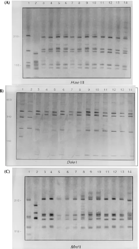
Fig. 2A, B and C shows the profiles obtained with HaeIII, MspI and MnlI respectively. The
174 174 174 174
174 Identification of Biomphalaria by PCR-RFLP Roberta L Caldeira et al.
Fig. 2A
Fig. 2B
Fig. 2C
Fig. 2: silver-stained polycrylamide gels (6%) showing the restriction fragment length polymorphism profiles obtained by digest-ing the rDNA internal trancriber spacer (ITS) with HaeIII (A), MspI (B)and MnlI (C) enzymes. In each gel the snails specimens were: Biomphalaria kuhniana lanes 1-2 San Casimiro, La Barquera Quebrada 2 (State of Aragua, Venezuela); 3-4 Villa de Cura, El Cortijo (State of Aragua, Venezuela); 5-6 Pao de Zárate (State of Aragua, Venezuela); 7-8 Laguna del Parque Recreacional, Sur de Valencia (State of Carabobo, Venezuela); 9-10 Villa de Cura (State of Aragua, Venezuela); 11-12 Tucuruí (State of Pará, Brazil). B. straminea lane 13 Belém (State of Pará, Brazil); 14 Picos (State of Piauí, Brazil); 15 Florianópolis (State of Santa Catarina, Brazil).
(A)
(B)
175 175175 175175 Mem Inst Oswaldo Cruz, Rio de Janeiro, Vol. 95(2), Mar./Apr. 2000
Fig. 3: silver-stained polycrylamide gels (6%) showing the restriction fragment length polymorphism profiles obtained by digesting of the rDNA internal trancriber spacer (ITS) with HaeIII (A), DdeI (B)and MnlI (C) enzymes. In each gel the snails specimens were: Biomphalaria havanensis: lane 1 Guatao (Cuba). B. obstructa: lane 2 Isla del Carmem (Mexico). B. prona: lanes 3-4 Lake de Valencia, Pan de Azúcar, Club Bahia Paraíso (State of Aragua Venezuela); 5-6 San Casimiro, El Loro Rio (State of Aragua, Venezuela); 7-8 Laguna del Parque Recreacional, Sur de Valencia (State of Carabobo, Venezuela); 9-10 Canal La Pista, Tinaquillo (State of Cojedes, Venezuela); 11-12 Represa El Guamo (State of Monagas, Venezuela); 13-14 San Carlos (State of Cojedes, Venezuela).
Fig. 3A
Fig. 3B
Fig. 3C (A)
(B)
176 176 176 176
176 Identification of Biomphalaria by PCR-RFLP Roberta L Caldeira et al.
files obtained with theses enzymes grouped to-gether the B. kuhniana from Venezuela and Brazil (lanes 1 to 12) and separated B. straminea from Brazil (lanes 13 to 15). Theses results confirmed the morphological identification and the molecu-lar data showed above with DdeI enzyme.
Fig. 3A, B and C shows profiles obtained with
HaeIII, DdeI and MnlI, respectively, which dem-onstrated that theses enzymes permit the separa-tion of B. havanensis (Cuba), B. obstructa (Mexico) of the B. prona populations from Venezuela.
DISCUSSION
The snails B. kuhniana and B. straminea present tenuous morphological differences, making the distinction of these two species the subject of di-verse taxonomic problems. Paraense (1988) cur-rently groups them with B. intermedia in the B. straminea complex. Caldeira et al. (1998) were able to separate these two species with the PCR-RFLP technique, with a small genetic distance (0.58) being observed between them in the several popu-lations studied. In the present study, popupopu-lations from Venezuela previously identified as B. straminea were identified as B. kuhniana based on their morphology and later confirmed by molecu-lar analysis. The data obtained by PCR-RFLP us-ing the enzymes DdeI, HaeIII, MnlI and MspI con-firmed the morphological findings. In fact, all the populations exhibited similar profiles to those of
B. kuhniana from Tucuruí (Brazil) and were dis-tinct from those of B. straminea from different re-gions of Brazil.
The only reference in the literature to the pres-ence of B. kuhniana in Venezuela was made by Baker in 1930. This record is noteworthy from an epidemiological viewpoint since B. kuhniana is considered to be refractory to S. mansoni (Floch & Fauran 1954) while B. straminea is an interme-diate host of this trematode.
Paraense and Deslandes (1958), found B. prona
(Martens 1873) among material originating from Lake Valencia (Venezuela). Despite the fact that Paraense et al. (1992) later found specimens with morphology and biotypes apparently distinct from the Lake Valencia population in other areas, sub-sequent morphological comparisons and isoen-zyme studies enabled all these snail populations to be identified as B. prona. Although the enzymes used in our PCR-RFLP analysis generated similar profiles for the populations and provide confirma-tion of this grouping it appears that this species possesses great phenotypic plasticity.
The enzymes HaeIII, MspI and MnlI presented complexes profiles, however they were distinct for each species.
From the technical point of view PCR-RFLP proved to be effective in the specific identification of these snails, facilitating distinction between
Biomphalaria species.
ACKNOWLEDGMENTS
To Dr W Lobato Paraense, Departament of Mala-cology, Instituto Oswaldo Cruz, Rio de Janeiro, for help in the identification of B. kuhniana and for providing B. obstructa. To Dr Gloria Perera Puga, Departament of Malacology of the Pedro Kori Institute, Havana, Cuba, for providing B. havanensis
REFERENCES
Baker HB 1930. The mollusca collected by the Univer-sity of Michigan Williamson Expedition in Venezu-ela. Occ Pap Mus Zool Univ Mich 210: 1-94. Balzán CB 1992. Un nuevo enfoque en la lucha contra
la esquistosomiasis, XXXIX Asamblea anual ordi-naria y Jornadas científicas Maracay, Aragua. Caldeira RL, Vidigal THDA, Paulinelli ST, Simpson
AJG, Carvalho OS 1998. Molecular identification of similar species of the genus Biomphalaria (Mol-lusca: Planorbidae) determined by a PCR-RFLP. Mem Inst Oswaldo Cruz 93: 219-225.
Chrosciechowski P 1968. Conocimiento actual de los caracoles de la familia planorbidae (Mollusca, Gas-tropoda, Pulmonata) de Venezuela. Bol Inform Direc Malariol Saneam Ambiental VIII: 3-10.
Chrosciechowski P 1988. Hospedador Intermediario, Venezuela, mimiog, 33 pp.
Deslandes N 1951. Técnica de dissecação e exame de planorbídeos. Rev Serv Esp Saúde Pub 4: 371-382. Floch H, Fauran P 1954. Essais infructueux d’infection expérimentale de Tropicorbis kuhnianus (Clessin) par Schistosoma mansoni. Bull Soc Pathol Exot 47: 452-459.
Hope M, McManus DP 1994. Genetic variations in geo-graphically isolated populations and subspecies of Oncomelania hupensis determined by a PCR-based RFLP method. Acta Trop 57: 75-82.
Hubendick B 1961. Studies on Venezuelan Planorbidae. Meddel Goteborgs Mus Zool Avdeln 132: 17-23. Kane RA, Rollison D 1994. Repetitive sequences in the
ribosomal DNA internal transcribed spacer of Schis-tosoma haematobium, SchisSchis-tosoma intercalatum and Schistosoma mattheii. Mol Biol Parasit 63: 153-156. Martens E 1873. Die Binnenmollusken Venezuela’s, p. 157-225. In Festschrift zur Feier des hundertjãhrigen Bestehens der Gesellschaft Naturforschender Freunde zu Berlin. Ferd. Dummlers Verlagsbuchhandlung, Harrwitz und Gossmann, Berlin, apud Hubendick B 1961. Studies on Ven-ezuelan Planorbidae. Meddel Goteborgs Mus Zool Avdeln 132: 17-23.
Paraense WL 1975. Estado atual da sistemática dos planorbídeos brasileiros. Arq Mus Nac 55: 105-128. Paraense WL 1976. A natural population of Helisoma
duryi in Brazil. Malacology 15: 360-376.
177 177177 177177 Mem Inst Oswaldo Cruz, Rio de Janeiro, Vol. 95(2), Mar./Apr. 2000
Paraense WL 1990. Biomphalaria obstructa (Morelet, 1849): a study of topotypic specimens (Mollusca: Pulmonta: Planrobidae). Mem Inst Oswaldo Cruz 85: 391-399.
Paraense WL, Deslandes N 1958. Observations on “Taphius havanensis” (Pulmonata, Planorbidae). Rev Bras Biol 18: 87-91.
Paraense WL, Pointier JP, Delay B, Pernot AF, Incani RN, Balzan C, Chrosciechowski P 1992. Biomphalaria prona (Gastropoda: Planorbidae): a morphological and biochemical study. Mem Inst Oswaldo Cruz 87: 171-179.
Sanguinetti CJ, Neto ED, Simpson AJG 1994. Rapid silver staining and recovery of PCR products sepa-rated on polyacrylamide gels. Biotechniques 17: 915-918.
Santos FR, Pena SDJ, Epplen TJ 1993. Genetic popula-tional study of a Y-linked tetranucleotide repeat DNA polymorphism. Human Genetics 90: 655-656. Spatz L, Vidigal THDA, Caldeira RL, Dias Neto E,
Cappa SMG, Carvalho OS 1998. Molecular study of similar Biomphalaria species. Mem Inst Oswaldo Cruz 93: 169-170.
Spatz L, Vidigal THDA, Caldeira RL, Dias Neto E, Cappa SMG, Carvalho OS 1999. Study of Biomphalaria tenagophila, B. t. guaibensis and B.
occidentalis by polymerase chain reaction amplifi-cation and restriction enzyme digestion of the ribo-somal RNA gene intergenic spacer. J Moll Stud 65: 143-149.
Stothard JR, Hughes S, Rollinson D 1996. Variation within the internal transcribed spacer (ITS) of ribossomal DNA genes of intermediate snail hosts within the genus Bulinus (Gastropoda: Planorbidae). Acta Trop 61:19-29.
Stothard JR, Rollinson D 1997. Molecular characteriza-tion of Bulinus globosus and B. nasutus on Zanzi-bar, and an investigation of their roles in the epide-miology of Schistosoma haematobium. Trans R Soc Trop Med Hyg 91: 353-357.
Vidigal THDA, Dias Neto E, Spatz L, Nunes ND, Pires RE, Simpson AJG, Carvalho OS 1998a. Genetic variability and identification of the intermediate snail hosts of Schistosoma mansoni. Mem Inst Oswaldo Cruz 93: 103-110.