online | memorias.ioc.fiocruz.br
Schistogram changes after administration of antischistosomal drugs
in mice at the early phase of
Schistosoma mansoni
infection
Andréa Cássia Simões Vimieiro1, Neusa Araújo2/+, Naftale Katz2,3, John Robert Kusel4, Paulo Marcos Zech Coelho2
1Hospital Alberto Cavalcanti, Fundação Hospitalar do Estado de Minas Gerais, Belo Horizonte, MG, Brasil 2Laboratório de Esquistossomose, Centro de Pesquisas René Rachou-Fiocruz, Belo Horizonte, MG, Brasil
3Academia Mineira de Medicina, Belo Horizonte, MG, Brasil 4University of Glasgow, Scotland, UK
Mice infected with Schistosoma mansoni were treated with oxamniquine, praziquantel, artesunate at the pre-pat-ent phase, aiming at observing schistogram alterations. Half of the animals were perfused five days post-treatmpre-pat-ent for counting and classification of immature worms, based on pre-established morphological criteria (schistogram); the remaining animals were evaluated 42 or 100 days after infection and perfusion of the portal-system was per-formed for collection and counting of adult worms and oogram. It was observed that oxamniquine and artesunate treatment administered at the pre-postural phase causes significant reduction in the number of immature and adult worms. However, there was little reduction with praziquantel when used at the dose of 400 mg/kg for treatments ad-ministered 14, 15, 21 or 23 days post-infection. Artesunate was responsible for significant alterations in development of young worms, as well as for a higher number of worms presenting intestinal damages. Immature adult worms were detected in mice treated with artesunate or oxamniquine at the pre-patent phase of infection and recovered by perfu-sion 100 days after infection. Schistogram proved to be a very useful tool for experimental evaluation of the activity of antischistosomal drugs and a good model to identify the most sensitive stages to drugs.
Key words: schistogram - Schistosoma mansoni - oxamniquine - praziquantel - artesunate - oogram
One of the major challenges of schistosomiasis che-motherapy is the efficacy of treatment at the initial phase of the disease. It is well known that Schistosoma man-soni development from the schistosomulum stage to the adult worm is accomplished through evolutive stages, with morphological changes and a large body growth. Faust et al. (1934) using experimental infection in rats, rabbits and monkeys observed that this development oc-curs in an asynchronous way.
Several authors have already demonstrated that at the initial stage of development (immature worms) S. mansoni
is less susceptible to chemotherapeutic action, including oxamniquine and praziquantel (Xiao et al. 1985, Sabah et al. 1986, Silva et al. 2003, Pica-Mattoccia & Cioli 2004, Botros et al. 2005, Grandière-Pérez et al. 2006).
Sabah et al. (1986) demonstrated that S. mansoni im-mature worm is less susceptible at least to six antischis-tosomal drugs: antimonium and potassium artesunate, hycanthone, oxamniquine, niridazol, amoscanato and praziquantel, used at doses considered curative against adult worms. The six tested drugs were ineffective against worms already housed in the portal system. It is noteworthy that hycanthone, oxamniquine, praziquantel and niridazol showed activity against schistosomula up
doi: 10.1590/0074-0276130135 Financial support: FIOCRUZ, CNPq
+ Corresponding author: araujon@cpqrr.fiocruz.br Received 17 May 2013
Accepted 12 June 2013
to two-week-old. It was observed that there was a greater activity against mature adult worms than in five-six-week-old worms. An important result was the total or partial activity on two-five-week-old worms.
Treatment of infection at the initial stages would bring a great advantage for vertebrate hosts, since it would avoid production of eggs and, consequently, for-mation of granulomas, which are the fundamental ele-ments of the pathology of the disease (Bogliolo 1959, Warren 1968, Enk et al. 2008, Coelho et al. 2009).
The fundamentals of the present study are based on the well established biological fact that immature worms, by reasons related to time required to reach the portal-hepatic system, coming from the lungs, present a marked evolutive asynchronism (Faust et al. 1934, Barbosa et al. 1978). This fact could explain, at least in part, the dif-ficulty of conventional antischistosomal drugs, admin-istered at the initial phase of infection, to eliminate with efficacy all the parasites, considering the difference of susceptibility to the drug regarding the mentioned stage of worm development.
The procedure related to counting of schistosomula recovered by perfusion of animals previously infected with S. mansoni cercariae and their classification based on morphological criteria (1st-6th stages), according to intestinal development generating the percentage related to each evolutive stage in the portal system, is called schistogram (Barbosa et al. 1978).
For treatment three antischistosomal drugs, which are currently used for schistosomiasis treatment, were selected oxamniquine, praziquantel and artesunate, ad-ministered alone or in association (Utzinger et al. 2002, Lu et al. 2004, Shaohong et al. 2006).
MATERIALS AND METHODS
Infection of mice - Swiss female mice, weighing
ap-proximately 20 g, infected with 200 ± 10 S. mansoni cer-cariae (LE strain), by subcutaneous route, or 50 cercar-iae when mice were sacrificed 100 days after infection, were used in this study.
Treatment of animals - Oxamniquine, praziquantel and artesunate were administered in monotherapy or in asso-ciation (praziquantel and artesunate), always with single dose and by oral route. The schedules utilised for treat-ments were as follows: oxamniquine - 200 mg/kg or 400 mg/kg weight in monotherapy, praziquantel - 400 mg/kg or 800 mg/kg in monotherapy or in association with 300 mg/kg or 600 mg/kg artesunate, which was also used in monotherapy with the same doses as the association.
The animals were treated 14, 15, 21 or 23 days post-infection and the perfusion of mice was performed five, 42 or 100 days after treatment for collection, counting and classification of worms.
Evaluation of schistosomicidal activity - In order to evaluate the activity of drugs at the pre-patent (pre-pos-tural) phase of infection, the animals were submitted to euthanasia by cervical fracture (experimental groups and controls in all the schedules). After that, the animals were submitted to perfusion of the mesenteric veins and liver, according to the technique prescribed by Pellegrino and Siqueira (1956) for worm recovery. After perfusion of the portal system, the recovered immature worms were counted and classified using a stereomicroscope. Morpho-logical criteria, based on the development of the digestive tract of the parasite after ingestion of blood, were used for its classification regarding its evolutive stage, constituting the schistogram (Barbosa et al. 1978).
For activity evaluation at the patent phase, perfu-sion of mesenteric veins and liver was performed aim-ing at recoveraim-ing and countaim-ing adult worms (Pellegrino & Siqueira 1956). The livers were crushed between slide and coverslip and observed under a stereomicroscope for counting of dead worms; fragments from the distal por-tion of the small intestine, measuring approximately 1 cm, were compressed between two plastic slides and observed under optical microscope for detection of S. mansoni eggs at different evolutive stages (Pellegrino et al. 1962).
Groups of untreated infected mice (control of infec-tion) were kept in all the schedules and were submitted to the same procedure for treated animals, except treatment.
Schistogram changes were observed taking into ac-count differences in the average of recovered imma-ture worms, at each evolutive stage after comparison to untreated control and percentage in reduction of re-covered adult worms of treated groups in relation to untreated control.
The average of recovered worms, distribution of worms in the liver and mesentery, percentage of dead worms in the liver and of oogram changes were indicators of drug activity on the adult worm (Pellegrino & Katz 1968).
Statistical analyses - All the results were submit-ted to normality test. Kruskal-Wallis test, followed by Dunn’s multiple comparison, were applied to non-para-metric data, whereas Pearson’s X2 test and ANOVA,
fol-lowed by Tukey’s multiple comparisons, were applied to parametric data.
Ethics - The guidelines of the Ethical Committee for the use of experimental animals of Oswaldo Cruz Foun-dation were followed (CEUA L-018/09).
RESULTS
The results obtained in all the experiments are pre-sented in Tables I-IV.
Except for praziquantel, administered at the dose of 400 mg/kg, 15 or 23 days post-infection, it was observed a significant decrease in the mean number of worms re-covered by perfusion in all the other therapeutic sched-ules used. Oxamniquine reduced from 70.2-57% the young worms at fourth evolutive stage, when treatment was administered 15 days after infection and it was ob-served a statistically significant increase in the percent-age of worms (4th evolutive stpercent-age) when the drug was used 23 days after infection; the drug also reduced 70% of the worms at the sixth evolutive stage and increased from 1% (control group) to 13.3% (oxamniquine 400 mg/ kg) the percentage of unidentified worms due to intestinal damages (NI), when treatment was administered 23 days post-infection. Treatment with artesunate was respon-sible for a significant decrease in the percentage of young worms at third stage (300 mg/kg, treatment on 15 days after infection), as well as an increase in the percentage of worms at second stage; there was also a decrease about 30% and 50% in the percentage of worms at third and fourth stages, respectively (dose of 600 mg/kg, treatment 15 days after infection) and diminished by almost half the percentage of worms at fourth, fifth and sixth evolutive stages (dose of 600 mg/kg, treatment 23 days after infec-tion). The percentage of immature worms, which could not be assigned to any of the six stages and were con-sidered as NI, was at most 4% in untreated mice. On the other hand, a significant increase in the percentage of NI could be observed, varying from 16.9% (animals treated 15 days post-infection with 300 mg/kg artesunate) to 38.8%, when artesunate (600 mg/kg) was administered 23 days after infection. In relation to the other two drugs, only oxamniquine (400 mg/kg, treatment 23 days after infection) showed a significant increase in the percentage
of NI (13%, p ≤ 0.05) (Table I).
TABLE I
Distribution of young worms recovered from mice infected with 200 Schistosoma mansoni cercariae treated with single dose by oral route as monotherapy and submitted to euthanasia (treated and control groups) five days after treatment
Animals (n)
Distribution of schistosomules per evolutive stage (%)
Treatment schedule (mg/kg)
Treated after infection
(days) Treated Examined
Mean of worm
(% reduction) 1 2 3 4 5 6 NI
Oxamniquine 200 15 13 13 12.5 (57)a 0.5 12.2 36.8 50 0 0 0.5
Artesunate 300 15 13 13 10.0 (65.6)a 3.8 8.5 10.8b 57.7 2.3 0 16.9b
Praziquantel 400 15 13 13 16.5 (43.3)a 0.9 6 24.2 67.5 0.9 0 0.5
Control - - 12 29.1 (0) 3.1 7.2 26.4 59 0.3 0 4
Oxamniquine 200 23 13 12 37.0 (31.2) 0 0 0.7 14.6 37.8 46.8 0
Artesunate 300 23 13 13 27.8 (48.3)a 0 0.3 0.3 13 31.6 54.8 0
Praziquantel 400 23 13 13 42.7 (20.6) 0 0 2 13.5 38.3 46.2 0
Control - - 11 53.8 (0) 0 0.3 1.2 8.9 32.9 56.7 0
Oxamniquine 400 15 13 9 33.3 (43.7)a 1.7 17 24.3 57a 0 0 6
Artesunate 600 15 13 12 24.1 (59.2)a 6.2 19a 12.1a 35.6a 0 0 27b
Praziquantel 800 15 13 9 28.9 (51.1)a 1.5 13.8 29.6 54.6a 0 0 0.4
Control - - 11 49.4 (16.4) 0.9 7.4 18.8 70.2 0.6 0 2.2
Oxamniquine 400 23 13 12 28.9 (51.1)a 0 0.3 4 27.7a 41.8 13a 13.3a
Artesunate 600 23 13 12 25.8 (56.3)a 0 0.3 0.3 4.9a 32c 23.6a 38.8b
Praziquantel 800 23 13 13 40.5 (31.6)a 0 0.6 1.4 10.5 61.2 25.8a 0.4
Control - - 12 59.1 (0) 0 0.1 1.3 10.4 42 45.1 1
a: statistically significant difference in relation to control (p ≤ 0.05); b: statistically significant difference of artesunate in rela -tion to praziquantel and oxamniquine (p ≤ 0.05); c: statistically significant difference of artesunate in relation to praziquantel
(p ≤ 0.05); NI: schistosomules with damaged intestines that did not allow their identification.
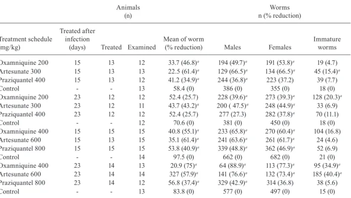
treated with 200 mg/kg or 400 mg/kg oxamniquine, or with 300 mg/kg or 600 mg/kg artesunate, 15 or 23 days after infection, showed a significant statistical reduc-tion in the number of collected male and female worms. Treatment with praziquantel resulted in significant re-duction only in the number of male worms, about 40% in reduction when doses of 400 mg/kg or 800 mg/kg were administered 23 or 15 days post-infection, respectively.
The percentage of immature worms collected reached a maximum of 2.4% in untreated mice of the control group. Treatment with 300 mg/kg artesunate, 15 days after infec-tion or 600 mg/kg 23 days after infecinfec-tion, significantly increased the percentage of immature worms (15.4% and 40.4%, respectively). Treatment with 200 mg/kg or 400 mg/kg oxamniquine, 23 days post-infection, significantly increased the percentage of recovered immature worms (20.3% and 34.9%, respectively). When treatment with 400 mg/kg oxamniquine was administered 15 days after infection, there was a significant statistical increase of 16.8% in the percentage of immature worms (Table II).
In the experiments using praziquantel and artesunate in monotherapy or in association, it was observed a sig-nificant reduction in the mean number of worms collect-ed by perfusion, except for the groups treatcollect-ed with 400 mg/kg praziquantel, 14 or 21 days after infection. When treatments performed in monotherapy were compared
with those ones using drugs in association, the results re-lated to decrease in the average of worms were similar. Praziquantel used in monotherapy significantly reduced the percentage of fourth stage worms (400 mg/kg and 800 mg/kg administered 21 days after infection); artesunate in monotherapy was more effective reducing the number of worms (3rd and 4th stages), when used at the doses of 300 mg/kg or 600 mg/kg, 21 days after infection. It was observed a significant increase in the number of young worms, which were not identified due to intestinal dam-ages in animals treated with artesunate in monotherapy or with other drug in association, but not in treatment schedules with only praziquantel (Table III).
TABLE II
Adult worm burden recovered from mice infected with 200 Schistosoma mansoni cercariae treated with single dose by oral route as monotherapy and submitted to euthanasia (treated and control groups) 42 days after treatment
Animals (n)
Worms n (% reduction)
Treatment schedule (mg/kg)
Treated after infection
(days) Treated Examined
Mean of worm
(% reduction) Males Females
Immature worms
Oxamniquine 200 15 13 12 33.7 (46.8)a 194 (49.7)a 191 (53.8)a 19 (4.7)
Artesunate 300 15 13 13 22.5 (61.4)a 129 (66.5)a 134 (66.5)a 45 (15.4)a
Praziquantel 400 15 13 12 41.2 (34.9)a 244 (36.8)a 223 (37.2) 39 (7.7)
Control - - 13 58.4 (0) 386 (0) 355 (0) 18 (0)
Oxamniquine 200 23 12 12 52.4 (25.7) 228 (39.6)a 273 (39.3)a 128 (20.3)a
Artesunate 300 23 12 11 43.7 (43.2)a 200 ( 47.5)a 248 (44.9)a 33 (6.9)
Praziquantel 400 23 12 12 52.4 (25.7) 277 (27.3) 282 (37.8)a 70 (11.1)
Control - - 12 70.6 (0) 381 (0) 450 (0) 18 (0)
Oxamniquine 400 15 15 15 40.8 (55.1)a 233 (65.8)a 270 (60.4)a 104 (16.8)
Artesunate 600 15 13 15 35.1 (61.4)a 241 (63.6)a 261 (61.7)a 24 (4.6)
Praziquantel 800 15 15 15 53.8 (40.9)a 339 (48.8)a 362 (46.9)a 52 (6.9)
Control - - 14 97.5 (0) 662 (0) 682 (0) 21 (0)
Oxamniquine 400 23 14 13 20.9 (75)a 64 (88.9)a 113 (77.3)a 95 (34.9)a
Artesunate 600 23 14 14 327 (57.9)a 141 (76.6)a 132 (73.4)a 185 (40.4)a
Praziquantel 800 23 14 12 56.8 (37.4)a 329 (42.9)a 314 (36.8) 38 (5.6)
Control - - 13 83.8 (0) 577 (0) 497 (0) 15 (0)
a: statistically significant difference in relation to control (p ≤ 0.05).
TABLE III
Distribution of young worms recovered from mice infected with 200 Schistosoma mansoni cercariae treated with single dose by oral route as monotherapy and combination of drugs and submitted to euthanasia
(treated and control groups) five days after treatment
Animals (n)
Distribution of schistosomules per evolutive stage
(%)
Treatment schedule (mg/kg)
Treated after infection
(days) Treated Examined
Mean of worm
(% reduction) 1 2 3 4 5 6 NI
Praziquantel 400 14 11 10 29.3 (32.6) 0.3 5 24 68 0.3 0 1.6
Praziquantel 800 14 11 11 23.5 (46)a 0.4 4.7 31 54.3a 6.6 0 3.1
Artesunate 300 14 11 10 17.9 (58.9)a 1.2 8.7 16.9 a53.5a 0.6 0 19.2a
Artesunate 600 14 11 9 7.7 (82.3)a 1.4 20.3a 7.2 a 34.8a 1.4 0 34.8a
Praziquantel 400 + artesunate 300 14 12 12 9.8 (77.5)a 2.5 16.9 29.7 a42.4a 0 0 8.5a Praziquantel 800 + artesunate 600 14 12 11 9.1(79.1)a 1 5.9 18.8 55.4a 1 0 17.8a
Control - - 12 43.5 (0) 0.6 3.8 18.6 73.9 2.3 0 0.8
Praziquantel 400 21 11 10 57.6 (5.6) 0 1.7 3.8 34.9 44.4 13.7a 1.4
Praziquantel 800 21 11 11 36.0 (41)a 0 0.3 9.6 34.1 45.4 9.6a 0.8
Artesunate 300 21 11 11 26.9 (55.9)a 0 0.3 1.4 10.1 29.4 14.5a 44.3a
Artesunate 600 21 12 12 20.6 (66.2)a 0 0.8 0 15.4 35.2 4.9a 43.7a
Praziquantel 400 + artesunate 300 21 12 11 29.3 (52)a 0 0.9 3.4 27 27.6 4.7a 36.6a Praziquantel 800 + artesunate 600 21 12 11 19.2 (68.5)a 0 0.5 4.3 19.9 38.9 8.1a 28.4a
Control - - 11 61 (0) 0 0.3 3.3 21.6 37.9 35.2 1.8
a: statistically significant difference in relation to control (p ≤ 0.05); NI: schistosomules with damaged intestines that did not
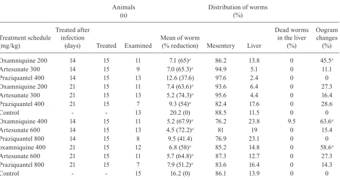
TABLE IV
Results obtained using mice experimentally infected with 50 Schistosoma mansoni cercariae treated
by oral route single dose as monotherapy 14 or 21 days after infection and sacrificed 100 days after infection
Animals (n)
Distribution of worms (%)
Treatment schedule (mg/kg)
Treated after infection
(days) Treated Examined
Mean of worm
(% reduction) Mesentery Liver
Dead worms in the liver
(%)
Oogram changes (%)
Oxamniquine 200 14 15 11 7.1 (65)a 86.2 13.8 0 45.5a
Artesunate 300 14 15 9 7.0 (65.3)a 94.9 5.1 0 11.1
Praziquantel 400 14 15 13 12.6 (37.6) 97.6 2.4 0 0
Oxamniquine 200 21 15 11 7.4 (63.6)a 93.6 6.4 0 27.3
Artesunate 300 21 15 13 5.2 (74.3)a 95.6 4.4 0 16.4
Praziquantel 400 21 15 7 9.3 (54)a 82.4 17.6 0 28.6
Control - - 13 20.2 (0) 88.5 11.5 0 0
Oxamniquine 400 14 15 11 5.2 (67.9)a 76.2 23.8 9.5 63.6a
Artesunate 600 14 15 13 4.5 (72.2)a 81 19 0 15.4
Praziquantel 800 14 15 8 9.5 (41.4) 76.9 23.1 0 0
oxamniquine 400 21 15 12 6.8 (58)a 85.2 14.8 0 58.6a
Artesunate 600 21 15 11 5.7 (64.8)a 87.3 12.7 0 27.3
Praziquantel 800 21 15 7 7.9 (51.2)a 83.6 16.4 0 14.3
Control - - 15 16.2 (0) 86.1 13.9 0 0
a: statistically significant difference in relation to untreated control (p ≤ 0.05).
It is important to emphasise that even performing perfusion 100 days after infection, a part of the recov-ered worms in treated groups (400 mg/kg oxamniquine and 600 mg/kg artesunate) remained small and with fragile aspect. Most of the females were with a clear-looking intestine, seeming to be female immature worms. In the group treated with artesunate (600 mg/ kg, 21 days post-infection) 16 out of 34 (47.1%) females presented immature characteristics, whereas 14 out of 36 (38.9%) females seemed to be immature in the group treated 14 days after infection. In the group treated with oxamniquine (400 mg/kg, 21 days post-infection) 22 out of 49 (44.9%) females showed an immature aspect, while eight out of 28 (28.6%) females showed an immature as-pect in the group treated 14 days after infection.
DISCUSSION
The asynchronous development of S. mansoni at the pre-patent phase was clearly observed in this study and the distribution of the immature worms recovered in the untreated control groups was similar to that reported by Barbosa et al. (1978).
These results here presented show that the drug in-terferes in the worm development, slowing or blocking the development of parasites, since significant percent-ages of worms with an immature aspect were detected up to 100 days after infection. This original finding has important implications in the fields of pathology, diag-nosis and epidemiology of schistosomiasis.
Oogram alteration was observed in worms of the group treated with oxamniquine (200 mg/kg or 400 mg/
kg), 14 or 21 days after infection, and submitted to eu-thanasia 42 days post-infection, indicating that treatment interfered with sexual maturation of the worms, since immature females were recovered.
In the animals treated with 300 mg/kg or 600 mg/ kg artesunate, 14 or 21 days post-infection, a small per-centage of oogram changes could be observed, although not statistically significant, is of relevance. According to Araújo et al. (1999), treatment with artesunate, at the postural phase, is capable of interrupting egg-laying only temporarily.
Praziquantel and derivatives of artesunate shown to be effective in different stages of worm development, be-ing suggested that these drugs would be more efficacious when administered in association than in monotherapy (Inyang-Etoh et al. 2008, Abdul-Ghani et al. 2009). Prom-ising results were obtained with praziquantel/artesunate in association, when administered in S. mansoni and
Schisto-soma japonicum infected mice, presenting immature and
The experimental chemotherapy at the pre-postural phase of S. mansoni is a field of great importance for de-velopment of antischistosomal drugs, due to its potential to eliminate the parasitism before lying of eggs, which cause the major injuries responsible for the disease - the granulomatous inflammatory reactions (Enk et al. 2008, Coelho et al. 2009). Thus, experimental chemotherapy is of the utmost importance and deserves a greater number of studies in this direction.
Worms treated at the early phase of infection (un-fertile worms), 100 days post-chemotherapy, could maintain an immunological response, as well as the presence of circulating antigens in blood, even in ab-sence of eggs in the faeces.
These findings represent a major cause of confusion regarding the interpretation of schistosomiasis mansoni diagnosis: the absence of eggs in the faeces, but mainte-nance of specific immune response, the presence of circu-lating antigens and genetic material of S. mansoni worms (detected by polymerase chain reaction techniques).
The results obtained allow us to conclude that the schistogram method used in this study proved to be a useful tool for experimental evaluation of the activity of antischistosomal drugs, at the early stages of infec-tion, besides being a good model to investigate which of the evolutive stages are more sensitive to antischis-tosomal drugs.
REFERENCES
Abdul-Ghani R, Loutfy N, Sahn AE, Hassan A 2009. Current che-motherapy arsenal for schistosomiasis mansoni: alternatives and challenges. Parasitol Res 104: 955-965.
Araújo N, Kohn A, Katz N 1999. Therapeutical evaluation of the arte-sunate in experimental Schistosoma mansoni. Rev Soc Bras Med Trop 32: 7-12.
Barbosa MA, Pellegrino J, Coelho PMZ, Sampaio IBM 1978. Quantita-tive aspects of the migration and evoluQuantita-tive asynchronism of Schis-tosoma mansoni in mice. Rev Inst Med Trop S Paulo 20: 121-132.
Bogliolo L 1959. Schistosomiasis mansoni. Pathology. Rev Bras Ma-lariol Doenças Trop 11: 359-424.
Botros S, Pica-Mattoccia L, William S, El-Lakkani N, Cioli D 2005. Effect of praziquantel on the immature stages of Schistosoma haematobium. Int J Parasitol 35: 1453-1457.
Coelho PMZ, Enk MJ, Katz N 2009. Treatment of clinical schisto-somiasis at the prepatent phase: an option? Trends Parasitol 25: 299-300.
De Clercq D, Vercruysse J, Verlé P, Kongs A, Diop M 2000. What is the effect of combining artesunate and praziquantel in the treatment of Schistosoma mansoni infections? Trop Med Inter Health 5: 744-746.
Enk MJ, Katz N, Coelho PMZ 2008. A case of Schistosoma mansoni infection treated during the prepatent period. Natur Clin Prac Gastroenterol Hepato 15: 112-115.
Faust EC, Jones CA, Hoffman WA 1934. Studies on schistosomiasis mansoni in Puerto Rico. III - Biological studies. 2. The mam-malian phase of the life cycle. Puerto Rico J Publ Healt Trop Med 10: 133-196.
Grandière-Pérez L, Ansart S, Paris L, Faussart A, Jaureguiberry S, Grivois JP, Klement E, Bricair F, Danis M, Caumes E 2006. Ef-ficacy of praziquantel during the incubation and invasive phase of Schistosoma haematobium schistosomiasis on 18 travelers. Am J Trop Med Hyg 74: 814-818.
Inyang-Etoh PC, Ejezie GC, Useh MF, Inyang-Etoh EC 2008. Effi-cacy of a combination of praziquantel and artesunate in the treat-ment of urinary schistosomiasis in Nigeria. Trans R Soc Trop Med Hyg 103: 38-44.
Lu SH, Yan XL, Li SW, Wu LJ, Shi JF, Liu X, Yan XH, Yang MJ, Lou LJ, Kumagai T, Wen LY, Ohta N 2004. Prophylactic effect of artesunate against experimental infection of Schistosoma mansoni. Chinese Journal of Parasitology & Parasitic Diseases 22: 20-23.
Pellegrino J, Katz N 1968. Experimental chemotherapy of Schisto-soma mansoni. Adv Parasitol 6: 233-291.
Pellegrino J, Siqueira AF 1956. Técnica de perfusão para colheita de Schistosoma mansoni em cobaias experimentalmente infectadas. Rev Bras Malariol Doencas Trop 8: 589-597.
Pellegrino J, Oliveira CA, Faria J, Cunha AS 1962. New approach to the screening of drugs in experimental schistosomiasis mansoni in mice. Amer J trop Med Hyg 11: 201-215.
Pica-Mattoccia L, Cioli D 2004. Sex and stage-related sensitivity of Schistosoma mansoni to in vivo and in vitro praziquantel treat-ment. Int J Parasitol 34: 527-533.
Sabah AA, Fletcher C, Webbe G, Doenhoff MJ 1986. Schistosoma mansoni: chemotherapy of infections of different ages. Exp Para-sitol 61: 294-303.
Shaohong L, Kumagai T, Qinghua A, Xiaolan Y, Ohmae H, Yabu Y, Sewen L, Liyong W, Maruyama H, Ohta N 2006. Evaluation of the anthelmintic effects of artesunate against experimental Schis-tosoma mansoni infection in mice using different treatment pro-tocols. Parasitol Int 55: 63-68.
Silva LM, Menezes RMC, Oliveira SA, Andrade ZA 2003. Chemo-therapeutic effects on larval stages of Schistosoma mansoni dur-ing infection and re-infection of mice. Rev Soc Bras Med Trop 36: 335-341.
Utzinger J, Chollet J, Tu Z, Xiao S, Tanner M 2002. Comparative study of the effects of artemether and artesunate on juvenile and adult Schistosoma mansoni in experimentally infected mice. Trans R Soc Trop Med Hyg 96: 318-323.
Utzinger J, Chollet J, You J, Mei J, Tanner M, Xiao S 2001. Effect of combined treatment with praziquantel and artesunate on Schis-tosoma japonicum and SchisSchis-tosoma mansoni in experimentally infected animals. Acta Trop 80: 9-18.
Warren S 1968. Pathophysiology and pathogenesis of hepatosplenic schistosomiasis mansoni. Bull N Y Acad Med 44: 280-294.