Sílvia Vilares Conde
FUNCTIONAL SIGNIFICANCE OF ADENOSINE IN
CAROTID BODY CHEMOSENSORY ACTIVITY IN
CONTROL AND CHRONICALLY HYPOXIC
ANIMALS
Dissertation presented to obtain the PhD degree in
“Ciências da Vida – Especialidade Farmacologia” at the Faculdade de Ciências
Médicas, Universidade Nova de Lisboa
and
Biotecnología: Aplicaciones Biomédicas at the Facultad de Medicina,
Universidad de Valladolid
Supervised by Profs. Constancio Gonzalez, Emília Monteiro and Ana
Realizado com o apoio da Fundação para a Ciência e Tecnologia,
SFRH/BD/14178/2003 financiada pelo POCI 2010 no âmbito da Formação
This work:
Originated the following publications:
- Conde S.V., Obeso A., Vicario I., Rigual R., Rocher A. and Gonzalez C.
(2006), Caffeine inhibition of rat carotid body chemoreceptors is mediated by
A2A and A2B adenosine receptors, J. Neurochem., 98, 616-628. (Chapter 3)
- Conde S.V. and Monteiro E.C. (2006), Activation of nicotinic ACh receptors
with α4 subunits induces adenosine release at the rat carotid body, Br. J. Pharmacol., 147, 783-789. (Chapter 2)
- Conde S.V. and Monteiro E.C. (2004), Hypoxia induces adenosine release
from the rat carotid body, J. Neurochem., 89 (5), 1148-1156. (Chapter 1)
- Conde S.V. and Monteiro E.C. (2003), Adenosine-acetylcholine interactions at
the rat carotid body, In: “Chemoreception: From cellular signalling to functional
plasticity”, JM Pequinot et al. (Eds), Klumer Academic Press London, 305-311.
(Chapter 2)
Won the following awards:
- 1st Prize “De Castro-Heymans–Neil Award” awarded by ISAC (International
Society for Arterial Chemoreception) to the best work presented by a Young
Researcher each triennial ISAC Meeting, 2002
- Pfizer Honor Young Researcher Prize awarded by the Portuguese Society of
INDEX
Page
List of Figures and Tables………... VII
Acknowledgements ……… XII
Abbreviations used………. XIV
Glossary………. XVII
Abstract ………. XX
Resumo……….. XXI
Resumen……… XXVI
1. GENERAL INTRODUCTION 1
1.1. The carotid body: morphology 4 1.2. Chemotransduction mechanisms at the carotid body: coupling stimulation and secretion 5
1.3. Function and control of the carotid body in resting conditions and in hypoxia 8 1.4. Function and control of the carotid body in hypercapnia and acidosis…… 11
1.5. Neurotransmission in the carotid body: role of adenosine, dopamine, ACh and ATP 13 1.5.1. Adenosine……….. 13
1.5.1.1. Metabolic pathways of adenosine formation and release……… 14
1.5.1.2. Adenosine receptors……….. 15
1.5.1.3. Physiological role of adenosine……….. 17
1.5.1.4. Adenosine in chemoreception in the carotid body ………... 18
1.5.2. Dopamine……….. 20
1.5.2.1. Catecholamines synthesis and metabolism……….. 20
1.5.2.2. Dopamine receptors……….. 22
1.5.2.3. Dopamine effects on chemoreception and ventilation…………. 23
1.5.3. ATP………. 26
1.5.3.1. Role of ATP in chemoreception in the carotid body………. 27
1.5.4. Acetylcholine……….. 28
1.5.4.1. Acetylcholine receptors………. 29
1.5.4.2. Effects of ACh on carotid body chemoreception………... 31
1.6.2. Neurochemical changes in the carotid body in response to
chronic sustained hypoxia………...
36
1.6.3. Chemoreceptor function blunting after prolonged hypoxia………... 39
1.7. Caffeine 40 1.7.1. Effects of acute caffeine treatment on ventilation………. 41
1.7.2. Chronic treatment with caffeine………... 42
2. GENERAL AND SPECIFIC AIMS 45 3. CHAPTER 1 – STUDY OF THE EFFECT OF HYPOXIA ON THE RELEASE OF ADENOSINE FROM THE RAT CAROTID BODY 46 3.1 Introduction and aim 46 3.2. Material and methods 46 3.2.1. Tissue preparation and experimental conditions……….. 46
3.2.2. Characterization of the content and release of adenosine in rat carotid bodies……… 47
3.2.3. Characterization of the release of adenosine from SCG and arterial tissue ……… 47
3.2.4. Effect of adenosine transporter inhibitors……….. 48
3.2.5. Metabolic pathways of adenosine production at the rat carotid body ………... 48
3.2.6. Nucleotide extraction ……… 48
3.2.7. HPLC analysis ……….. 49
3.2.8. Drugs……….. 50
3.2.9. Data analysis……….. 50
3.3. Results 51 3.3.1. Adenosine quantification ………. 51
3.3.2. Adenosine content and release from carotid body during hypoxia 52
3.3.3. Adenosine released by SCG and arterial tissue………... 53
3.3.4. Effect of adenosine transport inhibitors………. 55
3.3.5. Metabolic pathways of adenosine production at the rat carotid body……… 55
3.4. Discussion 56
4. CHAPTER 2 – EFFECT OF ACTIVATION OF NICOTINIC ACH
RECEPTORS WITH α4 SUBUNITS ON ADENOSINE RELEASE AT THE
RAT CAROTID BODY 61
4.2. Material and methods 61
4.2.1. Animals and surgical procedures……… 61
4.2.2. Effect of ACh nicotinic receptor agonists on adenosine released from carotid body……….. 62
4.2.3. Effect of ACh nicotinic receptor antagonists on adenosine released from carotid body……….. 62
4.2.4. Pharmacological demonstration of the involvement of neuronal nicotinic acetylcholine receptors………. 63
4.2.5. Effect of extracellular ATP catabolism inhibitor on the release of adenosine evoked by nicotine……… 63
4.2.6. Nucleotide extraction and HPLC analysis………. 63
4.2.7. Drugs and chemicals……… 63
4.2.8. Data analysis……….. 64
4.3. Results 64 4.3.1. Demonstration of the involvement of neuronal nicotinic acetylcholine receptors and characterization of nicotinic receptors that modulates the release of adenosine from rat carotid body ………... 64
4.3.2. Effect of inhibition of ATP catabolism on the release of adenosine evoked by nicotine on carotid body……… 68
4.4. Discussion 69 5. CHAPTER 3 – ACUTE EFFECTS OF CAFFEINE ON RAT CAROTID BODY CHEMORECEPTOR FUNCTION 73 5.1. Introduction and aim 73 5.2. Material and methods 73 5.2.1. Animals and surgical procedures……… 73
5.2.2. Labelling of catecholamines stores: release of 3 H-CA……… 74
5.2.3. Recording of carotid sinus nerve activity………... 75
5.2.4. Carotid body cell dissociation and culture, and immunocytochemistry……….. 76
5.2.5. Drugs and chemicals……… 77
5.2.6. Data analysis………. 78
5.3. Results 78 5.3.1. Effect of caffeine on the basal release of 3H-CA………. 78
5.3.3. Pharmacological characterisation of adenosine receptors
involved in the inhibitory effect of caffeine on the release of 3H-CA from
the carotid body………. 82
5.3.4. Immunocytochemical demonstration of A2B adenosine receptors
in chemoreceptor cells………. 86
5.3.5. Actions of caffeine on the carotid sinus nerve activity: a mixed
A2A and A2B receptors mediated effect……….. 87
5.4. Discussion 90
6. CHAPTER 4 – ADENOSINE MODULATES THE RELEASE OF
CATECHOLAMINES FROM RAT CAROTID BODY CHEMORECEPTORS
THROUGH AN INTERACTION BETWEEN D2 DOPAMINE RECEPTORS
AND A2B ADENOSINE RECEPTORS 95
6.1. Introduction and aim 95
6.2. Material and methods 95
6.2.1. Animals and surgical procedures……… 95
6.2.2. Labelling of catecholamines stores: release of 3
H-CA……… 96
6.2.3. Drugs and chemicals……… 96
6.2.4. Data analysis……….. 96
6.3. Results 97
6.3.1. Effect of NECA, an A2 agonist on the release of 3H-CA from rat
carotid body……….. 97
6.3.2. Reversion of the inhibitory effect of caffeine on the 3H-CA by a
D2 antagonist, sulpiride……… 98
6.3.3. Effect of D2 antagonists on the release of 3H-CA release by rat
carotid body and its potentiation with NECA……… 99
6.3.4. Inhibitory effect of the D2 agonist, propylnorapomorphine, on the
release of 3H-CA from carotid body and its reversion by NECA………... 102 6.3.5. Effect of ionomycin on the release of 3H-CA by carotid body
modified by norapropylapomorphine and NECA………. 104
6.4. Discussion 105
7 – CHAPTER 5 – EFFECT OF CHRONIC CAFFEINE INTAKE ON THE
CAROTID BODY CHEMOSENSORY ACTIVITY IN CONTROL AND
CHRONIC HYPOXIC RATS 112
7.1. Introduction and aim 112
7.2.1. Chronic caffeine intake in control and chronic hypoxic animals…. 112
7.2.2. Measurement of tissue catecholamines content……… 114
7.2.3. Labelling of CA stores to measure the rate of synthesis of 3H-CA 115 7.2.4. Endogenous release of ATP, adenosine and CA from carotid
body……… 116
7.2.5. Quantification of ATP by bioluminescence luciferine-luciferase
assay……….. 117
7.2.6. Western blot analysis of tyrosine hydroxylase expression in the
carotid body………... 117
7.2.7. Recording of carotid sinus nerve activity………... 118
7.2.8. Whole-body pletysmographic recordings of ventilatory
responses in response to hypoxia and hypercapnia ………. 118
7.2.9. Drugs and chemicals………. 120
7.2.10. Data analysis……… 120
7.3. Results 120
7.3.1. Effect of chronic caffeine intake on carotid body and superior
cervical ganglion weight in control and chronic hypoxic
rats……….. 121
7.3.2. Effect of chronic caffeine intake on CA content of the carotid
body in control and chronic hypoxic rats………... 122
7.3.3. Effect of chronic caffeine intake on CA synthesis and CA
turnover time in the carotid body of control and chronic hypoxic rats….. 124
7.3.4. Effect of chronic caffeine intake on CA content, CA synthesis
and CA turnover time in superior cervical ganglia of control and chronic
hypoxic rats ……….. 126
7.3.5. Effect of chronic caffeine intake on DA, ATP and adenosine
release evoked by acute hypoxia, from carotid body in control and
chronic hypoxic rats……….. 128
7.3.6. Effect of chronic caffeine intake on tyrosine hydroxylase
expression on the carotid body in control and chronic hypoxic rats……. 133
7.3.7. Effect of chronic caffeine intake on chemosensory activity in the
carotid sinus nerve evoked by hypoxia and hypercapnia in control and
chronic hypoxic rats………. 134
7.3.8. Effect of chronic caffeine intake on the ventilatory responses
induced by hypoxia and hypercania in control and chronic hypoxic rats. 137
7.4.1.Effects of chronic hypoxia on CB function………. 143
7.4.1.1. Effect of chronic hypoxia on morphology and
neurotransmitter dynamics in the carotid body………. 145
7.4.1.2. Effect of chronic hypoxia on carotid body output……… 146
7.4.2.Effects of chronic caffeine ingestion on rat CB function…………. 147
7.4.2.1. In control animals……… 147
7.4.2.2. In chronic hypoxic animals………. 148
8. GENERAL DISCUSSION 152
9. CONCLUSIONS 162
LIST OF FIGURES AND TABLES
Figures page
Figure 1 Hypoxia signalling pathway with the major adaptive responses to
acute and chronic hypoxia; 2
Figure 2 Respiratory and cardiovascular responses to carotid body activation; 3
Figure 3 Carotid artery bifurcation, cellular cluster of the carotid body and a
histological image of a slice of 10 µM of the carotid body; 4
Figure 4 Functional organization of carotid body (CB) chemoreceptors; 6
Figure 5 Transduction cascade of the hypoxic transduction mechanism in the
carotid body; 7
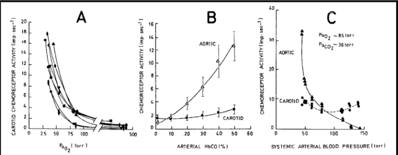
Figure 6 Graph that correlates the intensity of hypoxia with carotid sinus
nerve chemosensory activity; 9
Figure 7 Effect of several hypoxic intensities on minute ventilation (VE; l/min)
in a Nembutal-anesthetized cat before and after sectioning both carotid sinus
nerves; 10
Figure 8 The relationship between afferent chemosensory discharge and CO2
levels, recorded in carotid sinus nerve preparation; 12
Figure 9 Extra- and intracellular adenosine metabolism and the nucleotide
transporters that contribute to its release and uptake; 15
Figure 10 The pathways of synthesis and metabolism of dopamine (DA) in
humans; 21
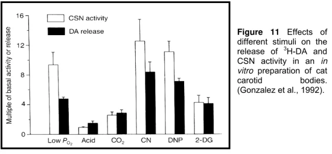
Figure 11 The effect of different stimuli on the release of 3H-DA and CSN activity in an in vitro preparation of cat carotid bodies; 24 Figure 12 Biosynthesis pathway of acetylcholine formation; 28
Figure 13 Staining of rat carotid body in control conditions and when rats
were exposed to 1 week of chronic hypoxia; 35
Figure 14 Caffeine effects on different biochemical targets in relation to its
levels in humans; 40
Figure 15 HPLC setup; 49
Figure 16 Chromatograms of adenosine obtained by reverse phase HPLC; 52
Figure 17 Effect of hypoxia (10% O2) on the content and release of adenosine
from the carotid bodies (CBs); 53
Figure 18 Time dependence of adenosine release in rat superior cervical
Figure 19 Effect of different hypoxic intensities on the release of adenosine
from superior cervical ganglia and arterial tissue (common carotid arteries); 54
Figure 20 Effects of nucleoside transport inhibitors, dipyridamole and NBTI,
on adenosine released by carotid bodies in normoxia and hypoxia; 55
Figure 21 Effect of an inhibitor of nucleoside transport systems, NBTI, and an
inhibitor of ecto-5’-nucleotidase, AOPCP, on adenosine release by carotid
bodies in normoxia and hypoxia; 56
Figure 22 Effect of acetylcholine on adenosine concentrations released from
rat carotid bodies in the presence of distinct concentrations of physostigmine; 65
Figure 23 Dose-response curves for the effects of nicotinic ACh receptor
agonists, cytosine, dimethylphenylpiperazinium (DMPP) and nicotine on
adenosine concentrations released from rat carotid bodies in normoxia; 65
Figure 24 Effects of nicotinic ACh receptor antagonists, α-bungarotoxin, d-tubocurarine, and di-hydro-β-erythroidine (DHβE) and of the allosteric inhibitor, mecamylamine, on the release of adenosine from rat CBs stimulated
by hypoxia; 67
Figure 25 Effect of the selective nicotinic receptor antagonist, di-hydro-β -erythroidine (DHβE) on the release of adenosine evoked by nicotine during
normoxia; 68
Figure 26 Effect of α,β-methylene ADP (AOPCP) on the release of adenosine
in CBs stimulated by 100 nM of nicotine in normoxia; 69
Figure 27 Setup for recording carotid sinus nerve chemosensory activity and
microphotography of carotid body-carotid sinus nerve preparation after
cleaning and digestion; 76
Figure 28 Effect of caffeine on the basal (normoxic) release of
catecholamines from the intact rat carotid body in vitro and its Ca2+
dependency; 79
Figure 29 Effect of 1mM of caffeine on the release of CA from rat carotid body
induced by hypoxia; 81
Figure 30 Effect of 1mM of caffeine on the release of CAs from rat carotid
body induced by 30 and 50 mM of extracellular K+; 82
Figure 31 Effects of 1 µM of NECA, an A2 adenosine receptor agonist, on the
release of 3H-CA from rat carotid body in basal and moderate hypoxic
conditions; 83
Figure 33 Effect of DPCPX (an A1 and A2B antagonist) and MRS1754 (an A2B
antagonist) on the release of 3H-CA evoked by 30 mM extracellular K+ in the
rat carotid body; 85
Figure 34 Immunocytochemical demonstration of A2B adenosine receptors in
dissociated CB cells in culture; 86
Figure 35 Effect of caffeine on the carotid sinus nerve activity elicited by acute
hypoxia (5% O2); 88
Figure 36 Kinetic and pharmacological analysis of the effects of caffeine on
the carotid sinus nerve response to hypoxia (5% O2); 89
Figure 37 Dose response curve for the effect of NECA, an adenosine A2
agonist, on the basal release of 3H-CA and effect of 1 µM of NECA on the
release evoked by 2% O2 from rat carotid body; 98
Figure 38 Effect of sulpiride (1 µM) on the release of 3H-CA evoked by 30 mM extracellular K+ and by 10% O2 and reversion of the inhibitory effect of caffeine
on the release of 3H-CA from rat carotid body; 99
Figure 39 Dose-response curves for the effect of domperidone and
haloperidol, D2 dopamine antagonists, on the release of 3H-CA from rat CB; 100 Figure 40 Potentiation of haloperidol, a D2 dopamine receptor antagonist,
effect on 3H-CA release from carotid body by NECA, an A2 adenosine receptor
agonist in normoxic conditions; 101
Figure 41 Effect of propylnorapomorphine (N-Apo) on the release of 3H-CA in normoxic conditions and in response to moderate hypoxia, and attenuation of
the inhibitory effect of N-Apo by NECA on the basal and low intensity stimulus
evoked release of 3H-CA from carotid body; 103
Figure 42 Inhibitory effect of propylnorapomorphine, a D2 agonist, on the
ionomycin evoked release of 3H-CA from CB and reversion of the inhibitory
effect by NECA; 105
Figure 43 Mechanisms of interaction between A2B and D2 receptors in
chemoreceptor cells that modulate the release of CA from rat carotid body; 109
Figure 44 Time courses and the paradigms to which rats were submitted and
the definition (name) applied to the distinct groups of animals; 113
Figure 45 Equipment used to produce a hypoxic atmosphere and the
chamber into which animals were inserted to be submitted to chronic hypoxia; 114
Figure 46 Effect of chronic caffeine intake, chronic hypoxia and both
treatments applied conjunctly on carotid body and superior cervical ganglion
Figure 47 Effect of chronic caffeine intake, chronic hypoxia and effect of both
treatments applied together on the levels of catecholamines and in the
dopamine/ norepinephrine ratios in the rat carotid body; 123
Figure 48 Effect of chronic caffeine intake, chronic hypoxia and both on the
synthesis rate of catecholamine, and accumulation of the natural precursor
tyrosine, and turnover time in the CB, in normoxic animals and in rats
submitted to different paradigms; 125
Figure 49 Effect of chronic caffeine intake, chronic hypoxia and effect of both
treatments applied together on the levels of catecholamines and in the
dopamine/norepinephrine ratio in rat superior cervical ganglion; 126
Figure 50 Effects of chronic caffeine intake, chronic hypoxia and both
treatments on the synthesis rate of catecholamines, and accumulation of the
natural precursor tyrosine and turnover time in the SCGs in normoxic animals
and in rats submitted to the distinct paradigms; 127
Figure 51 Effects of chronic caffeine intake, chronic hypoxia and effect of both
treatments applied together on the basal endogenous release of DA, ATP and
adenosine from rat carotid body; 129
Figure 52 Effect of chronic caffeine intake on the endogenous release of
dopamine from CB in response to acute hypoxia (7% and 2% O2) in normoxic
and chronically hypoxic rats; 130
Figure 53 Effect of chronic caffeine intake on the release of ATP from the
carotid body in response to acute hypoxia (7% and 2% O2) in normoxic and
chronic hypoxic rats; 131
Figure 54 Effect of chronic caffeine intake on the release of adenosine from
carotid body in response to acute hypoxia (7% and 2% O2) in normoxic and
chronically hypoxic rats; 132
Figure 55 Tyrosine hydroxylase (TH) immunoreactivity in the carotid body and
in superior cervical ganglion in control rats and in rats submitted to distinct
treatments: chronic caffeine intake, chronic hypoxic exposure and chronic
caffeine + chronic hypoxia; 134
Figure 56 Effect of chronic caffeine intake on the carotid sinus nerve activity
in normoxic and chronically hypoxic rats; 136
Figure 57 Effect of 8 and 15 days exposure to hypoxia (12% O2) on the
ventilatory responses to acute hypoxia (10 and 7% O2) and hypercapnia (5%
Figure 58 Effect of chronic caffeine intake on minute volume (VE) in response
to acute hypoxias of several intensities (12%, 10% and 7% O2) in normoxic
animals and in animals submitted to chronic hypoxia for 8 and 15 days; 140
Figure 59 Effect of chronic caffeine intake on minute volume in response to
hypercapnia (5%CO2) in normoxic rats and in rats exposed to an atmosphere
of 12% O2 for 8 and 15 days; 141
Figure 60 The possible mechanism of action of neurotransmitters involved in
the chemosensory response to acute hypoxia in the carotid body. 156
Tables page
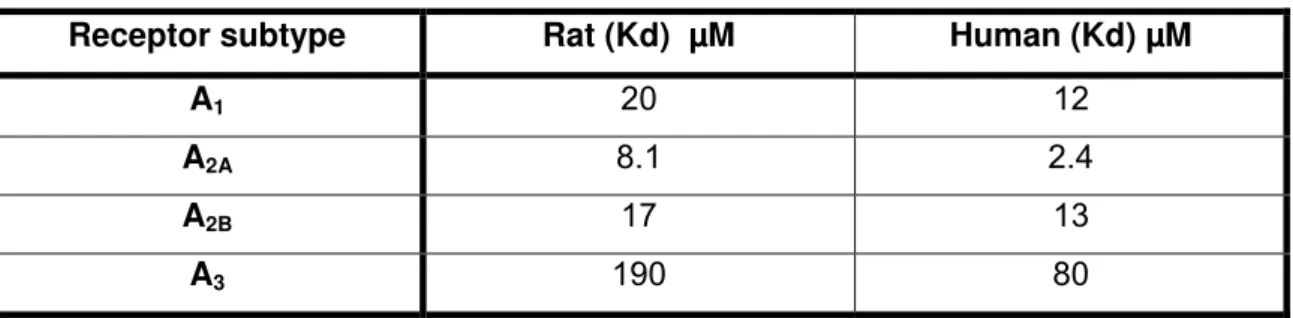
Table 1 G protein coupling of the four adenosine receptor subtypes; 16
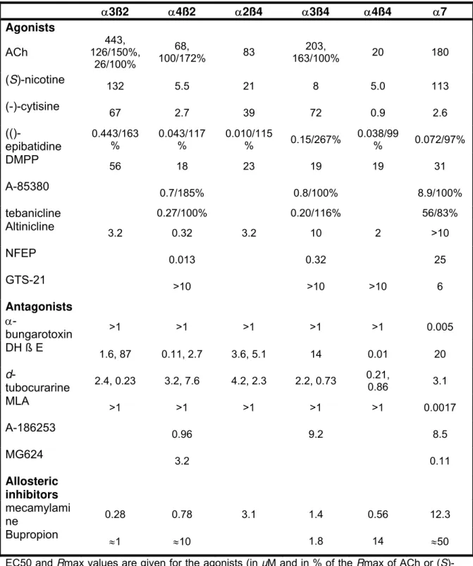
Table 2 Functional characteristics of selected nicotinic AChR ligands at
neuronal nicotinic ACh receptors; 30
Table 3 Potency of caffeine at rat and human adenosine receptor subtypes; 41
Table 4 Efficacy and potency of nicotinic ACh receptor agonists in stimulating
adenosine release at the carotid body; 66
Table 5 Efficacy and potency of nicotinic ACh antagonists in inhibiting the
release of adenosine in CBs stimulated by acute hypoxia; 67
Table 6 Effect of NECA on the efficacy and potency of haloperidol, a D2
antagonist, in stimulating the release of CA from rat carotid body; 102
Table 7 Effect of NECA on the efficacy and potency of propylnorapomorphine,
a D2 agonist, in inhibiting the release of CA from rat carotid body; 104 Table 8 Effects on neurochemical and physiological parameters that include
CB neurotransmitters, ATP, DA and adenosine dynamics (content, synthesis,
basal and evoked release), CSN activity and ventilation in rats submitted to
chronic caffeine intake (1g/l) during 15 days, in rats exposed a hypoxic
ACKNOWLEDGEMENTS
My first acknowledgment goes to Professor Emília Monteiro. It was she
who initiated me into the “chemoreception and hypoxic sensing” area some
years ago, who let me take my first steps in science, and without her this thesis
would never have been written. I would also like to thank her for all the support
and opportunities. She was also responsible for sending me to Valladolid to
meet Professor Constancio Gonzalez, giving me the opportunity to do part of
my PhD in his laboratory.
To Professor Constancio Gonzalez for all the knowledge, the scientific
discussions and for giving me the chance to learn so much “science” in his
laboratory. He has really contributed to my scientific journey in these last few
years, and to my personal development.
To Professor Ana Obeso for all the help in the laboratory, the advice and
because she was, so many times, my “Spanish mummy”.
My parents deserve a special acknowledgment - without them I would not
be here and this thesis would have never been done. Therefore to my parents,
and especially to my mother, for all the support and opportunities that they give
me in life. Thanks for being so supportive and for believing in me.
To my brothers and sister, Tiago, Pedro, António and Patrícia for all the
affection, friendship and all the funny “dinners”.
To Nuno, because without you, living in Spain for three and a half years
would probably have been much more difficult. Thanks!!!!
To all my friends, the Portuguese and Spanish ones, a special
acknowledgment. Thank you for being there when I needed you.
To “La Comuna” for all the friendship, support and good moments that
To all in the Department of Pharmacology, Faculty of Medical Sciences in
Lisbon, specially to Sofia and Joana for all the friendship, fellowship and for
being always available for me.
To all in my laboratory in the Faculty of Medicine in Valladolid, especially
to Jesus, Maria Llanos and Elena, for all the laughs, for all the sharing of good
and bad moments, for teaching me so many things, including Spanish,
particularly slang (Mamen this acknowledgment is for you!)
I would also like to thank the Fundação para a Ciência e Tecnologia
(FCT/MCTES) for giving me the PhD grant that allowed me to carry out this
work.
Finally, I have to express my gratitude to the Faculty of Medical Sciences
of the New University of Lisbon, to CEPR/FCT (Portugal), to the Department of
Biochemistry, Molecular Biology and Physiology of the Faculty of Medicine of
the University of Valladolid and to IBGM/CSIC (Spain) for all the financial
ABBREVIATIONS USED
5-HT- 5-hydoxytryptamine, serotonin
AADC - aromatic amino acid decarboxylase
AC – Adenylyl cyclase
ACh – Acetylcholine
AChE – Acetylcholinesterase
ADA – Adenosine deaminase
Ado – Adenosine
ADP- 5’-adenosine diphosphate
AK – Adenosine kinase
ANOVA – Analysis of variance
AMP – 5’-adenosine monophosphate
AOPCP - α,β-methylene ADP ATP – 5’-adenosine triphosphate
CA – Catecholamine
cAMP – cyclic 5’-adenosine monophosphate
CB - Carotid body
CGS 21680 –
2-p-(2-Carboxyethyl)phenethyl-amino-5’-N-ethylcaboxamido-adenosine hydrochloride
ChAT – Choline acetyltransferase
CNT - Concentrative nucleotide transport
COPD – Chronic obstructive pulmonary disease
CSH – Chronic sustained hypoxia
CSN - Carotid sinus nerve
Cyt - Cytisine
DA – Dopamine
DBH – Dopamine–ß–hydroxylase
DH ßE – dihydro-ß-erythroidine
DMEM – Dulbecco’s modified Eagle’s medium
DMPP - dimethylphenylpiperazinium
DMSO – dimethylsulfoxide
E - Epinephrine
EHNA – erythro-9-(2-hydroxy-3-nonyl)adenine
Emax – maximal increase
ENT – Equilibrative nucleoside transport
ENT1 – Type 1 equilibrative nucleoside transport system
ENT2 – Type 2 equilibrative nucleoside transport system
GABA - Gamma aminobutyric acid
HE-NECA – 2-hexynyl-NECA
HPLC – High performance liquid chromatography
HVR – Hypoxic ventilatory response
L-DOPA – 3, 4,dihydroxy-L-phenylalanine
ME – met-encephalin
MRS 1754 –
8-4-{[(4-cyanophenyl)carbamoylmetyl]-oxy}phenyl)-1,3-di(n-propyl)xanthine
N-Apo – Propylnorapomorphine
NBTI - nitrobenzylthioinosine
NE – norepinephrine
NECA – 5’-(N-ethylcarboxamido)adenosine
Nic - Nicotine
NO – Nitric oxide
NT – Neurotransmitter
OSA – Obstructive sleep apnoea
PaO2 – Partial arterial pressure of oxygen
PaCO2 - Partial arterial pressure of carbon dioxide
PBS – Phosphate buffered saline
PG – Petrosal ganglion
PO2 – Partial pressure of oxygen
RT-PCR – Real time polymerase chain reaction
SAH –S-adenosylhomocysteine
SAHH –S-adenosylhomocysteine hydrolase
SAM – S-adenosylmethionine
SEM – Standard error of the mean
SCG – Superior cervical ganglion
SP – Substance P
TH – Tyrosine hydroxylase
TTX – Tetradotoxin
UDP - uridine 5'-diphosphate
VAChT – Vesicular acetylcholine transporter
VAH – Ventilatory acclimatisation to hypoxia
VEGF – Vascular endothelial growth factor
GLOSSARY
1 mmHg = 1 Torr
α-bungarotoxin – Selective antagonist of nicotinic ACh receptors with α7 subunits AOPCP – Inhibitor of 5’-ectonucleotidase, inhibits ATP extracellular catabolism
Caffeine – Non-selective antagonist of adenosine receptors
CGS 21680 – Selective A2A adenosine receptor agonist
Cytisine – Selective nicotinic ACh receptor agonist
DHßE – Selective nicotinic ACh receptor antagonist
Domperidone - Selective antagonist of D2 dopamine receptors
DMPP - Selective nicotinic ACh receptor agonist
DPCPX – Agonist of A1 adenosine receptors in nM concentrations, and also of
A2B adenosine receptors in µM concentrations
d-tubocurarine - Non selective nicotinic ACh receptor antagonist
Dipyridamole – Inhibitor of equilibrative nucleotide transport system
EC50 - drug concentration that produces 50% of maximal effect
EHNA – Inhibitor of adenosine deaminase, inhibits adenosine deamination
Haloperidol – Selective antagonist of D2 dopamine receptors
HE-NECA - Selective A2A adenosine receptor agonist
IC50 - drug concentration that produces 50% of maximal inhibition
Ionomycin - Alters cell permeability, producing an increase in intracellular Ca2+
Mecamylamine – Non-selective allosteric inhibitor of nicotinic ACh receptors
MRS 1754 – Selective A2B adenosine receptor antagonist
NBTI - Inhibitor of equilibrative nucleotide transport system
NECA – Non-selective agonist of A2 adenosine receptors
Nicotine - Selective nicotinic ACh receptor agonist
Propylnorapomorphine – Selective D2 dopamine receptor agonist
SCH 58621 - Selective A2A adenosine receptor antagonist
Sulpiride – D2-like dopamine receptor antagonist
ZM 241385 – Selective antagonist of A2A and A2B adenosine receptors, with
ABSTRACT
Carotid bodies (CB) are peripheral chemoreceptor organs sensing
changes in arterial blood O2, CO2 and pH levels. Hypoxia and acidosis or
hypercapnia activates CB chemoreceptor cells, which respond by releasing
neurotransmitters in order to increase the action potential frequency in their
sensory nerve, the carotid sinus nerve (CSN). CSN activity is integrated in the
brainstem to induce a fan of cardiorespiratory reflex responses, aimed at
normalising the altered blood gases. Exogenously applied adenosine (Ado)
increases CSN chemosensory activity inducing hyperventilation through
activation of A2 receptors. The importance of the effects of adenosine in
chemoreception was reinforced by data obtained in humans, in which the
intravenous infusion of Ado causes hyperventilation and dyspnoea, an effect
that has been attributed to the activation of CB because Ado does not cross
blood-brain barrier and because the ventilatory effects are higher the closer to
the CB it is injected.
The present work was performed in order to establish the functional
significance of adenosine in chemoreception at the carotid body in control and
chronically hypoxic rats. To achieve this objective we investigated: 1) The
release of adenosine from a rat carotid body in vitro preparation in response to
moderate hypoxia and the specificity of this release. We also investigated the
metabolic pathways of adenosine production and release in the organ in
normoxia and hypoxia; 2) The modulation of adenosine/ATP release from rat
carotid body chemoreceptor cells by nicotinic ACh receptors; 3) The effects of
caffeine on peripheral control of breathing and the identity of the adenosine
receptors involved in adenosine and caffeine effects on carotid body
chemoreceptors; 4) The interactions between dopamine D2 receptors and
adenosine A2B receptors that modulate the release of catecholamines (CA) from
the rat carotid body; 5) The effect of chronic caffeine intake i.e. the continuous
blockage of adenosine receptors thereby simulating a caffeine dependence, on
the carotid body function in control and chronically hypoxic rats. The
methodologies used in this work included: molecular biology techniques (e.g.
immunocytochemistry and western-blot), biochemical techniques (e.g.
methods), electrophysiological techniques (e.g. action potential recordings) and
ventilatory recordings using whole-body plethysmography.
It was observed that: 1) CB chemoreceptor sensitivity to hypoxia could
be related to its low threshold for the release of adenosine because moderate
acute hypoxia (10% O2) increased adenosine concentrations released from the
CB by 44% but was not a strong enough stimulus to evoke adenosine release
from superior cervical ganglia and arterial tissue; 2) Acetylcholine (ACh)
modulates the release of adenosine/5’-adenosine triphosphate (ATP) from CB
in moderate hypoxia through the activation of nicotinic receptors with α4 and ß2
receptor subunits, suggesting that the excitatory role of ACh in chemosensory
activity includes indirect activation of purinergic receptors by adenosine and
ATP, which strongly supports the hypothesis that ATP/adenosine are important
mediators in chemotransduction; 3) adenosine increases the release of CA from
rat CB chemoreceptor cells via A2B receptors; 4) the inhibitory effects of caffeine
on CB chemoreceptors are mediated by antagonism of postsynaptic A2A and
presynaptic A2B adenosine receptors indicating that chemosensory activity
elicited by hypoxia is controlled by adenosine; 5) The release of CA from rat CB
chemoreceptor cells is modulated by adenosine through an antagonistic
interaction between A2B and D2 receptors, for the first time herein described; 6)
chronic caffeine treatment did not significantly alter the basal function of CB in
normoxic rats assessed as the dynamics of their neurotransmitters, dopamine,
ATP and adenosine, and the CSN chemosensory activity. In contrast, the
responses to hypoxia in these animals were facilitated by chronic caffeine
intake because it increased the ventilatory response, slightly increased CSN
chemosensory activity and increased dopamine (DA) and ATP release; 7) In
comparison with normoxic rats, chronically hypoxic rats exhibited an increase in
several parameters: ventilatory hypoxic response; basal and hypoxic CSN
activity; tyrosine hydroxylase expression, CA content, synthesis and release;
basal and hypoxic adenosine release; and in contrast a normal basal release
and diminished hypoxia-induced ATP release; 8) Finally, in contrast to
chronically hypoxic rats, chronic caffeine treatment did not alter the basal CSN
chemosensory activity. Nevertheless, the responses to mild and intense
hypoxia, and hypercapnia, were diminished. This inhibitory effect of chronic
ventilation parameter in basal conditions and in response to acute hypoxic
challenges remained unaltered in rats exposed to chronic hypoxia.
We can conclude that adenosine both in acute and chronically hypoxic
conditions have an excitatory role in the CB chemosensory activity, acting
directly on adenosine A2A receptors present postsynaptically in CSN, and acting
presynaptically via A2B receptors controlling the release of dopamine in
chemoreceptor cells. We suggest that A2B -D2 adenosine / dopamine
interactions at the CB could explain the increase in CA metabolism caused by
chronic ingestion of caffeine during chronic hypoxia. It was also concluded that
adenosine facilitates CB sensitisation to chronic hypoxia although this effect is
RESUMO
Os corpos carotídeos (CB) são pequenos orgãos emparelhados
localizados na bifurcação da artéria carótida comum. Estes órgãos são
sensíveis a variações na PaO2, PaCO2, pH e temperatura sendo responsáveis
pela hiperventilação que ocorre em resposta à hipóxia, contribuindo também
para a hiperventilação que acompanha a acidose metabólica e respiratória. As
células quimiorreceptoras (tipo I ou glómicas) do corpo carotídeo respondem às
variações de gases arteriais libertando neurotransmissores que activam as
terminações sensitivas do nervo do seio carotídeo (CSN) conduzindo a
informação ao centro respiratório central. Está ainda por esclarecer qual o
neurotransmissor (ou os neurotransmissores) responsável pela sinalização
hipóxica no corpo carotídeo. A adenosina é um neurotransmissor excitatório no
CB que aumenta a actividade eléctrica do CSN induzindo a hiperventilação
através da activação de receptores A2. A importância destes efeitos da
adenosina na quimiorrecepção, descritos em ratos e gatos, foi reforçada por
resultados obtidos em voluntários saudáveis onde a infusão intravenosa de
adenosina em induz hiperventilação e dispneia, efeito atribuído a uma
activação do CB uma vez que a adenosina não atravessa a barreira
hemato-encefálica e o efeito é quanto maior quanto mais perto do CB for a
administração de adenosina.
O presente trabalho foi realizado com o objectivo de esclarecer qual o
significado funcional da adenosina na quimiorrecepção no CB em animais
controlo e em animais submetidos a hipoxia crónica mantida. Para alcançar
este objectivo investigou-se: 1) o efeito da hipóxia moderada sobre a libertação
de adenosina numa preparação in vitro de CB e a especificidade desta mesma
libertação comparativamente com outros tecidos não quimiossensitivos, assim
como as vias metabólicas de produção e libertação de adenosina no CB em
normoxia e hipóxia; 2) a modulação da libertação de adenosina/ATP das
células quimiorreceptoras do CB por receptores nicotínicos de ACh; 3) os
efeitos da cafeína no controlo periférico da ventilação e a identidade dos
receptores de adenosina envolvidos nos efeitos da adenosina e da cafeína nos
quimiorreceptores do CB; 4) as interacções entre os receptores D2 de
catecolaminas (CA) no CB de rato e; 5) o efeito da ingestão crónica de cafeína,
isto é, o contínuo bloqueio e dos receptores de adenosina, simulando assim o
consumo crónico da cafeína, tal como ocorre na população humana mundial e
principalmente no ocidente, na função do corpo carotídeo em ratos controlo e
em ratos submetidos a hipoxia crónica.
Os métodos utilizados neste trabalho incluíram: técnicas de biologia
molecular como imunocitoquímica e western-blot; técnicas bioquímicas, tais
como a quantificação de neurotransmissores por HPLC, bioluminescência e
métodos radioisotópicos; técnicas electrofisiológicas como o registro de
potenciais eléctricos do nervo do seio carotídeo in vitro; e registros ventilatórios
in vivo em animais não anestesiados e em livre movimento (pletismografia).
Observou-se que: 1) a especificidade dos quimiorreceptores do CB
como sensores de O2 está correlacionada com o baixo limiar de libertação de
adenosina em resposta à hipóxia dado que a libertação de adenosina do CB
aumenta 44% em resposta a uma hipóxia moderada (10% O2), que no entanto
não é um estímulo suficientemente intenso para evocar a libertação de
adenosina do gânglio cervical superior ou do tecido arterial. Observou-se
também que aproximadamente 40% da adenosina libertada pelo CB provém do
catabolismo extracelular do ATP quer em normóxia quer em hipóxia moderada,
sendo que PO2 reduzidas induzem a libertação de adenosina via activação do
sistema de transporte equilibrativo ENT1.
2) a ACh modula a libertação de adenosina /ATP do CB em resposta à hipoxia
moderada sugerindo que o papel excitatório da ACh na actividade
quimiossensora inclui a activação indirecta de receptores purinérgicos pela
adenosina e ATP, indicando que a adenosina e o ATP poderiam actuar como
mediadores importantes no processo de quimiotransducção uma vez que: a) a
activação dos receptores nicotínicos de ACh no CB em normóxia estimula a
libertação de adenosina (max 36%) provindo aparentemente da degradação
extracelular do ATP. b) a caracterização farmacológica dos receptores
nicotínicos de ACh envolvidos na estimulação da libertação de adenosina do
CB revelou que os receptores nicotínicos de ACh envolvidos são constituídos
por subunidades α4ß2.
3) a adenosina modula a libertação de catecolaminas das células
a cafeína, um antagonista não selectivo dos receptores de adenosina, inibiu a
libertação de CA quer em normóxia quer em resposta a estímulos de baixa
intensidade sendo ineficaz na libertação induzida por estímulos de intensidade
superior; b) o DPCPX e do MRS1754 mimetizaram os efeitos da cafeína no CB
sendo o SCH58621 incapaz de induzir a libertação de CA indicando que os
efeitos da cafeína seriam mediados por receptores A2B de adenosina cuja
presença nas células quimiorreceptoras do CB demonstramos por
imunocitoquímica.
4) a aplicação aguda de cafeína inibiu em 52% a actividade quimiossensora do
CSN induzida pela hipóxia sendo este efeito mediado respectivamente por
receptores de adenosina A2A pós-sinápticos e A2B pré-sinápticos indicando que
a actividade quimiossensora induzida pela hipóxia é controlada pela adenosina.
5) existe uma interacção entre os receptores A2B e D2 que controla a libertação
de CA do corpo carotídeo de rato uma vez que: a) os antagonistas dos
receptores D2, domperidona e haloperidol, aumentaram a libertação basal e
evocada de CA das células quimiorreceptoras confirmando a presença de
autorreceptores D2 no CB de rato que controlam a libertação de CA através de
um mecanismo de feed-back negativo. b) o sulpiride, um antagonista dos
receptores D2, aumentou a libertação de CA das células quimiorreceptoras
revertendo o efeito inibitório da cafeína sobre esta mesma libertação; c) a
propilnorapomorfina, um agonista D2 inibiu a libertação basal e evocada de CA
sendo este efeito revertido pela NECA, um agonista dos receptores A2B. O
facto de a NECA potenciar o efeito do haloperidol na libertação de CA sugere
que a interacção entre os receptores D2 e A2B poderia também ocorrer ao nível
de segundos mensageiros, como o cAMP.
6) a ingestão crónica de cafeína em ratos controlo (normóxicos) não alterou
significativamente a função basal do CB medida como a dinâmica dos seus
neurotransmissores, dopamina, ATP e adenosina e como actividade
quimiossensora do CSN. Contrariamente aos efeitos basais, a ingestão crónica
de cafeína facilitou a resposta à hipóxia, dado que aumentou o efeito no
volume minuto respiratórioapresentando-se também uma clara tendência para
aumentar a actividade quimiossensora do CSN e aumentar a libertação de ATP
7) após um período de 15 dias de hipóxia crónica era evidente o fenómeno de
aclimatização dado que as respostas ventilatórias à hipóxia se encontram
aumentadas, assim como a actividade quimiossensora do CSN basal e
induzida pela hipóxia. As alterações observadas no metabolismo da dopamina,
assim como na libertação basal de dopamina e de adenosina poderiam
contribuir para a aclimatização durante a hipoxia crónica. A libertação
aumentada de adenosina em resposta à hipóxia aguda em ratos hipóxicos
crónicos sugere um papel da adenosina na manutenção/aumento das
respostas ventilatórias à hipóxia aguda durante a hipóxia crónica. Observou-se
também que a libertação de ATP induzida pela hipóxia aguda se encontra
diminuída em hipóxia crónica, contudo a ingestão crónica de cafeína reverteu
este efeito para valores similares aos valores controlo, sugerindo que a
adenosina possa modular a libertação de ATP em hipóxia crónica.
8) a ingestão crónica de cafeína em ratos hipóxicos crónicos induziu o aumento
do metabolismo de CA no CB, medido como expressão de tirosina hidroxilase,
conteúdo, síntese e libertação de CA.
9) a ingestão crónica de cafeína não provocou quaisquer alterações na
actividade quimiossensora do CSN em ratos hipóxicos crónicos no entanto, as
respostas do CSN à hipóxia aguda intensa e moderada e à hipercapnia
encontram-se diminuídas. Este efeito inibitório que provém da ingestão crónica
de cafeína parece ser compensado ao nível dos quimiorreceptores centrais
dado que os parâmetros ventilatórios em condições basais e em resposta à
hipoxia aguda não se encontram modificados em ratos expostos durante 15
dias a uma atmosfera hipóxica.
Resumindo podemos assim concluir que a adenosina quer em situações
de hipoxia aguda quer em condições de hipoxia crónica tem um papel
excitatório na actividade quimiossensora do CB actuando directamente nos
receptores A2A presentes pós-sinapticamente no CSN, assim como facilitando a
libertação de dopamina pré-sinapticamente via receptores A2B presentes nas
células quimiorreceptoras. A interacção negativa entre os receptores A2B e D2
observadas nas células quimiorreceptoras do CB poderia explicar o aumento
do metabolismo de CA observado após a ingestão crónica de cafeína em
animais hipóxicos. Conclui-se ainda que durante a aclimatização à hipóxia a
quimiorreceptores periféricos é compensada pelos efeitos excitatórios desta
RESUMEN
Los cuerpos carotídeos (CB) son órganos emparejados que están
localizados en la bifurcación de la arteria carótida común. Estos órganos son
sensibles a variaciones en la PaO2, en la PaCO2, pH y temperatura siendo
responsables de la hiperventilación que ocurre en respuesta a la hipoxia,
contribuyendo también a la hiperventilación que acompaña a la acidosis
metabólica y respiratoria. Las células quimiorreceptoras (tipo I o glómicas) del
cuerpo carotídeo responden a las variaciones de gases arteriales liberando
neurotransmissores que activan las terminaciones sensitivas del nervio del
seno carotídeo (CSN) llevando la información al centro respiratorio central.
Todavía esta por clarificar cual el neurotransmisor (o neurotransmisores)
responsable por la señalización hipóxica en el CB. La adenosina es un
neurotransmisor excitatório en el CB ya que aumenta la actividad del CSN e
induce la hiperventilación a través de la activación de receptores de adenosina
del subtipo A2. La importancia de estos efectos de la adenosina en la
quimiorrecepción, descritos en ratas y gatos, ha sido fuertemente reforzada por
resultados obtenidos en voluntarios sanos en los que la infusión intravenosa de
adenosina induce hiperventilación y dispnea, efectos estés que han sido
atribuidos a una activación del CB ya que la adenosina no cruza la barrera
hemato-encefalica y el efecto es tanto más grande cuanto más cercana del CB
es la administración.
Este trabajo ha sido realizado con el objetivo de investigar cual el
significado funcional de la adenosina en la quimiorrecepción en el CB en
animales controlo y en animales sometidos a hipoxia crónica sostenida. Para
alcanzar este objetivo se ha estudiado: 1) el efecto de la hipoxia moderada en
la liberación de adenosina en una preparación in vitro de CB y la especificidad
de esta liberación en comparación con otros tejidos no-quimiosensitivos, así
como las vías metabólicas de producción y liberación de adenosina del órgano
en normoxia y hipoxia; 2) la modulación de la liberación de adenosina/ATP de
las células quimiorreceptoras del CB por receptores nicotínicos de ACh; 3) los
efectos de la cafeína en el controlo periférico de la ventilación y la identidad de
cafeína en los quimiorreceptores del CB; 4) las interacciones entre los
receptores D2 de dopamina y los receptores A2B de adenosina que modulan la
liberación de catecolaminas (CA) en el CB de rata y; 5) el efecto de la ingestión
crónica de cafeína, es decir, el bloqueo sostenido de los receptores de
adenosina, simulando la dependencia de cafeína observada en la populación
mundial del occidente, en la función del CB en ratas controlo y sometidas a
hipoxia crónica sostenida.
Los métodos utilizados en este trabajo incluirán: técnicas de biología
molecular como imunocitoquímica y western-blot; técnicas bioquímicas, tales
como la cuantificación de neurotransmissores por HPLC, bioluminescencia y
métodos radioisotópicos; técnicas electrofisiológicas como el registro de
potenciales eléctricos del nervio do seno carotídeo in vitro; y registros
ventilatórios in vivo en animales no anestesiados y en libre movimiento
(pletismografia).
Se observó que: 1) la sensibilidad de los quimiorreceptores de CB esta
correlacionada con un bajo umbral de liberación de adenosina en respuesta a
la hipoxia ya que en respuesta a una hipoxia moderada (10% O2) la liberación
de adenosina en el CB aumenta un 44%, sin embargo esta PaO2 no es un
estimulo suficientemente fuerte para inducir la liberación de adenosina del
ganglio cervical superior o del tejido arterial; se observó también que
aproximadamente 40% de la adenosina liberada del CB proviene del
catabolismo extracelular del ATP en normoxia y en hipoxia moderada, y que
bajas PO2 inducen la liberación de adenosina vía activación del sistema de
transporte equilibrativo ENT1.
2) la ACh modula la liberación de adenosina /ATP del CB en respuesta a la
hipóxia moderada lo que sugiere que el papel excitatório de la ACh en la
actividad quimiosensora incluye la activación indirecta de receptores
purinérgicos por la adenosina y el ATP, indicando que la adenosina y el ATP
pueden actuar como mediadores importantes en el proceso de
quimiotransducción ya que: a) la activación de los receptores nicotínicos de
ACh en el CB en normoxia estimula la liberación de adenosina (max 36%) que
aparentemente proviene de la degradación extracelular del ATP. Se observó
también que este aumento de adenosina en el CB en hipoxia ha sido
caracterización farmacológica de los receptores nicotínicos de ACh
involucrados en la estimulación de la liberación de adenosina del CB ha
revelado que los receptores nicotínicos de ACh involucrados son constituidos
por sub-unidades α4ß2.
3) la adenosina modula la liberación de CA de las células quimiorreceptoras del
CB a través de receptores de adenosina A2B ya que: a) la cafeína, un
antagonista no selectivo de los receptores de adenosina, ha inhibido la
liberación de CA en normoxia y en respuesta a estímulos de baja intensidad
siendo ineficaz en la liberación inducida por estímulos de intensidad superior;
b) el DPCPX y el MRS1754 ha mimetizado los efectos de la cafeína en el CB y
el SCH58621 ha sido incapaz de inducir la liberación de CA lo que sugiere que
los efectos de la cafeína son mediados por receptores A2B de adenosina que
están localizados pré-sinapticamente en las células quimiorreceptoras del CB.
4) la aplicación aguda de cafeína ha inhibido en 52% la actividad
quimiosensora del CSN inducida por la hipoxia siendo este efecto mediado
respectivamente por receptores de adenosina A2A pós-sinápticos y A2B
pré-sinápticos lo que indica que la actividad quimiosensora inducida por la hipoxia
es controlada por la adenosina.
5) existe una interacción entre los receptores A2B y D2 que controla la liberación
de CA del CB de rata ya que: a) el sulpiride, un antagonista de los receptores
D2, ha aumentado la liberación de CA de las células quimiorreceptoras
revertiendo el efecto inhibitorio de la cafeína sobre esta misma liberación; b) los
antagonistas de los receptores D2, domperidona y haloperidol, han aumentado
la liberación basal e evocada de CA de las células quimiorreceptoras
confirmando la presencia de autorreceptores D2 en el CB de rata que controlan
la liberación de CA a través de un mecanismo de feed-back negativo; c) la
propilnorapomorfina, un agonista D2, ha inhibido la liberación basal e evocada
de CA sendo este efecto revertido por la NECA, un agonista de los receptores
A2B. Ya que la NECA potencia el efecto del haloperidol en la liberación de CA la
interacción entre los D2 y A2B puede también ocurrir al nivel de segundos
mensajeros, como el cAMP.
6) la ingestión crónica de cafeína en ratas controlo (normóxicas) no ha
cambiado significativamente la función basal del CB medida como la dinámica
quimiosensora del CSN. Al revés de lo que pasa con los efectos básales, la
ingestión crónica de cafeína facilitó la respuesta a la hipóxia, ya que ha
aumentado la respuesta ventilatória medida como volumen minuto presentando
también una clara tendencia para aumentar la actividad quimiosensora del CSN
y aumentar la liberación de ATP y dopamina.
7. Después de un período de 15 días de hipoxia crónica se puede observar el
fenómeno de climatización ya que las respuestas ventilatórias a la hipoxia
están aumentadas, así como la actividad quimiosensora del CSN basal e
inducida por la hipoxia. Los cambios observados en el metabolismo de la
dopamina, así como en la liberación basal de dopamina y de adenosina
podrían contribuir para la climatización en hipoxia crónica. El aumento en la
liberación de adenosina en respuesta a la hipoxia aguda en ratas sometidas a
hipoxia crónica sugiere un papel para la adenosina en el
mantenimiento/aumento de las respuestas ventilatórias a la hipoxia aguda en
hipoxia crónica sostenida. Se ha observado también que la liberación de ATP
inducida por la hipoxia aguda está disminuida en hipoxia crónica y que la
ingestión crónica de cafeína reverte este efecto para valores similares a los
valores controlo, sugiriendo que la adenosina podría modular la liberación de
ATP en hipoxia crónica.
8. la ingestión crónica de cafeína ha inducido el aumento del metabolismo de
CA en el CB en ratas hipóxicas crónicas, medido como expresión de la tirosina
hidroxilase, contenido, síntesis y liberación de CA.
9. la ingestión crónica de cafeína no ha inducido cambios en la actividad
quimiosensora del CSN en ratas hipóxicas crónicas sin embargo las respuestas
do CSN a una hipoxia intensa y moderada y a la hipercapnia están
disminuidas. Este efecto inhibitorio que es debido a la ingestión crónica de
cafeína es compensado al nivel de los quimiorreceptores centrales ya que los
parámetros ventilatórios en condiciones básales y en respuesta a la hipoxia
aguda no están modificados en ratas expuestas durante 15 días a una
atmósfera hipóxica.
Resumiendo se puede concluir que la adenosina en situaciones de
hipoxia aguda así como en hipoxia crónica tiene un papel excitatório en la
actividad quimiosensora del CB actuando directamente en los receptores A2A
de dopamina pré-sinaptica vía receptores A2B localizados en las células
quimiorreceptoras. Las interacciones entre los receptores A2B y D2 observadas
en las células quimiorreceptoras del CB podrían explicar el aumento del
metabolismo de CA observado después de la ingestión crónica de cafeína en
animales hipóxicos. Por fin, pero no menos importante se puede concluir que
durante la climatización a la hipoxia la acción inhibitoria de la cafeína, medida
como respuesta ventilatória, mediada por los quimiorreceptores periféricos es
compensada por los efectos excitatórios de esta xantina al nivel de los
1. GENERAL INTRODUCTION
Oxygen is used in cells by cellular respiration to oxidize nutrients and to
obtain the energy (ATP) required for cell function, originating CO2 and water as
final products. The capture of oxygen from the atmosphere is done by the
respiratory and circulatory systems. The process of breathing allows the
continuous delivery of O2-rich fresh air into the lungs and the alveoli. When
atmospheric air enters into the alveoli an exchange of gases is produced. The
exchange of gases (O2 and CO2) between the alveoli and the blood occurs by
simple diffusion: O2 diffusing from the alveoli into the blood and CO2 from the
blood into the alveoli. Since diffusion requires a concentration gradient, the
concentration (or pressure) of O2 in the alveoli must be kept at a higher level
than in the blood and the concentration (or pressure) of CO2 in the alveoli must
be kept at a lower level than in the blood. This gaseous exchange and mixing
results in a partial arterial pressure of oxygen (PaO2) of 100 mmHg and a partial
arterial pressure of carbon dioxide (PaCO2) of 35-45 mmHg.
If O2 delivery to the cells is insufficient to perform all cell functions i.e.
when cells do not have enough oxygen to function properly, we are in a
situation of hypoxia. In its extreme form, when O2 is entirely absent, the
condition is called anoxia. There are four types of hypoxia: hypoxic hypoxia,
which is the most common and is caused by decreased oxygen in air or by the
inability of oxygen to diffuse across the lungs walls; hypoxemic (anaemic)
hypoxia that is caused by the reduction of the oxygen carrying capacity of the
blood (low haemoglobin); ischemic (stagnant) hypoxia that is caused by
reduced cardiac output and resultant slower blood circulation rate; and
histotoxic hypoxia that occurs when cells are incapable of using O2.
Physiologically, only hypoxic hypoxia could occur in a healthy animal, as all the
other hypoxias would be pathological. Pathologically, several cardiorespiratory
diseases result in chronic sustained or intermittent hypoxia. These include e.g.
sleep apnoea, congestive heart failure, emphysema, chronic obstructive
pulmonary disease (COPD) and sudden infant death syndrome (SIDS). Chronic
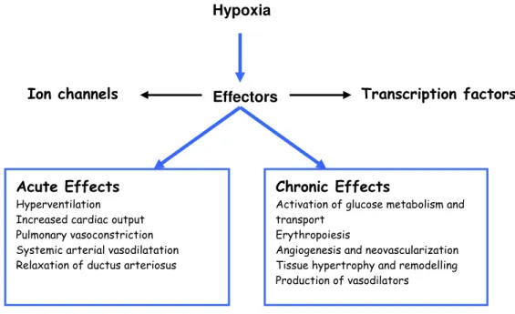
exposure to hypoxia regulates the expression of numerous genes, encoding
histological modifications to reduce the cellular need of, and dependence on, O2
and increase the O2 supply to the tissues (figure 1).
Hypoxia
Ion channels Effectors Transcription factors
Acute Effects
Hyperventilation Increased cardiac output Pulmonary vasoconstriction Systemic arterial vasodilatation Relaxation of ductus arteriosus
Chronic Effects
Activation of glucose metabolism and transport
Erythropoiesis
Angiogenesis and neovascularization Tissue hypertrophy and remodelling Production of vasodilators
Figure 1 Hypoxia signalling pathway with the major adaptive responses to acute and chronic hypoxia (adapted from Lopez-Barneo et al., 2004).
Mammals possess regulatory mechanisms that, acting on the respiratory
and cardiovascular systems, try to minimize hypoxia and to prevent its
deleterious effects. The arterial chemoreceptors, aortic bodies and in particular
the carotid bodies (CB) are the activators of those regulatory mechanisms. The
CBs are located near the carotid artery bifurcations and sense arterial PO2
(PaO2), arterial PCO2 (PaCO2), pH and temperature, being responsible for the
greatest part of the hyperventilation observed during hypoxia and contributing to
the hyperventilation that accompanies respiratory or metabolic acidosis. The
remaining respiratory drive is due to aortic bodies in the case of hypoxia and to
central chemoreceptors in the case of acidosis (Richerson and Boron, 2005).
CB chemoreceptor cells are sensors that, in response to low PO2, high
PCO2 and pH, release neurotransmitters that modify, increasing or inhibiting,
the frequency of sensory fibres of the carotid sinus nerve (CSN). Central
projections of the CSN terminate in the brain stem, where the firing frequency is
integrated by the respiratory central control system, generating the
Stimulation of the CB
Cardiovascular responses
Respiratory responses
Figure 2 Respiratory and cardiovascular responses to carotid body activation. Adapted from Fitzgerald, 2000.
Chemoreceptors also exert some actions on the cardiovascular system,
namely the heart and the resistance and capacitance of vessels (Figure 2) (for a
review see Gonzalez et al., 1994).
The CBs have been known as sensory organs since 1928 (De Castro,
1928) and in 1938 the Nobel Prize was awarded to Heymans for the discovery
of “the aortic and sinus mechanisms of respiration”. Over the subsequent
decades the CB was extensively studied in terms of reflex systemic responses
resulting from CB stimulation and in terms of the physiology and pharmacology
of neural mechanisms responsible for the action potentials generated at CSN
level and integrated at the brain stem, producing systemic responses. Several
theories were postulated, aimed at explaining the chemoreception process, the
“mechanism and the substances involved” that contribute to the
stimulus-generated neural activity in the CSN.
Adenosine is a mediator in the central and peripheral nervous system
and it is known that it stimulates ventilation in several mammalian species,
proposed at the CB and in this context we decided to investigate the functional
significance of this mediator in carotid body chemosensory activity, in control
and chronically hypoxic animals.
1.1. The carotid body: morphology
The CB parenchyma is characterized by the existence of islets of cells,
called clusters, glomeruli or glomoids enveloped by an extensive net of
fenestrated capillaries and dispersed in a stroma of fibrocytes and collagen
fibres (Figure 3C) (for a review see e.g. Verna, 1997). Each glomerulus
contains two cell types: glomic cells (Type I cells or chemoreceptor cells) and
Type II cells or sustentacular cells, and the surrounding capillary networks
(Figure 3).
C
Ultrastructural studies have demonstrated that chemoreceptor cells have
characteristics of active secretory cells (Verna, 1979). Chemoreceptor cells are
derived from the neural crest and possess a heterogeneous population of
synaptic vesicles in their cytoplasm that contain some of the putative
neurotransmitters. The neuroactive agents present in the chemoreceptors
include acetylcholine (ACh), dopamine (DA), norepinephrine (NE) and serotonin
(5-HT) and also several neuropeptides, like substance P (SP) and
met-encephalin (ME) and ATP (Gonzalez et al., 1994; Zhang et al., 2000; Buttigieg
and Nurse, 2004; Conde and Monteiro, 2006). Type II cells have a glial nature
and lack specialized organelles (Gonzalez et al., 1992) (Figure 3B).
The proximity of capillaries to chemoreceptor cells (less than 20 µM)
minimizes the diffusion pathway of bloodborn stimuli.
The CSN sensory fibres come from the petrosal ganglion (PG) neurons
and penetrate the glomeruli, ending in synaptic apposition to the chemoreceptor
cells (Figure 3B). Beyond this sensory innervation, the CB receives sympathetic
and parasympathetic innervation in the vessels that is originated in the neurons
present in the superior cervical ganglion (SCG) and at the surface of the CB
and in other neurons dispersed through CSN. These fibres are noradrenergic
and cholinergic and control the blood rate to the CB, i.e. its functional activity
(Verna, 1997).
1.2. Chemotransduction mechanisms at the CB: coupling stimulation and
secretion
It is universally accepted that chemoreceptor cells are the initial (first)
transducers of sensorial stimulus releasing neurotransmitters that generate
action potentials in CSN sensitive nerve fibres, this activity being integrated at
the brain stem to induce ventilation (Figure 4).
At normal blood gas pressures and pH, chemoreceptor cells possess a
basal activity that can be measured as basal release of neurotransmitters or as
basal CSN electrical activity. An important role has been attributed to CB
chemoreceptors in maintaining the resting ventilation since CB resection or
parameters, like a decrease in minute ventilation and a moderate increase of
PaCO2 (8 - 10 mmHg) (Bisgard et al., 1976; Bisgard and Vogel, 1971; Eugenin
et al., 1989; Feustel et al., 1981). The PaO2 threshold to hypoxia corresponds to
70-75 mmHg (Biscoe et al. 1970; Obeso et al.; 1997b) and below this PaO2 the
slope between PO2 and CSN discharges and minute ventilation increase
exponentially (for a review see Gonzalez et al., 1994, Figure 6).
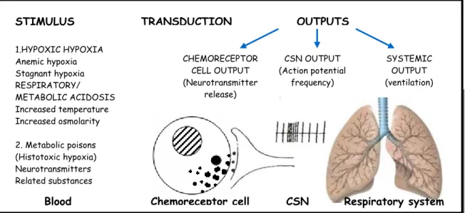
CSN Respiratory system STIMULUS 1.HYPOXIC HYPOXIA Anemic hypoxia Stagnant hypoxia RESPIRATORY/ METABOLIC ACIDOSIS Increased temperature Increased osmolarity
2. Metabolic poisons (Histotoxic hypoxia) Neurotransmitters Related substances
Blood
TRANSDUCTION OUTPUTS
CHEMORECEPTOR CELL OUTPUT (Neurotransmitter release) CSN OUTPUT (Action potential frequency) SYSTEMIC OUTPUT (ventilation) Chemoreceptor cell
Figure 4 Functional organization of carotid body (CB) chemoreceptors. (Adapted from Gonzalez et al., 1994)
However, for the hypercapnic stimulus the relation between the increase
in PaCO2 and CSN discharges and release of neurotransmitters is different,
being linear between 20-100 mmHg (Rigual et al., 1991). These relationships
suggest that the transduction mechanisms are different for the two stimuli.
Several studies have shown that DA is released in hypoxia and
hypercapnia, this release being proportional to stimulus intensity and to CSN
chemosensory activity increase and dependent on extracellular Ca2+ (Fidone et
al., 1982; Obeso et al., 1985, 1986, 1992, 1999; Rigual et al., 1986, 1991, 2002;
Rocher et al., 1991; Vicario et al., 2000a; Sanz-Alfayate, 2001). The same
effect was observed with the response of CB to high K+ (depolarizing stimulus)
(Almaraz et al., 1986). The response to hypoxia and to high K+ was sensitive to
dependent), however the acidic-stimulus response was dihydropyridine
insensitive (Obeso et al., 1992). Soon after, it was demonstrated that
veratridine, activator of voltage-dependent Na+ channels, increases the release
of DA, the release being dependent on Na+ and Ca2+ and sensitive to
tetradotoxin (TTX); in addition it was shown that the release of DA induced by
hypoxia was also TTX-sensitive. All these findings suggest that hypoxia
depolarizes chemoreceptor cells inducing Na+ and Ca2+ entrance.
PO2 diminution
O2 sensor
Diminution of the channels opening probability of K+ channels sensitive to PO2
Depolarization
Activation of Na+and Ca2+channels
Increase of intracellular Ca2+
Neurotransmitter release Coupling mechanisms
Increase in CSN discharges
High extracellular K+
Integration in the brain stem
Modification of respiratory and cardiovascular parameters
Figure 5 Hypoxic transduction mechanism at the carotid body. In red are the steps of the cascade not completely defined (established) (adapted from Gonzalez et al., 1992).
Next, K+ currents sensitive to PO2 that were inhibited by hypoxia were
found in the CB chemoreceptor cells as well as voltage-dependent Na+ and