A Natural Protective
Mechanism Against
Malaria
The Role of Gut Flora
Bahtiyar YILMAZ
Dissertation presented to obtain the Ph.D degree in Immunology
Instituto de Tecnologia Química e Biológica | Universidade Nova de LisboaBahtiyar YILMAZ
A Natural Protective Mechanism Against Malaria – The Role of Gut Flora
A Natural Protective
Mechanism Against
Malaria
The Role of Gut Flora
Bahtiyar YILMAZ
Dissertation presented to obtain the Ph.D degree in Immunology
Instituto de Tecnologia Química e Biológica | Universidade Nova de LisboaResearch work coordinated by:
The study described in this Ph.D. thesis was performed at Instituto Gulbenkian de Ciência (IGC), Oeiras, Portugal and was financially supported by Fundação para a Ciência e a Tecnologia (FCT), Portugal (SFRH/BD/51176/2010).
The work on this thesis resulted in the following publication and some written content of the thesis was introduced into the following publication:
Yilmaz, B., Portugal, S., Tran, T. M., Gozzelino, R., Ramos, S., Gomes, J., et
Acknowledgements
The work obtained during this amazing journey only exists due to the amazing people and their significant amounts of help -no matter it is scientific or not.
First all, my father… Still I remember the day when I was struggling on making a decision to go to Portugal. Your enthusiasm led me here to do this amazing study. Hope you are watching from up there -your true success story.
That makes you the greatest! Thank you very much for everything you have done. In addition, I enjoy every small coffee break that I had with you. I will miss that. Raffaella Gozzelino, the master of the Western blots… Your contribution on this study is incredibly crucial. Thanks a lot for your all help and incredibly cheerful friendship. Every lab needs one CC of you. Silvia Cardoso and Sofia Rebelo taught me how to work with murine model. Your patience to me when I was training mice cannot be described. Hope I can teach the same way as you did to me to the future scientists. Thank you guys! Rasmus Larsen… It was a pleasure to work with you. Thank you very much for your initial guidance on gut flora part of the story and the fruitful discussions on that. Virginia de Oliveira Marques, Andreia Cunha and Ivo Marguti… At the beginning of this project, they helped me for all immunological experiments. Guys, I appreciated it all your efforts. Sebastian Weis (a.k.a. Dr. Weis)… I am sorry that our never-ending, fruitful discussions will be over soon. I learned a lot from you about the infectious diseases and I enjoyed our collaborative efforts on a project that is not the part of this thesis. We should definitely repeat this collaboration in future. Lastly, I’d like to thank rest of the past and present Inflammation Group members, specifically Ana Rita Carlos, Jerzy Kotlinowski, Birte Blankenhaus and Ana Ribeiro. You made my days full of joy. I will miss your enjoyable and supportive chats. It was a pleasure to work with all of you.
Crompton… They designed and performed human studies to support our hypothesis. I’d like to thank Peter J. Cowan for providing us the knockout
mice and Anita S. Chong for anti-α-gal hybridomas. Here it gives me an opportunity to show my sincere gratefulness to Ana Regalado from Antibody Service at IGC. I owed you a lot. With monoclonal antibodies you produced for us, we did amazing job. Thank you very much, Ana.
Getting through this long journey, it does not only require scientific supports, but it also requires emotional support. I’d like to thank my
friends, especially Ali Altintaş, for listening and tolerating me over the past
years, although we were away from each other. Ali, hope one day we have a chance to work together again. And of course, Ali Ozgur Argunsah…
Hope you learned a lot about malaria and anti-α-gal antibodies through our never ending chat at the end of the tiring days. Thanks a lot for your suggestions and naïve but critical questions.
My Pires… Marta Marialva… You have been in my life more than two years. No words is enough to describe your part in my journey, here in Portugal. I felt your love and support every second. Among all these, I should thank you for your patience regarding to the moments when I was talking about my project without breathing. Life is beautiful with you.
Finally, I wish to express my love and gratitude to my beloved family; for their endless support and love. They taught me the values that have helped me to become the individual that I am today. They also taught me the importance of an education and did everything possible to help me to attain the best education possible.
Table of Contents
Preface 11
Summary 13
Resumo 15
Abbreviations and Acronyms 17
Chapter I 19
Chapter II 111
Chapter III 139
Chapter IV 179
Chapter V 205
Chapter VI 229
Preface
This dissertation assembles data obtained during my Ph.D research project, developed at Instituto Gulbenkian de Ciência (IGC) under the supervision of Miguel Che P. Soares from May 2010 to September 2014.
This thesis is structured in 6 chapters, preceded by a summary written in English and Portuguese, outlining the aims of the projects, results and outcomes of the project.
The first chapter provides information about general knowledge on malaria,
the importance of naturally occurring anti-α-gal antibodies and immunological aspects of these antibodies. This chapter also includes the specific aims of this work. The second chapter describes the detection of
α-gal on the surface of Plasmodium sporozoites. Moreover, it shows that
Plasmodium spp. express α-gal during the exo-erythrocytic stage and erythrocytic stage of the infection in mammals. The third chapter provides
insight about the fate of Plasmodium sporozoites when facing anti-α-gal
antibodies in the dermis of their mammalian host. The fourth chapter provides information about the role of complement system in this
protective mechanism driven by anti-α-gal antibodies. The fifth chapter
hopefully opens new doors in the field by presenting the role of gut flora in triggering this natural protective mechanism against malaria transmission. The last chapter provides an overall discussions and conclusions of this study. It also contains the future perspectives of the work developed during my Ph.D studies.
Summary
Malaria is an infectious disease of humans and other animals including
birds, reptiles and most mammals. It is transmitted via the inoculation of
Plasmodium sporozoites into the skin through the bite of an infected female Anopheles mosquito. Although every year, around 700.000 lives are
perished, mainly children under the age of 3-5 years old, to Plasmodium
infection this deadly parasite has a relatively low efficiency of transmission from mosquitoes into humans. This argues for a natural protective
mechanism, which presumably acts during the early stages of Plasmodium
infection in the human host. Using a rodent model of Plasmodium infection
to mimic the transmission of disease in humans, this PhD thesis demonstrates that such a mechanism does exists and is mediated through
the action of natural antibodies recognizing specifically the
Galα1-3Galb1-4GlcNAc-R (α‑gal) carbohydrate. It was shown that these T-cell dependent
circulating anti-α-gal antibodies can be produced upon antigenic exposure
by α-gal expressing specific components of the intestinal microbiota and
target Plasmodium sporozoites in the skin, immediately after their
inoculation by Anopheles mosquitoes in mice. Additionally, anti-α-gal
antibodies are associated with protection against P. falciparum infection in
humans, as shown for individuals from a malaria endemic area. Complement and polymorphonuclear (PMN) cell-dependent mechanisms contribute to the mechanism via which these antibodies prevent the migration of sporozoite from the skin into the liver and inhibit hepatocyte invasion, suppressing the establishment of liver stage of infection. Our findings also reveal that this natural host defense mechanism reduces the rate of disease transmission and provides sterile protection against malaria.
Importantly, vaccination against α-gal protects mice against malaria
Resumo
A Malária é uma doença infecciosa que afecta humanos e animais, como
aves, répteis e mamíferos. Causada por parasitas do género Plasmodium,
esta doença é transmitida por inoculação de esporozoítos na pele, através
de uma picada de um mosquito fêmea, do género Anopheles. Apesar de
anualmente serem perdidas cerca de 700,000 vidas, em particular crianças
de idade inferior a 3-5 anos, devido a infecções por Plasmodium, esta
infecção muitas vezes mortal, apresenta uma eficiência de transmissão relativamente baixa, o que argumenta em favor de um mecanismo natural de protecção do hospedeiro, que presumivelmente actua durante os primeiros estadios da infecção. Nesta tese de Doutoramento, recorrendo a
um modelo de infecção por Plasmodium em roedores, foi demonstrado que
tal mecanismo é mediado por anticorpos naturais que reconhecem especificamente um epitopo existente em carbohidratos, denominado
Galα1-3Galβ1-4GlcNAc-R (α‑gal). Este epitopo está presente na estrutura
celular do parasita, nas diversas fases do ciclo de vida, mas ausente em células humanas, permitindo assim a produção de elevados níveis de
anticorpos anti-α-gal, o que ocorre naturalmente e de forma dependente de
células T. Anticorpos anti-α-gal do tipo IgM e IgG, tais como os presentes em indivíduos adultos residentes em áreas endémicas de malária, inibem
os esporozoítos de Plasmodium na pele, imediatamente após a inoculação
por mosquitos Anopheles. Estes anticorpos naturais estão também
associados à protecção contra a infecção por Plasmodium falciparum em
anticorpos naturais conferem protecção estéril contra a malária, reduzindo
a taxa de transmissão da doença. De salientar que a vacinação contra α-gal
protege ratinhos deficientes em α1,3Gt contra a malária, sugerindo que
Abbreviations and Acronyms
α‑gal Galα1-3Galβ1-4GlcNAc-R
α1,3Gt UDP-Galactose:β-galactoside-α1-3-galactosyltransferase
ACP alternative complement pathway
ALUM aluminum hydroxide Cɣ
BSI-IB4 lectin from Bandeiraea simplicifolia
CFA complete Freund’s adjuvant
CSP circumsporozoite protein
CT cell traversal
EEF exo-erythrocytic form
GF germ-free
GFP green fluorescent protein
GI gastrointestinal
IFA incomplete Freund’s adjuvant
mAb monoclonal antibodies
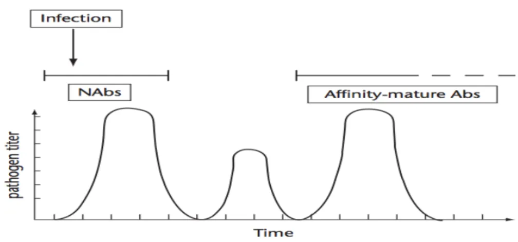
NAbs naturally occurring antibodies
NK natural killer
ODN oligodeoxynucleotide
PV parasitophorous vacuole
PBS phosphate buffered saline
PbA Plasmodium berghei ANKA
Pf Plasmodium falciparum
Py Plasmodium yoelii 17XNL
PMN polymorphonuclear cells
RBC red blood cell
rRBCM rabbit RBC membrane
General Introduction
1.1 Malaria: An Ancient Disease
1.1.1 History of the disease
Malaria is one of the most anciently described diseases, reported originally in a Chinese document as early as 2700 BC, in which fever and enlarged
spleen were recorded as the major clinical symptoms1. Later on,
Mesopotamian clay tablets from 2000 BC, Egyptian papyri from 1570 BC and Hindu texts from the 500 BC, provided more medical information
about this disease1,2. Knowledge related to malaria continued to increase in
the early Ancient Greece with Homer (~850 BC), Empedocles of Agrigentum (~550 BC) and Hippocrates (~400 BC) describing its clinical signs (i.e. poor health, intermittent fever and enlarged spleen in people living in marshy places), holding the first clear recordings of malaria
infection in ancient times3. However, many other studies were required in
order to identify the causative agent(s) of the disease, i.e. Plasmodium.
The various reports4 on malaria throughout the history of medicine,
spanning more than 4,000 years5 made it clear that the manifestations and
the causes of disease, including fevers and other symptoms were to be
considered as miasmas rising from swamps 1 . Modern scientific
understanding of malaria however, began only to emerge by the end of the 19th century with the germ theory and the beginning of microbiological studies. The very first understanding of malaria transmission dates back to 1880 with the detection of parasites in the blood of an infected patient,
reported by Charles Louis Alphonse Laveran4. Meticulous examination of
blood samples collected from more than 200 patients revealed that in 150 of those there was a clear detection of “crescentic bodies” that were never detected in healthy individuals. This led Charles Louis Alphonse to
conclude that a parasitic protozoan, which he named as Oscillaria malariae
was the causative agent of the disease called malaria4. These observations
!!!!!!!!!!!!!!!!!!!!!!!!!!!!!!!!!!!!!!!!!!!!!!!!!!!!!!!!
1
!
made possible the advance of scientific discoveries by Ronald Ross, a British officer of the Indian Medical Service, in 1897, who demonstrated that avian malaria is a disease transmitted via mosquito biting,
incriminating for the first time mosquitoes in this vector-borne disease4.
Ronald Ross described how parasites develop within the midgut of
mosquitoes, from where they eventually migrate to the salivary glands6. He
was able to successfully transmit the disease to healthy birds from an inoculum isolated from infected mosquitoes, thus inferring for the first time that transmission of avian malaria occurred via the bite of an infected mosquito. This was subsequently confirmed in a study carried out with a
human parasite by Battista Grassi and colleagues7. These scientists also
identified female Anopheles mosquitoes as the main vector of human
malaria. In 1890, Grassi and colleagues reported for the first time, that two
malaria parasites, named Plasmodium vivax and Plasmodium malariae, are
able to infect humans4. This finding was based on a simple experiment in
which mosquitoes fed on an infected patient in Rome were sent to London
where they were able to transmit the disease to two healthy volunteers8. In
1897, William H. Welch identified a third malaria parasite, named Plasmodium falciparum and in 1922, John William Watson Stephens, working in West Africa, described yet another human malaria parasite, which
resembled P. vivax and was named Plasmodium ovale9. Thirty more years
passed until Plasmodium knowlesi was identified as another Plasmodium
parasite able to infect humans10.
Despite the discovery of several life threatening Plasmodium strains,
information on the mechanisms by which Plasmodium infects and
proliferates in humans was still lagging until the beginning of 20th century.
In 1902, Friedrich Schaudinn described that Plasmodium vivax infective
sporozoites contained in the salivary glands of mosquitoes could infect
human red blood cells (RBCs)4. This was confirmed in 1948 by Cyril
mosquitoes, which carried Plasmodium cynomolgi parasite. The authors
demonstrated that when transmitted by mosquitoes, Plasmodium
sporozoites undergo a first round of multiplication, differentiation and
maturation in the liver before being released into the bloodstream11. These
steps represent the first evidence for the occurrence of a ‘tissue phase’ of
Plasmodium infection as formally demonstrated in 194811. An American clinician, Wojciech Krotoski showed that in some strains of Plasmodium vivax, the liver stage of infection could remain dormant for several months, called as hypnozoites1. Seventy years passed until the major human tissues
targeted by Plasmodium were finally identified (discussed later on in this
chapter).
1.1.2 Malaria outcome – current situation
Malaria is a global health problem, affecting more than 100 countries with an estimated 3.4 billion people at risk of developing the disease, i.e. half of
the world population. Malaria acts as the 10th leading cause of mortality
worldwide1213, driven by severe anemia, respiratory distress, multi-organs
failure and cerebral malaria. World Health Organization (WHO)14 reported
in 2012 that 219 million individuals were infected with Plasmodium parasite
(uncertainty range between 135 to 287 million) of which 660 000 succumbed to malaria (uncertainty range between 473 000 to 789 000). In 2013, the WHO Malaria Report showed that ongoing malaria transmission occurs in
97 countries and territories14. Moreover, 7 countries in which Plasmodium
transmission started to be observed again are subjected to preventive
measures and this makes to a total of 104 countries to be considered
endemic to malaria (Figure 1.1). Another striking fact related to this disease
is that 90% of the deaths occur in children and pregnant women. In other
Figure 1.1 Worldwide malaria distribution and burden. Although mortality among infected individuals is “low” (0.3-04%), malaria remains a major health problem and one of the largest scourges in many tropical and subtropical regions.
Adapted from15.
According to the WHO Malaria Report of 2012, the death toll imposed by malaria was reduced by 20-25% from 2000-2010, (a decrease of 33% in Africa), presumably due to the introduction of malaria control programs by
the WHO14. However, this decrease is not due to lower parasite viability,
given the emergence of drug resistance strains of Plasmodium. So far, there
is no efficient licensed vaccine, which will eventually act as a
1.2 Biology and Life Cycle of
Plasmodium
1.2.1
Plasmodium spp
.
Plasmodium is a heteroxenous protozoan parasite, i.e. the completion of its
life cycle involves a vertebrate host and an arthropod vector. It belongs to
the Apicomplexa phylum, the less phylogenetically well-known taxonomic
group (only 0.1% of the all species named to date)16. The genus Plasmodium
was first described in 1885 by Ettore Marchiafava and Angelo Celli4,17 and
today, it is known to be infective to a variety of possible hosts, including
reptiles, birds and mammals18. There are four described human infectious
Plasmodium species: P. falciparum, vivax, malariae, and ovale. In addition, P. knowlesi19, which infects macaques that inhabit the forested areas of Southeast Asia, has recently developed the ability to infect humans and is considered as the fifth causative agent of human malaria. These Plasmodium
species differ regarding morphology, life cycle and pathologic outcomes
imposed to their hosts. Differences include a) blood stage morphology
(erythrocytic schizogongy consists of 3-5 rounds), b) characteristics associated with liver stage (a dormant phase called hypnozoite observed in P. vivax and P. ovale), c) sequestration of trophozoite- and schizont-infected
RBC in microvasculature (exclusive to P. falciparum) and clinical
manifestations with prolonged pre-patent period for P. vivax, ovale, and
malariae up to several months, d) intensity of febrile attacks or e) duration of clinical symptoms.
P. falciparum is by far the most virulent species to mankind, which accounts for ≈ 85% of all malaria infections, and virtually all malaria-associated morbidity and mortality. The high pathogenicity of this parasite hints that
P. falciparum may be a recent human parasite transferred from a nonhuman host20,21.
In 1948, Ignace Vincke and Marcel Lips identified and isolated the first
tree rat) in Central Africa4. Since then, three other strains, i.e. P. yoelii, P. vinckei and P. chabaudi were identified, isolated and successfully transferred/adapted to laboratory rodents, thus allowing the establishment
of rodent models of the human disease4,22,23. The life cycle, physiology and
morphologic structure of these parasites resembles in many aspects the
human infecting Plasmodium spp. and therefore, rodent parasites are
considered valuable surrogate models of human malaria4,22,23. However,
there are reasonable concerns as to what extent the rodent models of malaria mimic disease pathogenesis and immunity in humans. In any case, the spectrum of murine malaria models obtained when using different hosts and parasite strains reflects to a large extent the diversity of human disease with specific parasite-host combinations capturing most if not all the specific aspects of the human disease.
1.2.2 Life-cycle of
Plasmodium
in mammals and mosquitoes
The life-cycle of Plasmodium can be extremely complex as it involves
different stages occurring in disparate host species such as an arthropod
vector and a vertebrate host. Sincethe original description of the different
stages of Plasmodium infection, scientists have been trying to understand
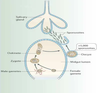
the life-cycle of Plasmodium in further detail. Briefly, the life-cycle in mammals consists of a pre-erythrocytic, erythrocytic and sporogonic phase (Figure 1.2). Each of these is explained in detail above. The pre-erythrocytic stage (Skin and Liver Stage) begins when a female Anopheles mosquito
injects sporozoites, i.e. the infectious form of Plasmodium parasites, into the
skin of a vertebrate host. Parasites spend a certain amount of time in the dermis (from 15 minutes to a couple of hours) before reaching a dermal microvascular blood vessel, through which they are transported via the
blood stream, into the liver. At this point, Plasmodium sporozoites invade
exoerythrocytic forms (EEFs), and subsequent release of infectious merozoites into the bloodstream. RBC invasion occurs within 1-2 minutes and then parasites undergo rounds of asexual multiplication leading to RBC disruption and to the clinical outcomes of the disease. In the sporogonic stage (Mosquito Stage), during a blood meal a mosquito
ingests, differentiated male and female Plasmodium gametocytes from an
infected host. Fertilization occurs in the mosquito midgut and the resulting zygotes develop into ookinetes that mature into oocysts, where thousands of sporozoites develop. This final form of the parasites in the mosquito vector migrates to the salivary glands, where it stays until it is injected into another vertebrate host. Each phase of infection is explained in further detail in the next paragraphs.
The skin stage of infection is a rapid, clinically silent and apparently
immunologically innocuous phase of Plasmodium infection. This initial step
is critical for the establishment of the infection in the mammalian host24.
Most of the injected sporozoites, in average 15-123 sporozoites per blood
meal, are inoculated into the extravascular matrix of the dermis25,26 and not
directly into the bloodstream as initially thought. This was first demonstrated by Boyd and Kitchen, in 1938, who found the presence of
Plasmodium sporozoites in human dermal tissue27, finding as well that
removal or heat treatment28 of the skin area targeted by the mosquito
dramatically decreased Plasmodium infection29. It argues strongly that
sporozoites are first deposited into the dermis, while the mosquito probes for a blood vessel and salivates. In further support of this notion, when the mosquito ingests blood it actually re-ingest a proportion of its sporozoites driven by the inward blood pressure, which overrides outwards salivation
pressure30,31. The development of intravital imaging techniques fully
Mosquito bites are also associated with the ejection of saliva that contains vasodilators like tachykinins and anti-coagulants like thrombin- and Fxa -directed molecules, which increase blood flow and inhibit platelet aggregation32, presumably facilitating blood ingestion33. During a mosquito bite, approximately ~1–2.5 sporozoites are released per second through the proboscis. In malaria endemic regions, individuals can be bitten by 1-100
mosquitoes during one single night corresponding to 0-4 infective bites34.
Considering around 4-5 minutes of mosquito salivation, an Anopheles
mosquito injects on average about 300 sporozoites per bite in a human host35,36.
When delivered into the dermis Plasmodium sporozoites show significant
level of motility before reaching the extracellular matrix of the blood
vasculature and contacting with vascular endothelial cells28,33. Sporozoites
glide in three-dimension with an average velocity of ~1–2 µm per second during the first 30 minutes after injection. While they continue to move for more than one hour, their speed decreases significantly over time. Once
Plasmodium sporozoites start migrating randomly in the cutaneous tissue, one of four different scenarios will decide their fate. These are dictated by many factors, including blood vessel density at the biting site, saliva components released by the mosquito and the parasite species itself. In any case, it should be noted that out of the ejection of 70-100 Plasmodium
sporozoites in mice during salivation only 15-25% of those deposited in the
skin will manage to successfully establish infection35. Under the first
scenario, once deposited into the skin sporozoites move at high speed by
gliding motility37 and traverse38 host cells, piercing host membranes until they find a blood vessel as a way to reach the liver, a process that lasts a
couple of minutes29,39,40. However, a small proportion of Plasmodium
the first hours after mosquito bite40,42. The development is arrested after a
short period of time and small EEFs are cleared from the host. Moreover,
this processes can initiate the immune response against Plasmodium
infection as suggested by the appearance of sporozoite specific cytotoxic T lymphocytes (CTLs) that can accumulate in the skin-draining lymph nodes, as early as two days after the intradermal injection of irradiated
sporozoites42. The other possible scenario is when the deposited Plasmodium
sporozoites fail to reach a draining blood or lymphatic micro-vessel in the dermis, as it occurs for approximately 50% of the cases. It is estimated that 10% of these initiate the development of EEFs in the skin hair follicles, which can persist for dayswithout leading to infection43,44. The last scenario,
as observed by Kebaier and Vanderberg, is when Plasmodium sporozoites are re-ingested by mosquitoes30, with 90% of the mosquitoes re-ingest a
highly variable number of sporozoites (1-400 sporozoites) while actively imbibing blood. Among these four scenarios only the first one allows for successful establishment of an infection.
The mechanism via which Plasmodium sporozoites manage to proceed with
microvascular blood vessel invasion is still not fully understood. Surface phospholipase is important to breach host cell membranes during sporozoite migration and in the absence of this protein, sporozoites’ ability to cross through vascular endothelial cells is impaired, with significant
reduction of infection45. This reveals that transit from the dermis
extracellular matrix, where sporozoites are initially deposited, to the blood microcirculation is a tightly regulated process presumably involving several gene products encoded by sporozoites as well as by the host. Transit from the dermis extracellular matrix into blood vessels is associated
with decreased Plasmodium sporozoite gliding speed, triggered upon
contact with endothelial cells, which are then crossed within few minutes46.
Figure 1.2 The life cycle of Plasmodium. A female mosquito transmits a motile Plasmodium sporozoite into the skin of a vertebrate, acting as the transmission
vector of malaria40,43. When sporozoites reach blood vessels, they are arrested in the
liver, invading hepatocytes, where thousands of merozoites are produced38,47.
Upon maturation, merozomes are release into blood circulation, reach the lungs48
and infect RBC, initiating a series of asexual multiplication cycles. Then, an
uninfected mosquito bites an infected host, taking up Plasmodium gametocytes
along with the blood meal35. Fertilization and maturation take place in the
mosquito midgut and when fully developed, Plasmodium sporozoites migrate to
the salivary glands.
Liver stage is the second short-lived and innocuous (asymptomatic) stage of Plasmodium infection in vertebrates, characterized by sporozoites migration through the liver sinusoids as to gain access to hepatocytes. During this process that occurs via an unknown mechanism, sporozoites switch from migrating into invasive mode and settle down in a final hepatocyte within two minutes after reaching blood vessels. The tropism of
suggests the existence of a selective process between parasite- and
host-encoded molecules. One example of a Plasmodium encoded-protein
involved in this process is the circumsporozoite protein (CSP), a molecule
distinctive of the Plasmodium genus that accounts for the predominant
surface protein covering the entire surface of Plasmodium sporozoites, not
found in any other Apicomplexa parasite49,50. Switching to invasive mode
correlates with CSP interaction with host heparan sulphate proteoglycans
(HSPGs) expressed at the surface of hepatocytes and hepatic stellate cells.
These present a significantly higher degree of sulphation when compared to other tissues51, which may explain the initial tropism of Plasmodium
sporozoites towards the liver. An early study by Victor Nussenzweig’s
group showed that CSP binds to the hepatocyte microvilli within the space2
of Disse52,53, migrating along fenestrated endothelial cells that allow for direct contact with hepatocytes. Presumably, this allows for sinusoidal barrier crossing and migration into the hepatic parenchyma, where
sporozoites traverse several hepatocytes before invading the final one,
where they differentiate47. During hepatocyte invasion38, each Plasmodium
sporozoite forms a parasitophorous vacuole (PV)47 and resides in it during
the liver stage of infection54.
Survival of sporozoites in the liver requires cell traversal (CT) motility, a
wounding and crossing process via which Plasmodium parasite passes
through host cell plasma membranes and migrates freely in the cytoplasm of host cells, including hepatocytes. During this process, hepatocytes are
wounded, releasing so called infection-susceptibility-inducing factors55,56,
e.g. hepatocyte growth factor (HGF)57, required to promote final hepatocyte
infection38. In support of this notion CT-deficient sporozoites display highly
impaired liver infection in vivo, even though normal hepatocyte invasion
!!!!!!!!!!!!!!!!!!!!!!!!!!!!!!!!!!!!!!!!!!!!!!!!!!!!!!!!
2
!
and development are observed in vitro58,59. While pre-erythrocytic stage proteins such as the ones essential for cell traversal (SPECT), cell traversal protein for ookinete and sporozoite (CelTOS) or phospholipase (PL) were thought to be essential for cell migration, sporozoites deficient in the genes encoding these proteins can still invade hepatocytes and complete a liver stage development similarly to wild type sporozoites58,59. Little is known about the mechanism by which Plasmodium sporozoite switches from migration through hepatocytes to invasion of a hepatocyte.
Once inside hepatocytes, sporozoites can develop and replicate within the
PV, generating EEFs60 via asexual multiplication. This culminates in the
presence of tens of thousands of infectious merozoites in ~2 days, which
ultimately will proceed to the blood stage of infection61. However, little is
known about the cellular and molecular interactions between parasite and host during this stage of the Plasmodium life cycle.
The final important step of Plasmodium development in the liver is the exit from the hepatocyte involving the PV lysis and the release of newly formed
merozoites contained in vesicles, called merozomes62. These bud off from
hepatocytes into the sinusoidal lumen, remaining covered by hepatocyte– derived membrane, which is thought to be important initially to afford protection against host innate immune system. Once released into the host circulation, merozomes are ruptured and the hepatic merozoites are free to invade the red blood cells.
The blood stage of infection is associated with all the clinical symptoms of malaria. After the release of merozomes into the blood circulation, shear forces inside the hepatic and other larger veins break down merozomes into smaller units called merozoites. Later, merozomes reach the lung microvasculature, in areas of low macrophage density and reduced shear
ruptured in the lung microvasculature, merozoites are released and upon first contact with RBC membranes invade RBC in a short period of time
(27-28s). RBC invasion occurs through several phases (Figure 1.3) that
involve receptor–ligand interactions involving more than 50 surface and
secreted proteins63. First, merozoites attach to the RBC surfaces (Phase 1,
Pre-Invasion). Then apical reorientation of the parasites occurs, which allows for a tight-junction formation between merozoites and RBC surfaces. This provides an entrance mechanism into the RBC through a movement that propels the merozoites into the host cells (Phase 2, Invasion). A final process allowing for the release of the RBC membrane is associated with potassium and chloride efflux resulting in RBC water loss (Phase 3,
Echinocytosis) and the initiation of parasite maturation in RBC begins64.
Figure 1.3Kinetics of RBC invasion. Merozoite achieves infection of RBC in three
phases. In Phase 1, merozoite attaches to RBC and causes dramatic RBC deformation and recovery process, afterwards. In Phase 2, merozoite is internalized within the RBC and later on, in Phase 3, dehydration-type morphology occurs in infected RBC (iRBC) and parasite maturation in RBC begins.
Adapted from64.
After RBC invasion, parasites progress through a ring and a mature trophozoite stage, dividing during the subsequent schizont stage and giving rise to 8-32 daughter merozoites contained within a single RBC.
Eventually, RBC ruptures and releases the newly formed merozoites65.
are partially hidden from immune recognition inside RBC, which lacks the expression of major histocompatibility complex (MHC) molecule recognized by antigen receptors of the host adaptive immune system. RBC also exposes some proteins at the surface, enabling cyto-adherence to different endothelial receptors and favoring sequestrations in organs while reducing splenic clearance by hemophagocytic monocyte/macrophages
(Mø)66.
Figure 1.4 Mosquito Stage of Plasmodium. In mosquito gut, gametocytes released
from burst of iRBC develop further into mature gametes. After the nuclei fusion and fertilization in the mosquito gut, actively moving ookinetes form oocysts. Inside the oocyst, the ookinete produces thousands of sporozoites by cell division,
releasing sporozoites that reach the salivary glands. Adapted from68.
After several asexual cycles inside RBC, a fraction of intraerythrocytic parasites divert from the asexual multiplication pathway towards sexual differentiation, developing highly specialized gametocyte forms (either males or females). These circulate in the bloodstream and are taken up
infected host is called gametocytogenesis and is triggered by genetic and
environmental factors67.
Mosquito stage is the last step of the life cycle of Plasmodium infection that begins with the uptake of iRBC from an infected vertebrate. This occurs via the bite of a female mosquito, which ingests both male and female gametocytes that arise during each erythrocytic cycle. Once inside the mosquito midgut, gametocytes undergo fertilization, zygote formation and
development into the motile ookinete69 generating new infectious
sporozoites.
Male gametocytes undergo the most drastic transformation while replicating their own genome. They are segregated into eight flagellated male microgametes, whereas female gametocytes transform into
macrogametes69,70. Exflagellation is a vibratory movement activity that
allows the release of male gametes from the gametocytes, enabling them to find female gametes. Subsequently, this process leads to the formation of a zygote, which undergoes meiosis and develops within 24 hours into an ookinete. During the differentiation process and development into a vegetative oocyst, the ookinete rests between the midgut epithelium and the luminal side of the basement membrane of the infected mosquito. Following multiple rounds of nuclear division that occur over 10–14 days, hundreds of sporozoites develop and are released into the mosquito
haemolymph70,71. From there, sporozoites are carried away to the salivary
glands in which they reside until transmission into their next host (Figure
1.4). Even though most sporozoites reside in salivary glands, large numbers
the next blood meal35. Plasmodium parasites can travel between the distal
part of the salivary duct system and the tip of the proboscis in just a few
seconds and the speed of ejection can reach approximately 500µm/sec. Surprisingly, the initial period of salivation is extremely important, as
many mosquitoes were found to eject sporozoites during this period26,35.
1.3 The Biology of
Plasmodium
Sporozoite
While Plasmodium sporozoites are lower eukaryotes, they have a genetic complexity that is higher than the one found in any bacteria family. This genetic complexity is combined with a considerable number of polymorphic genes in the haploid genome, which generate antigenic
diversity72. Therefore, Plasmodium sporozoites have various and highly
optimized adaptation mechanisms, which allow to exploit their hosts in extremely changing situations. This may well explain why the parasite has been so successful in surviving throughout the evolutionary selection
process73. Plasmodium sporozoites also present an active mechanism for
cellular motility, which is essential to reach and invade host cells (See Section 1.3.2) and also utilize their own locomotion for extracellular
migration through different host tissues31.
1.3.1 Morphology
of
Plasmodium
sporozoites
Sporozoites are highly polarized and usually elongated cells (defined by
the presence of an apical complex at their anterior pole) of about 10-15 µm
in length and 1µm in wide74. They have a thin outer membrane and a
double inner membrane under which lie the subpelicular microtubules anchored to the apical polar ring elongating underneath the inner membrane complexes (IMC), forming networks of closely apposed
flattened vesicles. Plasmodium sporozoites have 3 polar rings extending half
the length of the body. A morphological hallmark of Plasmodium
sporozoites is the secretory organelles at the apical tip, called micronemes
recognition and attachment to the host hepatocytes, generation of the non-phagosomal parasitophorous vacuole and remodeling of the host cell
following invasion75. Sporozoite morphology is established during the
development within oocysts, which is dependent on CSP expression76. The
sporozoite nucleus is divided into several sub-compartments including the nucleolus and the nuclear periphery, presumably involved in transcription control. Mitochondrion, apicoplast and microtubules are linked to the parasite pellicle via long tethering proteins. This defined structure is most
likely important to maintain the sporozoite shape77.
Figure 1.5 Structural illustration of a Plasmodium sporozoite. The major compartments such as apicoplast, rhoptries, micronemes and visible structures in a
Plasmodium sporozoite with its anterior ending towards the right. Adapted from78.
Plasmodium sporozoites express so called “invasion proteins” such as CSP that localizes mainly on the outer surface and in the micronemes and thrombospondin-related anonymous protein (TRAP), which localizes exclusively in the micronemes and plasma membrane of sporozoites with an irregular distribution. These proteins are often targeted as immunogens
for pre-erythrocytic malarial vaccines74,78,79.
1.3.2 Different modes of sporozoite motility
Single-cellular or multicellular organisms possess distinct features that support cellular locomotion, which are important for reorientation, homing and migration. These provide advantages for development and survival in a variety of complex and stressful environments and are essential to
from the liver into the blood. They can occur via three modes: gliding, cell
traversal and invasion motility, as supported by in vitro and in vivo studies
(Figure 1.6).
Plasmodium sporozoites share with the rest of the Apicomplexa phylum a
rather unique and mysterious process of locomotion, named gliding
motility was first described in Toxoplasma and later, in other apicomplexans
i.e. Eimeria, Plasmodium and Cryptosporidium80-82. Given the apparent
absence of specialized locomotive structures like cilia, flagella or pseudopods in Plasmodium sporozoites, crawling via gliding activity provides a mode of locomotion37at rapid and variable speeds ranging from
1–10 µm/s (Figure 1.7). This mode of locomotion underlies essential
processes in the life cycle of Plasmodium sporozoites, such as extracellular motility, associated with crawling type movement via a substrate-dependent locomotion on surfaces, as well as intracellular invasion.
Figure 1.6 Three modes of Plasmodium locomotion. Sporozoites deposited into
the skin by a female Anopheles mosquito exhibit flexing, turning, twisting motions
and circular gliding, i.e. gliding motility. Plasmodium sporozoites can pass through
cells in the skin and enter the bloodstream, reaching the liver and migrating through several hepatocytes before they invade the final one. This wounding movement through hepatocytes is defined as cell traversal and invasion of the last hepatocytes with formation of a parasitophorous vacuole occurs via invasion
Although, the main components in the core of the machinery regulating gliding motility are known, the exact mechanism by which gliding motility
occurs is not fully understood. Gliding motility relies on the activity of an
unconventional class of molecules referred to as “neckless” non-muscle myosin (type XIV). These are anchored to the inner membrane complex (IMC) of the pellicle via actin filaments. Pharmacological disruption of microfilaments via inhibition of actin polymerization, using cytochalasins or latrunculins, inhibits gliding motility and host cell invasion, arguing that
Plasmodium motility requires actin filaments polymerization and depolymerization for infection84. Although gliding motility mechanisms are
not fully identified, it is likely that the sheer force of polymerizing/depolymerizing actin filaments together with the surface molecules spanning the plasma membrane cluster activate a motor powered by actin-myosin interactions85.
Figure 1.7 Gliding of sporozoites on a glass slide in the presence of albumin.
CSP containing trails are deposited by
sporozoites via on-going secretion from
their trailing ends. Adapted from86.
The very first step towards
understanding the mechanisms
underlying gliding motility steam from the observation by Dubremetz
and Ferreira in 1978, who
discovered that cationized ferritin
binds the entire surface of Eimeria sporozoites and accumulates rapidly at
the posterior pole87. Based on this basic observation, a model has been
proposed for Plasmodium gliding motility, which relies on the ‘capping
movement of this complex that allows the sporozoite to move forward80,88.
Similar observation was done by C. A. King, who demonstrated that small
latex beads are actively transported across the surface of Eimeria and
Plasmodium sporozoites, accumulating at the posterior end from which they
are shed80. Notably, despite scarce experimental evidence of King’s model
regarding motor activity required for gliding motility and cell invasion, this model turned out to be essentially correct. In fact, Vanderberg’s group was not only able to confirm King’s model, but also start elucidating which
Plasmodium surface membrane components are crucial for gliding motility. He reported that gliding motility relies on a backward redistribution of
CSP89, in which CSP is secreted at the apical end of the parasite and then
translocated towards the posterior end, along the sporozoites’ surface, by
an actin-dependent and cytochalasin-sensitive process86. Thus, rearward
movement of this complex drives the parasite forward, releasing surface
proteins on the substrate, giving rise to the characteristic spiral trails visualized by CSP staining. The gliding activity can also be inhibited by incubation of sporozoites with anti-CSP monoclonal antibodies90.
Hepatocytes invasion by Plasmodium sporozoites is achieved via a process
involving cell traversal motility. This type of locomotion is defined as the
migration of Plasmodium sporozoites and ookinetes through the plasma
membrane of host cells without forming a PV. This is indispensable for
Plasmodium to pass through host cellular barriers and reach a hepatocyte as
to develop into the next invasive stage. CT activity was first described in
vitro in 1990 by Vanderberg’s group91, which showed that Plasmodium
sporozoites can repeatedly enter and exit host monocyte/Mø without being phagocytosed. Migration through host cells is not restricted to Mø and is associated with host cell membrane disruption, with the parasite moving into the cytoplasm and subsequently exiting from the host cells. Afterwards, Maria M. Mota and colleagues (2001) performed the first
can traverse hepatocytes38, before it matures in the vicinity of the traversed
hepatocytes, but never inside these injured cells. This observation suggests that sporozoites traverse several hepatocytes sequentially before settling in
a final one38. CT motility is also essential for sporozoites to reach the blood
circulation after being deposited in the dermis, thus facilitating access to
the blood vessel wall, as demonstrated by Robert Menard and colleagues58.
Finally, CT motility is also though to play an important role in avoiding sporozoite destruction by Mø as well as by non-professional phagocytes,
such as dermal fibroblasts92 or hair follicles43.
Invasion motility allows hepatocyte infection. Cell invasion begins with a
process that involves tight contact between the apical tip of Plasmodium and
the hepatocyte cell membrane. Parasite secretes microneme and rhoptry contents, which form junction-like structure, shaped as “bridges” between the host cell cytoskeleton. This allows the parasite motor system to anchor
to the host cell93 forming a ring shape structure, which is associated with
the movement of the parasite required to enter the host cell. Intracellular sporozoites form a PV in the hepatocytes cytoplasm connecting the vacuole membrane to the host plasma membrane. The cell membrane of the invaded hepatocyte is not wounded, allowing the parasite to develop into
the EEF94,95.
The mechanism switching CT to invasion motility remains unclear. During the cell invasion process, CSP, which has two conserved 5 amino acid sequence at N-terminus and a cell-adhesive motif repeats, called the type I thrombospondin repeat (TSR) at the C-terminus, is cleaved. Afterwards, the TSR domain is masked by the N-terminus at the skin stage and is
unmasked thereafter in the liver stage96. This is associated with cell
contact-dependent sporozoite activation, turning off CT motility, which is important to avoid lysis of the PV membrane. Sporozoite microneme
sporozoites that do not have CT activity, readily invade the host cells92.
While high levels of sulphation of liver HSPGs are likely to contribute to
mechanism switching CT to invasion motility55, cleavage of
glycosaminoglycan chains from mouse hepatocytes does not affect
sporozoite invasion in vitro97. Thus, the mechanism switching from CT to
invasion requires further investigation to define all the players involved in this process.
1.4 Glycans & Natural Antibodies
1.4.1 The immune system at a glance
All living organisms encounter thought their life cycle disease-causing agents and have developed protective mechanisms against those. This occurs even in single cell organisms such as bacteria, which have dedicated systems of protection against viral infections. These protective mechanisms became more complex and sophisticated as the complexity of organisms increases over evolution. Animals have evolved protective systems in which soluble molecules, cells and tissues have evolved to deal with infections, i.e. the immune system.
Pathogenic microbes can gain systemic access into a potential host at different sites including at epithelial surfaces, e.g. skin, respiratory, gastrointestinal and the genitourinary tract, or via parenteral routes such as those associated with wounds occurring upon injection of pathogens by a
vector98. The process of microbial invasion, i.e. infection, is countered by
Initially, host can avoid contact with potential pathogens through a crude
first line of defense called avoidance99. Upon detection of the potential
pathogenic organisms in the environment, the putative host can trigger emotional and cognitive responses that prevent the contact with pathogens
or with infected individuals99,100. This ancestral behavioral defense
mechanism relies often on olfactory and gustatory systems, as well as
visual signals for some species99.
Pathogens that escape from host avoidance mechanisms encounter epithelial surfaces that are physical boundaries and protect the host from systemic access. These physical barriers have a significant role in slowing down or blocking pathogenic invasions and are composed of epithelial cells juxtaposed via tight junctions and forming mono or multilayer structures. These barriers have a significant role in slowing down or blocking pathogenic invasions. The physical barriers of the host are composed of epithelial cells, which form mono- or multilayer structures, via tight junctions. Epithelial barriers include the skin and mucosal membranes such as in the digestive, reproductive, respiratory and genitourinary tracts. In the skin, epithelial cells that are held together by tight junctions provide a seal against the invaders. There is also a complex
population of normal skin flora, which can also exclude invaders101.
Additionally, epithelial surfaces can produce microbicidal chemical substances such as lysozymes in tears and saliva that damage bacterial cell walls. The acidity of the stomach is another example, in which host chemical substances inhibit the growth of pathogens outside barrier tissues. Digestive enzymes in the gastrointestinal (GI) tract also create a substantial
chemical barrier to infection102. Furthermore, resident bacterial flora in most
epithelial surfaces including not only the skin but also the intestinal tract can also provide a protective layer of defense against potential invaders via
mechanism that control many aspects of host immunity101. Moreover, the
covered by mucous membranes that contain enzymes (lysozymes), antimicrobial peptides, metabolites against invaders and cilia to filter out the substances.
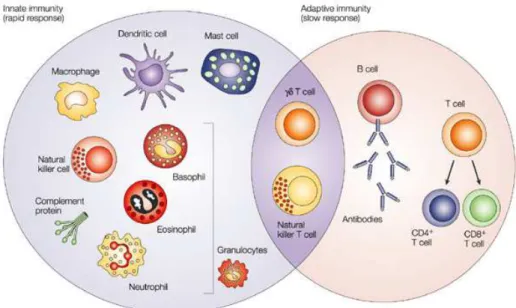
Figure 1.8 Components of immune system. The innate and adaptive immunity are
two inter-related defense systems103. Innate immunity provides a fast response,
which relies on soluble molecules and cell types such as basophils, eosinophils, neutrophils, mast cells, Mø, dendritic cells and natural killer cells. The adaptive immune response provides a slower response but is highly specific and encompasses memory. It is mediated by soluble molecules, e.g. antibodies (Abs), as
well B and T lymphocytes. Natural killer T cells and γδT cells are cytotoxic
lymphocytes that straddle the interface of innate and adaptive immunity. Adapted
from104.
When pathogens cross epithelial barriers, they are confronted by the host
immune system, composed of two inter-related sub-systems (Figure 1.8)103.
to the same challenge, i.e. microorganisms, although this notion has been
put into question more recently105. The second layer of the immune system,
referred as the adaptive immune system, provides a more specific and robust defense mechanism activated via antigen receptors, which trigger clonal expansion of antigen-specific effector cells. Contrary to PRRs, antigen receptors can refine and increase their specificities and affinities towards antigens via a complex process involving gene recombination and
allowing for affinity maturation102. These two lines of defense serve the
same purpose - to recognize and eliminate or expel pathogens.
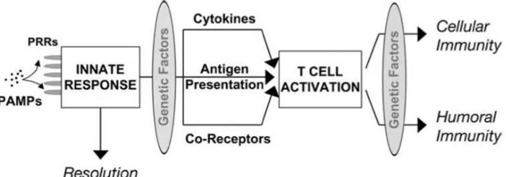
As microorganisms breach epithelial barriers, they are sensed by a
restricted number of PRRs106,107 that include Toll-like receptors (TLRs),
soluble receptors (i.e. lipopolysaccharide binding protein and soluble CD14 receptor, mannan binding lectin, C-reactive protein), scavenger receptors (i.e. macrophage scavenger receptor, dectin-1, mannose receptor), complement/Fc receptors and intracellular receptor (i.e. NOD-like receptor 2, protein kinase R and oligoadenylate synthetases). It is estimated that the
innate immune system can recognize about 103 PAMPs108.
PRRs do not undergo affinity maturation via gene recombination, in clear distinction with antigen receptors of adaptive immunity. Engagement of PRRs on DCs, monocyte/Mø, polymorphonuclear (PMN) cells, e.g. neutrophils, eosinophils, as well as natural killer (NK) cells and lymphokine-activated killer cells, triggers innate resistance mechanisms
that expel or destroy pathogens109. Innate resistance mechanisms include
microbial opsonization and production of intracellular anti-microbial
molecules110 as well as granuloma formation103. Other key component of
innate immunity are soluble anti-microbial peptides111, including defensins
as well as the complement system that trigger the lysis of microbial cell membranes via a proteolytic cascade that forms membrane pores while
In addition, innate immune cells express intracellular enzymes such as lysozyme that initiate the digestion of bacterial cell walls. Production of free radicals via the NADPH oxidase (NOX) family of reactive oxygen species (ROS) generating enzymes is another common resistance mechanism used by innate immune cells. This cytotoxic mechanisms can be further enhanced by PRR-driven nitric oxidase production, which occurs via the induction of the inducible form of nitric oxide synthase (iNOS/NOS2) as well as by mechanisms regulation intracellular iron
metabolism, which impact on the production of free radicals114,115. PRR are
also expressed on cells of epithelial barriers where they trigger the production of anti-microbial defense mechanisms while maintaining the functional integrity of tight junctions underlying barrier function integrity.
The inflammatory response triggered by soluble and cellular components of the innate immune system must be tightly regulated so that it targets microorganisms as soon as these gain systemic access. One of the constraints is that soluble and cellular components of innate immunity
must migrate towards the exact site of infection109. This occurs via the
expression of a variety of so-called pro-inflammatory genes encoding cytokines as well as other small proteins involved in cell-cell
communication such as chemokines116. This immediate response
encompasses local increase of microvascular permeability, increased expression of adhesion molecules by microvascular endothelial cells, which promote leukocytes recruitment at the site of infection. The adhesive and chemotactic gradient generated towards the site of infection recruits phagocytic cells, i.e. Mø and PMN cells that uptake and destroy extracellular pathogens. Cytokines and chemokines also alert other components of the adaptive immune system for the occurrence of infection.
Mø or PMN cells. These intracellular pathogens can be sensed by cytosolic PRRs, expressed in Mø and other innate immune cells such as PMN cells. PRR signaling is critical to trigger cell-autonomous immunity underlying host resistance to such intracellular pathogens. These intracellular PRRs include Nod-like receptors family members governing transcriptional responses (NOD1 and NOD2) and the activation of inflammasomes (NLRP3, NLRC4, NAIP, NLRP6 and NLRP12) among others. The activation of cytosolic PRRs triggers that cell-autonomous immune response that cell-autonomous immune response restricts intracellular pathogen replication and promotes pathogen clearance. This is assisted by non-cell autonomous responses involving NK cells activated in response to
proinflammatory cytokine i.e. IFN-γ and macrophage-derived cytokines to
promote cytolytic and proinflammatory mechanisms102,108. Activated NK
cells release cytotoxic granules such as perforin that forms a pore on cytosolic membranes and proteases known as granzymes resulting in programmed cell death of infected cells.
When pathogens are not eliminated via innate immune defense mechanisms, they can still be detected and eliminated by resistance mechanisms mediated by adaptive immunity, encompassing so called immunological memory that allows rapid and swift responses upon
re-infection102,117. This second layer of the immune system evolved
approximately 500 million years ago in early vertebrates118 and affords a
receptors are generated through random somatic chromosomal rearrangements and can bind a huge variety of antigens. However, PRRs play an important role in shaping adaptive immunity since the capacity of a given antigen to elicit an immune response relies strictly on antigen presentation by called innate antigen presenting cells that are activated via
the engagement of PRR, upon the recognition of PAMPs103,119 (Figure 1.9).
This is achieved essentially, but not exclusively, by DCs as well as Mø. They sample the environment for the presence of pathogens and recognize, engulf and digest antigenic peptides expressed by those pathogens. These are subsequently loaded into newly synthesized major histocompatibility complex (MHC) molecules to be presented to T cells. The engagement of PRRs on DCs and other antigen presenting cells is essential to induce the activation of a genetic program underlying the activation of antigen presenting cell (APC). This process is required so that APC become immunogenic, that is, are capable of triggering an immune response against specific antigens expressed by pathogens and loaded into the MHC class I and II molecules. This process that links innate and adaptive immunity allows for an integrated response of the host immune system,
which provides protection against infection119.
Figure 1.9 Interaction between innate immunity and adaptive immunity. Innate
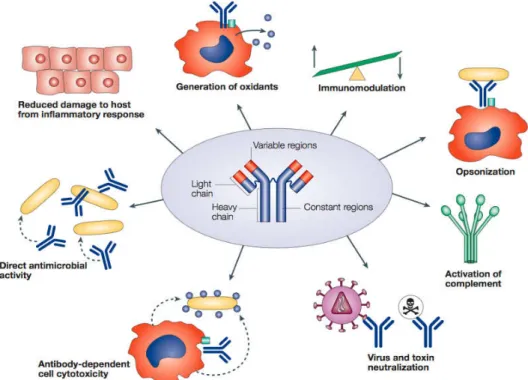
There are two main components of the adaptive immune system. One drives cellular immune response mediated by T cells and the other humoral immune response mediated by soluble antibodies (Abs) produced
by B cells (Figure 1.9). Major determinants of adaptive immune activation
are dependent on whether the pathogen resides on cytosol, intracellular
endosomes or extracellular environment102. Main components of
cell-mediated immune system include cytotoxic T cells (CD8+) that recognize
antigens expressed by intracellular pathogens loaded into MHC class I. T
helper cells (CD4+) recognize antigens expressed by extracellular pathogens,
loaded into MHC class II molecules. When sensed by specialized tissue resident APCs, pathogens are taken up and digested by the phagolysosome
system102. This process achieves the destruction of microbial organisms
while processing and loading antigens onto MHC class I or II molecules. Activated, tissue resident APC migrate to secondary lymphoid organism, such as lymph nodes and the spleen, in which they interact with
antigen-specific naive T cells102. Antigen recognition in the context of MHC class I
and II by TCR is essential for T cell activation and differentiation into specific effector functions.
Peptide structures derived from extracellular bacteria and toxins are
presented to CD4+ T cells via MHC class II molecules and can trigger the
differentiation of naïve TH cells into different effector functions that include
TH1 and TH2 cells. TH1 cells produce the cytokine IFN-γ whereas IL-4, IL-5
and IL-10 are the signature cytokines of TH2. There is also a tight
relationship between the TH cell activation and T cell dependent Ab
responses by B cells. Activation of TH cells by DCs as well as by B cells,
towards specific effector functions, such as TH1 or TH2, can drive the
production of different classes of Abs via cytokine dependent mechanisms regulating Ab class-switch recombination. The key transcription factors in
TH1 differentiation are signal transducer and activator of transcription 4