online | memorias.ioc.fiocruz.br
Microbiological and host features associated with corynebacteriosis
in cancer patients: a five-year study
CAS Martins1, 3, LMD Faria1, MC Souza3, TCF Camello3, E Velasco1, R Hirata Jr3, LCS Thuler2, AL Mattos-Guaraldi3/+
1Instituto Nacional do Câncer, Ministério da Saúde, Rio de Janeiro, RJ, Brasil 2Universidade Federal do Rio de Janeiro, Rio de Janeiro, RJ, Brasil 3Disciplina de Microbiologia e Imunologia, Faculdade de Ciências Médicas, Universidade do Estado do Rio de Janeiro, Av. 28 de
Setembro 87 fundos, 3º andar, 20551-030 Rio de Janeiro, RJ, Brasil
During a five-year period, 932 clinical isolates from cancer patients treated in a Brazilian reference centre were identified as corynebacteria; 86% of the cultures came from patients who had been clinically and microbiologically classified as infected and 77.1% of these patients had been hospitalised (71.1% from surgical wards). The adult solid tumour was the most common underlying malignant disease (66.7%). The univariate and multivariate analyses showed that hospitalised patients had a six-fold greater risk (OR = 5.5, 95% CI = 1.15-26.30 p = 0.033) related to 30-day mor-tality. The predominant species were Corynebacterium amycolatum (44.7%), Corynebacterium minutissimum (18.3%) and Corynebacterium pseudodiphtheriticum (8.5%). The upper urinary tracts, surgical wounds, lower respiratory tracts, ulcerated tumours and indwelling venous catheters were the most frequent sources of C. amycolatum strains.
Corynebacterium jeikeiuminfection occurred primarily in neutropenic patients who have used venous catheters, while infection caused by C. amycolatum and other species emerged mainly in patients with solid tumours.
Key words: Corynebacterium amycolatum - Corynebacterium infection - cancer
Financial support: INCA, FAPERJ, CNPq, CAPES, SR-2/UERJ, PRONEX
+ Corresponding author: [email protected] Received 15 May 2009
Accepted 20 August 2009
Corynebacterium species have recently been recog-nised as important pathogens that infect immunocom-promised patients. Despite the increase in the number of reports of severe infection caused by non-diphtheria corynebacteria, these organisms are still frequently dismissed as contaminants. Corynebacterium spphave been cited with an increased frequency as pathogens of nosocomial infections associated with septicaemias, en-docarditis, infections of surgical wounds, prostheses and infections related to the central venous catheter. Geo-graphical variations in the frequency of isolated species and variations in the natural and acquired antimicrobial resistance have been described (Osterwalder et al. 1986, Rozdzinski et al. 1991, van der Lelie et al. 1995).
Recent advances in Corynebacterium identification have shown that the genus exhibits considerable taxo-nomic complexity and that the phenotypic markers used in the past for its identification can be ambiguous (Bar-reau et al. 1993, Brandenburg et al. 1996, Oteo et al. 2001, Camello et al. 2003, Meyer & Reboli 2005, Otsuka et al. 2006). When these infections are linked to multi-resistant species, they are difficult to treat (Bodey 1995, Funke et al. 1996, Camello et al. 2003, Funke & Bernard 2003). Multiple resistance to antimicrobial agents has been described among some species (Lagrou et al. 1998,
Zalas et al. 2004); for example, mutations in the gyrase genes are determinants of the resistance to quinolones in
Corynebacterium striatum and Corynebacterium amy-colatum strains (Sierra et al. 2005).
In Rio de Janeiro, Brazil, recent studies conducted at a teaching hospital showed that 68% of the clinical iso-lates of corynebacteria species corresponded to Coryne-bacterium pseudodiphtheriticum, C. amycolatum, Corynebacterium propinquum and Corynebacterium minutissimum. The urinary tract and the venous access were the sites most commonly affected. In the blood and the respiratory tract, there was a predominance of
C. pseudodiphtheriticum and C. propinquum, while
Corynebacterium xerosis and C. amycolatum were the most commonly observed species in central venous catheters. C. propinquum and C. minutissimum were the most frequently observed species in surgical wounds. Data indicated the occurrence of multi-resistant pheno-types and the possibility of severe infections due to C. pseudodiphtheriticum, a pathogen usually overlooked in emerging countries (Camello et al. 2003, 2009).
are also not completely safe for the treatment of gram pos-itive life-threatening infections partially due to the emer-gence of microbial resistance (Boucher et al. 2000, Dobbs et al. 2006, Schoen et al. 2009).
The aim of this descriptive study was to assess the microbiological and clinical aspects as well as factors related to 30-day mortality in cancer patients with corynebacteriosis.
PATIENTS, MATERIALS AND METHODS
Study setting - In this descriptive study, we retro-spectively reviewed clinical and microbiological data over a five-year period (from January 2000-December 2004). The patient information originated from one of the units (Cancer Hospital I with 200 beds, Cancer Hospital II with 90 beds and Cancer Hospital III with 60 beds) of the National Cancer Institute (INCA) in Rio de Janeiro, Brazil.
The analysis included bacteriological data from 88,541 cultures and 1,100 irregular gram-positive rods isolated from cancer patients. The operational units in-cluded the Laboratory of Microbiology and the Hospital Infection Control Committee (HICC) at the INCA and the Laboratory of Diphtheria and Corynebacteria, State University of Rio de Janeiro (UERJ), Brazil.
During the period of the study, there were no chang-es in the technical team who procchang-essed the material for bacteriological cultures or in the medical team who assisted the patients.
Ethics - This paper was submitted and approved by the Ethical Research Committee at INCA (CEP 008/06) and complies with the Brazilian Government’s Ethical Guidelines for research involving human beings (resolu-tion of the Na(resolu-tional Health Council/Ministry of Health).
Diagnostic measures - The study analysed the clini-cal characteristics of 315 patients with Corynebacterium
isolates who had malignant diseases or underwent bone marrow transplantation in the last two years of the study. Data assessment was performed on the basis of a medi-cal records review of patients.
The hospitalised patients were monitored by at least one member of the HICC as a part of the antimicrobial vigilance routine through daily microbiology labora-tory charts. Ambulalabora-tory patients were identified by the dressing nurses committee, catheter ambulatory (both for children and adults) and bacteriological charts.
In addition to the physicians’ experiences of treating patients with cancer in INCA, the diagnosis of bacterial infection was staged according to the Centers for Disease Control and Prevention (CDC) classification (CDC 1992).
The patients with positive cultures were interpreted as infected when these cultures were derived from a nor-mally sterile site associated with a febrile illness. Alter-natively, patients with positive cultures were interpreted as infected when a Corynebacterium spp was isolated from two or more non-sterile sites in which there was a suspicion of infection and the physician considered it clinically significant to immediately start a specific an-timicrobial therapy. A positive urinary culture was con-sidered as significant in the presence of local (dysuria,
polyuria) or systemic signs of infection; pyuria was only required in non-neutropenic patients. Tracheal secretions in intubated patients, sputum or tracheal aspirations in non-intubated patients were considered as positive if they were associated with clinical or radiological signs that indicated an infection. Protected sampling was not performed in bronchoscopy (Garner et al. 1988, Hughes et al. 1996, Berghmans et al. 2003).
Bacterial strains and identification - We retrospec-tively reviewed 932 strains identified as Corynebacte-rium spp that were recovered from representative clini-cal sites of cancer patients with signs and symptoms of bacterial infection. Corynebacterium-like colonies were selected for further identification when they were grown in any quantity from normally sterile body fluid or when they were isolated in significant numbers or in pure culture from other specimens obtained at clini-cal sites in which infection was suspected (Funke & Bernard 2003). All clinical samples yielding more than three organisms were regarded as contaminated and discarded (Thomson 2007).
The Maki’s semi-quantitative method was used to distinguish infection (> 15 colonies) from contamination of catheter-tips (Maki et al. 1977). For quantitative BAL fluid cultures, a colony count > 103 colony-forming units (CFU) mL-1 of potential pathogens was considered posi-tive. Isolation of two or three species of microorganisms was regarded as a polymicrobial infection for catheter tips and the lower respiratory tract, respectively. Mi-croorganisms were identified from the urine cultures in cystine lactose electrolytes deficient agar (CLED; Mer-ck, Darmstadt, Germany) and were considered to be po-tential pathogens when the growth was > 104 CFU mL-1 as the only isolate or > 105 CFU mL-1 as the predominant isolate; > 103 CFU mL-1, in cases of nephropathies, was also considered a potential pathogen. Blood cultures were always obtained in pairs, wherein at least one of the samples was collected through the central venous catheter, if present. Blood specimens were inoculated in Bactec Plus anaerobic⁄aerobic vials and processed in a Bactec 9240 continuous-monitoring system (Becton-Dickinson Microbiology System, Cockeysville, MD, USA). Other clinical specimens were inoculated onto a Columbia agar base with the addition of 5% sheep’s blood and incubated at 37oC in 3-5% CO
2 atmosphere and monitored for 72 h.
In addition to Gram staining, colonial morphology, pigmentation and haemolysis, Corynebacterium-like colonies were characterised using the API-Coryne Sys-tem (BioMérrieux, Lyon, France) (Freney et al. 1991). Microorganisms were also submitted to the following conventional biochemical assays: catalase, pyrazinami-dase, lypophilic activities, motility, nitrate reduction, hydrolysis of urea and esculin, acidification of glucose, maltose, sucrose, mannitol and xylose, as well as oxida-tion-fermentation and CAMP reaction tests (Camello et al. 2003, Funke & Bernard 2003).
stan-dard (150 x 106 CFU mL-1 by direct colonial suspension),
adjusted for optical density at λ = 550 nm (Vitek colo -rimeter Durham, NC, USA). The plates were incubated at 35ºC in ambient air for 24 h and reconfirmed at 48 h in a cation-adjusted Mueller-Hinton Agar with the addition of 5% sheep’s blood (Funke et al. 1997). As there is not yet a defined standard by the Clinical and Laboratory Standards Institute for interpreting the results of disk diffusion tests (CLSI 2007), we used the breakpoint for penicillin suggested for Staphylococcus.
For the other antimicrobial agents, we used the breakpoints for other microorganisms but not Haemo-philus spp or Neisseria gonorrhoeae, which had been validated by previous studies (Martinez-Martinez et al. 1995, Weiss et al. 1996, Zinkernagel et al. 1996, Funke et al. 1997).
The intermediate results were included as resistant. Microorganisms were tested against 15 antimicrobial agents, according to the clinical criteria for empirical therapy in patients with underlying malignancy and infection. The in vitro antimicrobial susceptibility test associated with the analysis of clinical relevance used the interpretation criteria suggested by the Sanford Guide (Gilbert et al. 2006): (+) usually clinically ef-fective or > 60% susceptible, (±) clinical trials lacking or 30-60% susceptible and (0) not clinically effective or < 30% susceptible.
Statistical analysis - The variables were compared using a Pearson Chi square or Fisher exact test. For statistical significance, the value was established at p < 0.05. A logistic regression model was developed to identify the variables independently associated with the 30-day mortality. This model included every variable that showed a statistically significant association (p <
0.05) in the univariate analysis. The softwares used for the statistical analysis were Epi-Info 2000 for Windows, version 3.3.2 and SPSS, version 14.
RESULTS
During a five-year period (from January 2000-Decem-ber 2004), the clinical microbiology laboratory at INCA conducted 88,541 cultures, of which 25,173 (28.4%) were positive (n = 36,199). Gram-positive rods represented 1,436 (4%) of the clinical isolates. The genus Corynebac-terium corresponded with 932 (84.7%) of the total (1,100 strains) number of irregular gram-positive rods (Table I).
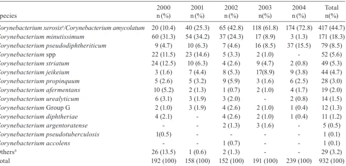
The Corynebacterium species isolated from cancer patients presenting signs and symptoms of infection are displayed in Table II. Most of the isolates (44.7%) were recognised as C. amycolatum, followed by C. minutis-simum (18.3%) and C. pseudodiphtheriticum (8.5%).
C. jeikeium was the sixth in frequency among the iso-lates (4.7%). The species Corynebacterium ulcerans and
Corynebacterium pseudotuberculosis, which are usually associated with zoonoses, were absent or observed in a very low incidence in our population, respectively.
Taking into account the retrospective analysis of our study from 2000-2004, the isolation of C. amycolatum/ xerosis increased from 10.4-72.8%. On the other hand,
C. minutissimum decreased from 31.3-1.3%; the percent-age of Corynebacterium spp (unidentified species)went from 11.5-0% and C. striatum from 12.5-0.8%. C. xe-rosis comprised 20 isolates in 2000 and six in 2001 and was absent from new isolates in the subsequent years.
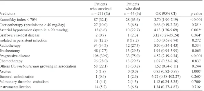
During the second phase of the study (from Janu-ary 2003-December 2004), 10 Corynebacterium species were observed in 425 clinical isolates obtained from 315 cancer patients. C. amycolatum was the most frequent clinical isolate, followed by C. pseudodiphtheriticum, C. jeikeium and C. minutissimum (Table III). When only bloodstream infections were considered, catheter-related infections were frequently observed. C. amycolatum was widely distributed in all topographies. The upper urinary tract, surgical wounds, lower respiratory tract, ulcerated tumours and indwelling venous catheters were the most
TABLE I
Strains of irregular gram-positive rods isolated from cancer patients between 2000-2004
Genus
2000 n(%)
2001 n(%)
2002 n(%)
2003 n(%)
2004 n(%)
Total n(%)
Corynebacterium 192 (80.3) 158 (83.2) 152 (78.8) 191 (88.0) 239 (91.6) 932 (84.7)
Arcanobacterium 16 (6.7) 4 (2.1) 8 (4.1) 8 (3.7) 5 (1.9) 41 (3.7)
Brevibacterium 13 (5.4) 1 (0.5) 7 (3.6) 7 (3.2) 4 (1.5) 32 (2.9)
Actinomyces 6 (2.5) 5 (2.6) 11 (5.7) 1 (0.5) 2 (0.8) 25 (2.3)
Aureobacterium 2 (0.8) 10 (5.3) 8 (4.1) 1 (0.5) 2 (0.8) 23 (2.1)
Arthrobacter - 7 (3.7) 3 (1.6) 4 (1.8) 7 (2.6) 21 (1.9)
Leifsonia 8 (3.3) 4 (2.1) 3 (1.6) 1 (0.5) 1 (0.4) 17 (1.5)
Propionibacterium 1 (0.5) 1 (0.5) - 3 (1.3) 1 (0.4) 6 (0.6)
Rothia 1 (0.5) - 1 (0.5) 1 (0.5) - 3 (0.3)
Total 239 (100) 190 (100) 193 (100) 217 (100) 261 (100) 1,100 (100)
the strains identified as gram-positive regular rods [Lactobacillus (213), Bacillus (94), Rhodococcus (17), Listeria (9), Nocardia
frequent sources of C. amycolatum strains. C. pseudo-diphtheriticum strains were mainly isolated from the lower and upper respiratory tract and C. jeikeium from the intravenous sites and skin lesions. The antimicrobial spectra of microorganisms are exhibited in Table IV.
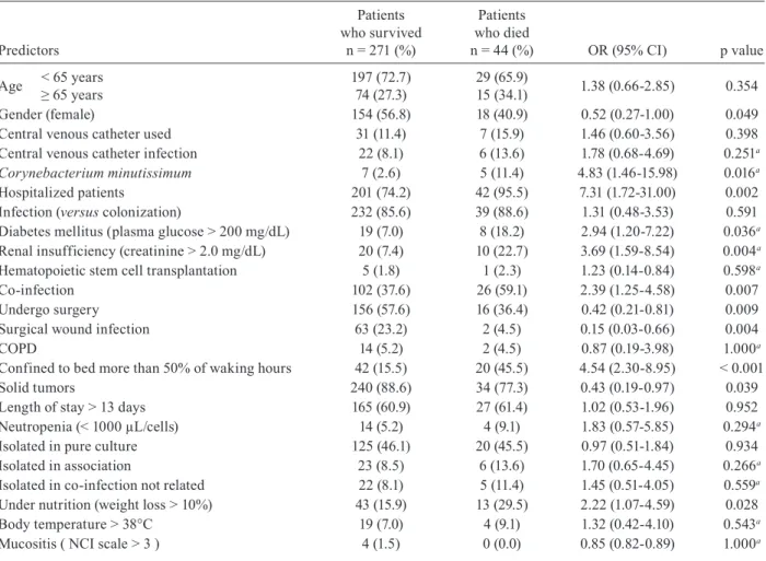
Clinical data of the 315 cancer patients with fever and/or other signs of infection were retrieved. The epi-demiological characteristics, the topographies that were involved and the factors that predisposed cancer patients to corynebacteria infection are depicted in Table V. The findings were interpreted as infection in 86% of patients and specific treatments were initiated. Corynebacterium
strains were observed as the only microorganism in 46% of the cultures. Bacterial colonisation was confirmed in 44 cases (14%), 35 of which originated in the respiratory tract (25 rhinosinusal, 3 lower respiratory and 7 upper respiratory), four cases were in central venous catheters (Maki method negative), two cases were in skin/tumours and one case was in a surgical wound; two cases were isolated from the digestive tract.
The main underlying malignant diseases were 66.7% adult solid tumours, followed by tumours of the central nervous system and paediatric solid tumours (10.8% and 9.2%, respectively). The patients from surgical wards (71.1%) were previously submitted to head and neck (25.1%), gynaecological (13.3%) and abdominal-pelvic (9.5%) surgeries. Surgical wound infections comprised 32.7% of these patients. The patients from clinical wards were from pediatrics (28.9%), oncology (12.1%) and hae-matology (7.3%). The majority of patients had been hos-pitalised (77.1%) and exposed to a hospital environment for a long term (median = 13 days).
The clinical-epidemiological characteristics of 315 patients with 30-day mortality involved a uni and multi-variate analysis of risk factors for corynebacteria-asso-ciated infection (Table IV).
DISCUSSION
Identification of non-diphtheria bacteria at the spe-cies level is often problematic. Recent advances in iden-tification have shown that the genus exhibits consider-able taxonomic complexity and the phenotypic markers that have been used in the past for its identification can be ambiguous. Even when sent to a reference labora-tory, 30-50% of coryneform bacteria isolates cannot be reliably identified at the species level. Consequently, there is a low rate of identification from clinical isolates (Schiff et al. 2004).
Variations in the occurrence of Corynebacterium
species during the course of the study were probably due to improvements in the taxonomy and laboratory diagnosis of the genus Corynebacterium. The substan-tial increase in the number of C. amycolatum isolates was partially due to the disappearance of C. xerosis and the significant reduction of C. striatum and C. minutis-simum, which reflects the progress in the diagnosis of
Corynebacterium species and consequently, the vari-ability in antimicrobial sensitivity patterns. The former
Corynebacterium CDC F-2 and CDC I-2 groups have been reclassified as C. amycolatum.
Due to imprecise diagnosis at the species level, data from the first three years (507 strains) were not included in the analysis of the bacteriological and clinical charac-teristics of cancer patients with corynebacteriosis. TABLE II
Corynebacterium species isolated from cancer patients between 2000-2004
Species
2000 n(%)
2001 n(%)
2002 n(%)
2003 n(%)
2004 n(%)
Total n(%)
Corynebacterium xerosisa/Corynebacterium amycolatum 20 (10.4) 40 (25.3) 65 (42.8) 118 (61.8) 174 (72.8) 417 (44.7)
Corynebacterium minutissimum 60 (31.3) 54 (34.2) 37 (24.3) 17 (8.9) 3 (1.3) 171 (18.3)
Corynebacterium pseudodiphtheriticum 9 (4.7) 10 (6.3) 7 (4.6) 16 (8.5) 37 (15.5) 79 (8.5)
Corynebacterium spp 22 (11.5) 23 (14.6) 5 (3.3) 2 (1.0) - 52 (5.6)
Corynebacterium striatum 24 (12.5) 10 (6.3) 4 (2.6) 9 (4.7) 2 (0.8) 49 (5.3)
Corynebacterium jeikeium 3 (1.6) 7 (4.4) 8 (5.3) 17(8.9) 9 (3.8) 44 (4.7)
Corynebacterium propinquum 5 (2.6) 5 (3.2) 9 (5.9) 3 (1.6) 6 (2.5) 28 (3.0)
Corynebacterium afermentans 10 (5.2) 2 (1.3) 1 (0.7) 2 (1.0) 4 (1.7) 19 (2.0)
Corynebacterium urealyticum 6 (3.1) 3 (1.9) 3 (2.0) - 2 (0.8) 14 (1.5)
Corynebacterium Group G 2 (1.0) 3 (1.9) 4 (2.6) 2 (1.0) 1 (0.4) 12 (1.3)
Corynebacterium diphtheriae 4 (2.1) - 4 (2.6) 2 (1.0) 1 (0.4) 11 (1.2)
Corynebacterium argentoratense - - 2 (1.3) 3 (1.6) - 5 (0.5)
Corynebacterium pseudotuberculosis 1(0.5) - - - - 1 (0.1)
Corynebacterium accolens - - 1 (0.7) - - 1 (0.1)
Othersb 26 (13.5) 1 (0.6) 2 (1.3) - - 29 (3.2)
Total 192 (100) 158 (100) 152 (100) 191 (100) 239 (100) 932 (100)
Many non-diphtheria corynebacteria-caused ac-cess infections can be effectively treated by antibiotics and local care. Susceptibility testing of corynebacteria is highly recommended to establish a specific therapy (CLSI 2007). Except for the unvarying activity of van-comycin against corynebacteria, the variability in resis-tance to other classes of antimicrobial agents emphasises the need for the continuous surveillance of their resis-tance patterns. Resisresis-tance to oxacillin was observed for all corynebacteria species isolated from cancer patients at INCA. The data emphasise the relevance of suscep-tibility testing of Corynebacterium isolates during the diagnosis and treatment of infections in cancer patients.
A growing number of reports have confirmed the importance of C. amycolatum in the aetiology of a variety of infectious processes. In the reviewed litera-ture, many C. amycolatum clinical isolates were at first identified in different laboratories as C. xerosis (Funke et al. 1996) and C. minutissimum (Zinkernagel et al. 1996). After the mid-1990s, with the advent of the taxo-nomical characterisation of C. amycolatum, this spe-cieshas been described as a causal agent of infections with high morbidity and mortality rates. Multi-resis-tant C. amycolatum strains were isolated from patients with septicaemia (Berner et al. 1997, de Miguel et al. 1999, Camello et al. 2003), septic arthritis after vascu-lar transplants (Cvascu-larke et al. 1999), cardiac electrode implantation (Vaneechoutte et al. 1998), infections in vascular prostheses and open fractures (von Graevenitz et al. 1998), peritonitis in patients undergoing perito-neal dialysis (Chiu et al. 2005), infectious endocardi-tis accompanied by aorto-atrial fistula (Daniels et al. 2003), nosocomial endocarditis (Knox & Holmes 2002) and septicaemia in leukemic patients (de Miguel et al. 1999). These reports drew attention to the clinical sig-nificance of C. amycolatum strains in different clinical materials (Esteban et al. 1999).
The clinical characteristics of the patients with fa-vourable outcomes and documented corynebacteriosis could not be compared with those patients who died (controls). Due to the nature of the present research, which was a retrospective study, we could not investi-gate the pathogenicity of the isolates in relation to 30-day mortality in cancer patients. For the same reason, it was impossible to establish prognostic factors in these patients considering their descriptive characters. Anoth-er limitation of this study was the exclusion of isolates from 2000-2002 in the complete analysis due to major changes in taxonomical classification. Nevertheless, the sampling procedures adopted in this study allowed us to detect some patterns and characteristics of the popula-tion under investigapopula-tion.
The univariate and multivariate analyses showed that hospitalised patients had a six-fold greater risk (OR = 5.5, 95% CI = 1.15-26.30 p = 0.033) related to 30-day mortality. Also of statistical significance were patients bedridden for longer than 50% of the day with neoplastic disease in progress or diabetes mellitus.
The study revealed that corynebacteria are increas-ingly being recognised as a cause of infections in cancer patients. These organisms have been underreported, but
T A B L E I II S tr a in s o f n o n d ip h th e ri al C o ry n eb ac te riu m sp ec ie s i so la te d f ro m v ar ie d c lin ic al s ou rc es i n c an ce r p at ie nt s (
n = 3
15 ) O rga n is m T ot al n ( % ) R e sp ir at o ry t ra ct in fe ct io n s S u rg ic al w o u n d in fe ct io n s U ri n a ry t ra ct in fe ct io n s In tra v e n o us si te s Sk in / tu m ou r R h in o si n u sa l M is c U /L U /L C at h eter B lo o d C o ry n e b a ct e ri u m a m yc o la tu m 2 3 0 ( 7 3 .0 ) 7/ 4 8 56 38 /0 19 4 9/ 2 2 3 24 C o ry n e b a ct e ri u m p se u d o d ip h th e ri ti c u m 3 9 ( 1 2 .4 ) 3/1 1 4 0 /0 0 0 0 /1 19 1 C o ry n e b a ct e ri u m j e ik e iu m 1 3 ( 4 .1 ) 1 /1 2 0 /0 4 1 1/ 3 0 0 C o ry n eb ac te riu m m in u ti ss im u m 1 2 ( 3 .8 ) 0/ 2 1 0/ 3 1 0 2 /1 0 2 C o ry n eb ac te riu m p ro p in q u u m 8 ( 2 .5 ) 0/ 3 0 1/ 0 1 0 0 /0 2 1 C o ry n eb ac te riu m s tr ia tu m 6 ( 1 .9 ) 1/ 3 2 0 /0 0 0 0 /0 0 0 C o ry n eb ac te riu m a fe rm e n ta n s 3 ( 1 .0 ) 0 /1 0 0 /0 1 1 0 /0 0 0 C o ry n e b a ct e ri u m a rg e n to ra te n se 2 ( 0 .7 ) 0 /0 0 0 /0 1 1 0 /0 0 0 C o ry n eb ac te riu m G ro u p G 1 ( 0 .3 ) 0 /0 0 0 /0 0 0 0 /0 1 0 C o ry n e b a ct e ri u m u re a ly ti c u m 1 ( 0 .3 ) 0 /0 0 1/ 0 0 0 0 /0 0 0 T ot al 31 5 ( 10 0 ) 12 /6 9 65 4 0/ 3 27 7 12 /2 7 25 28 L : lo w e r; M is c: m is ce ll a n e o u s [g e n it al tr a ct : 8 ( C . a m yc o la tu m ), g a st ro in te st in al tr a ct : 5 [ C . a m yc o la tu m : 4; C . m in u ti ss im u m : 1) , n e p h ro st o m y : 3 ( C . a m yc o la tu m ), ey e: 3 ( C . a m yc o -la tu m : 2 ; C . p ro p in q u u m : 1) , b o n e: 2 ( C . a m yc o la tu m ), o ro p h a ry n x : 2 ( C . a m yc o la tu m : 1 ; C . m in u ti ss im u m : 1) , ab sc e ss : 2 ( C . a m yc o la tu m ); si n u s: 2 ( C. a my c o la tu m : 1 ; C . p se u d o d ip h -th e rit ic u m : 1 ), b io p sy
: 1 (
TABLE V
Risk factors to mortality related to Corynebacterium spp infection in 315 cancer patients
Predictors
Patients who survived
n= 271 (%)
Patients who died
n = 44 (%) OR (95% CI) pvalue
Age < 65 years
≥ 65 years 197 (72.7)74 (27.3)
29 (65.9)
15 (34.1) 1.38 (0.66-2.85) 0.354
Gender (female) 154 (56.8) 18 (40.9) 0.52 (0.27-1.00) 0.049
Central venous catheter used 31 (11.4) 7 (15.9) 1.46 (0.60-3.56) 0.398
Central venous catheter infection 22 (8.1) 6 (13.6) 1.78 (0.68-4.69) 0.251a
Corynebacterium minutissimum 7 (2.6) 5 (11.4) 4.83 (1.46-15.98) 0.016a
Hospitalized patients 201 (74.2) 42 (95.5) 7.31 (1.72-31.00) 0.002
Infection (versus colonization) 232 (85.6) 39 (88.6) 1.31 (0.48-3.53) 0.591
Diabetes mellitus (plasma glucose > 200 mg/dL) 19 (7.0) 8 (18.2) 2.94 (1.20-7.22) 0.036a
Renal insufficiency (creatinine > 2.0 mg/dL) 20 (7.4) 10 (22.7) 3.69 (1.59-8.54) 0.004a
Hematopoietic stem cell transplantation 5 (1.8) 1 (2.3) 1.23 (0.14-0.84) 0.598a
Co-infection 102 (37.6) 26 (59.1) 2.39 (1.25-4.58) 0.007
Undergo surgery 156 (57.6) 16 (36.4) 0.42 (0.21-0.81) 0.009
Surgical woundinfection 63 (23.2) 2 (4.5) 0.15 (0.03-0.66) 0.004
COPD 14 (5.2) 2 (4.5) 0.87 (0.19-3.98) 1.000a
Confined to bed more than 50% of waking hours 42 (15.5) 20 (45.5) 4.54 (2.30-8.95) < 0.001
Solid tumors 240 (88.6) 34 (77.3) 0.43 (0.19-0.97) 0.039
Length of stay > 13 days 165 (60.9) 27 (61.4) 1.02 (0.53-1.96) 0.952
Neutropenia (< 1000 µL/cells) 14 (5.2) 4 (9.1) 1.83 (0.57-5.85) 0.294a
Isolated in pure culture 125 (46.1) 20 (45.5) 0.97 (0.51-1.84) 0.934
Isolated in association 23 (8.5) 6 (13.6) 1.70 (0.65-4.45) 0.266a
Isolated in co-infection not related 22 (8.1) 5 (11.4) 1.45 (0.51-4.05) 0.559a
Under nutrition (weight loss > 10%) 43 (15.9) 13 (29.5) 2.22 (1.07-4.59) 0.028
Body temperature > 38°C 19 (7.0) 4 (9.1) 1.32 (0.42-4.10) 0.543a
Mucositis ( NCI scale > 3 ) 4 (1.5) 0 (0.0) 0.85 (0.82-0.89) 1.000a
TABLE IV
Comparison of antimicrobial spectra of Corynebacterium species (315 patients/425 strains)
Species (samples) Van
c
o
m
y
ci
n
A
m
ik
a
ci
n
T
o
b
ra
m
y
ci
n
C
ef
a
z
ol
in
C
ef
tr
ia
x
o
ne
C
ef
ta
z
id
im
e
C
efe
p
im
e
Im
ip
e
n
em
P
e
n
ic
il
li
n G
A
m
p
ic
il
li
n
O
x
ac
il
li
n
C
ip
rof
lo
x
a
ci
n
O
fl
ox
a
ci
n
E
ry
th
ro
m
yc
in
C
h
lo
ra
m
p
h
e
n
ic
ol
Corynebacterium amycolatum (292) + + + + + 0 + + + + 0 ± ± ± ±
Corynebacterium pseudodiphtheriticum (53) + + + + + + + + + + + + + ± +
Corynebacterium jeikeium (26) + 0 0 0 0 0 0 0 0 0 0 0 0 0 0
Corynebacterium minutissimum (20) + + + + + 0 + + ± ± 0 ± ± ± ±
Corynebacterium striatum (11) + + + + + 0 + + + + 0 ± ± + 0
Corynebacterium propinquum (9) + + + + + ± + + + + ± + + ± +
Corynebacterium afermentans (6) + + + + + ± + + ± ± ± + + ± +
Corynebacterium Group G (3) + + + + + 0 + + + + 0 + + + +
Corynebacterium argentoratense (3) + + + + + ± + + + + 0 + + + +
they may account for approximately 4% of the cases of infection in patients presenting solid tumours. The data reflects a tendency towards infection or colonisation by
Corynebacterium in cancer patients who have undergone surgical procedures and a long subsequent hospitalisa-tion period. From the study data, we also emphasise the need for the rigorous identification of Corynebacterium
species isolates from different sites, especially of inva-sive strains in patients with clinical conditions of per-sistent fever and no other attributable site of infection.
C. jeikeium infection occurs primarily in neutropenic patients who have used venous catheters (Rozdzinski et al.1991), while C. amycolatum infection appears mainly in patients with solid tumours.
REFERENCES
Barreau C, Bimet F, Kiredjian M, Rouillon N, Bizet C 1993. Compar-ative chemotaxonomic studies of mycolic acid-free coryneform bacteria of human origin. J Clin Microbiol31: 2085-2090.
Berghmans T, Sculier JP, Klastersky J 2003. A prospective study of infections in lung cancer patients admitted to the hospital. Chest 124: 114-120.
Berner R, Pelz K, Wilhelm C, Funke A, Leititis JU, Brandis M 1997. Fatal sepsis caused by Corynebacterium amycolatum in a prema-ture infant. J Clin Microbiol35: 1011-1012.
Bodey GP 1995. Emerging antimicrobial-resistant pathogens in the immunocompromised host. Curr Opin Infect Dis8: 411-414.
Boucher HW, Wennersten CB, Eliopoulos GM 2000. In vitro
activi-Predictors
Patients who survived
n= 271 (%)
Patients who died
n = 44 (%) OR (95% CI) pvalue
Karnofsky index < 70% 87 (32.1) 28 (63.6) 3.70 (1.90-7.19) < 0.001
Corticotherapy (prednisone > 40 mg/day) 27 (10.0) 3 (6.8) 0.66 (0.19-2.28) 0.781a
Arterial hypotension (systolic < 90 mm/hg) 18 (6.6) 10 (22.7) 4.13 (1.76-9.69) 0.002a
Graft-versus-host disease 2 (0.7) 1 (2.3) 3.12 (0.27-35.24) 0.364a
Isolated in persistent infection 33 (12.2) 8 (18.2) 1.60 (0.68-3.74) 0.272
Radiotherapy 94 (34.7) 12 (27.3) 0.70 (0.34-1.43) 0.334
Tracheotomy 48 (17.7) 13 (29.5) 1.94 (0.94-3.99) 0.065
Progressive disease 108 (39.9) 33 (75.0) 4.52 (2.19-9.34) < 0.001
Chemotherapy 76 (28.0) 13 (29.5) 1.07 (0.53-2.16) 0.837
Others Corynebacterium growing in association 58 (22.1) 13 (30.2) 1.52 (0.74-3.11) 0.244
Ascites 5 (1.8) 0 (0.0) 0.85 (0.82-0.89) 1.000a
Tumoral embolization 1 (0.4) 1 (2.3) 6.27 (0.38-102.27) 0.260a
Pulmonary thrombo-embolism 11 (4.1) 2 (4.5) 1.12 (0.24-5.25) 0.700a
Instrumentalization 14 (5.2) 3 (6.8) 1.34 (0.37-4.87) 0.716a
a:Fisher exact test
TABLE VI
Multivariate analysis of independent risk factors for 30-day mortality
Variable OR 95% CI p value
Hospitalized patients 5.500 1.150 26.304 0.033
Confined to bed more than 50% of waking hours 3.621 1.173 11.179 0.025
Corynebacterium minutissimum 3.560 0.681 18.625 0.133
Progressive disease 3.167 1.346 7.453 0.008
Diabetes mellitus (plasma glucose > 200 mg/dL) 3.106 1.015 9.504 0.047
Renal insufficiency (creatinine > 2.0 mg/dL) 2.710 0.951 7.722 0.062
Arterial hypotension (systolic < 90 mm/hg) 2.429 0.851 6.934 0.097
Undernutrition (weight loss > 10%) 1.842 0.710 4.779 0.209
Isolated in co-infection no related 1.260 0.572 2.773 0.566
Solid tumors 0.762 0.268 2.169 0.610
Gender (male) 0.656 0.295 1.457 0.301
Undergo surgery 0.441 0.192 1.012 0.053
ties of the glycylcycline GAR-936 against gram-positive bacteria. Antimicrob Agents Chemother 44: 2225-2229.
Brandenburg AH, van Belkum A, van Pelt C, Bruining HA, Mou-ton JW, Verbrugh HA 1996. Patient-to-patient spread of a single strain of Corynebacterium striatum causing infections in a surgi-cal intensive care unit. J Clin Microbiol34: 2089-2094.
Camello TCF, Mattos-Guaraldi AL, Formiga LCD, Marques EA 2003. Nondiphtherial Corynebacterium species isolated from clinical specimens of patients in a University Hospital, Rio de Janeiro, Brazil. Brazilian J Microbiol 34: 39-44.
Camello TCF, Souza MC, Martins CAS, Damasco PV, Marques EA, Pimenta FP, Pereira GA, Hirata Jr R, Mattos-Guaraldi AL 2009. Corynebacterium pseudodiphtheriticum isolated from rel-evant clinical sites of infection: a human pathogen overlooked in emerging countries. Lett App Microbiol 48: 458-464.
CDC - Center for Disease Control and Prevention 1992. 1993 revised classification system for HIV infection and expanded surveil-lance case definition for AIDS among adolescents and adults. Morb Mortal Wkly Rep Recomm Rep41: 1-19.
Chiu YL, Wu VC, Wun KD, Hsueh PR 2005. Recurrent peritonitis caused by Corynebacterium amycolatum in a patient undergo-ing continuous ambulatory peritoneal dialysis. Clin Nephrol 63: 241-242.
Clarke R, Qamruddin A, Taylor M, Panigrahi H 1999. Septic arthri-tis caused by Corynebacterium amycolatum following vascular graft sepsis. J Infect38: 126-127.
CLSI - Clinical and Laboratory Standards Institute 2007. Methods for antimicrobial dilution and disk susceptibility testing of infre-quently isolated or fastidious bacteria. Approved Standard Docu-ment M45-A, CLSI, Wayne, p. 4-6.
Daniels C, Schoors D, Van Camp G 2003. Native valve endocarditis with aorta-to-left atrial fistula due to Corynebacterium amycola-tum.Eur J Echocardiogr4: 68-70.
de Miguel I, Rodriguez E, Martin AM 1999. Corynebacterium amy-colatum: sepsis in hematologic patients. Enferm Infecc Microbiol Clin17: 340-341.
Dobbs TE, Patel M, Waites KB, Moser SA, Stamm AM, Hoesley CJ 2006. Nosocomial spread of Enterococcus faecium resistant to vancomycin and linezolid in a tertiary care medical center. J Clin Microbiol44: 3368-3370.
Esteban J, Nieto E, Calvo R, Fernandez-Robals R, Valero-Guillen PL, Soriano F 1999. Microbiological characterization and clinical significance of Corynebacterium amycolatum strains. Eur J Clin Microbiol Infect Dis18: 518-521.
Freney J, Duperron MT, Courtier C, Hansen W, Allard F, Boeufgras JM, Monget D, Fleurette J 1991. Evaluation of API Coryne in comparison with conventional methods for identifying coryne-form bacteria. J Clin Microbiol 29: 38-41.
Funke G, Bernard KA 2003. Coryneform gram-positive rods. In: PR Murray, EJ Barron, JH Jorgensen, MA Pfaller, RH Yolken, Manual of Clinical Microbiology, 8th ed., ASM Press Washing-ton DC, p. 472-501.
Funke G, Punter V, von Graevenitz A 1996. Antimicrobial suscepti-bility patterns of some recently established coryneform bacteria. Antimicrob Agents Chemother40: 2874-2878.
Funke G, von Graevenitz A, Clarridge JE, Bernard KA 1997. Clini-cal microbiology of coryneform bacteria. Clin Microbiol Rev 10: 125-159.
Furtado GH, Mendes RE, Pignatari AC, Wey SB, Medeiros EA 2006. Risk factors for vancomycin-resistant Enterococcus faecalis bac-teremia in hospitalised patients: an analysis of two case-control studies. Am J Infect Control34: 447-451.
Garner IS, Iarvis WR, Emeri TG, Horan TC, Hughes IM 1988. CDC definitions for nosocomial infections. Am J Infect Contr 16: 128-140.
Gilbert DN, Moellering RC, Eliopoulos GM, Sande MA 2006. Com-parison of antimicrobial spectra. In DN Gilbert et al. The Sanford guide to antimicrobial therapy, 36th ed., Antimicrobial Therapy Inc, Hyde Park, p. 53-55.
Hughes WT, Flynn PM, Williams BG 1996. Nosocomial infection in patients with neoplastic diseases. In: CG Mayall, Hospital epide-miology and infection control, Williams & Wilkins, Baltimore, p. 618-631.
Knox KL, Holmes AH 2002. Nosocomial endocarditis caused by Corynebacterium amycolatum and other nondiphtheriae coryne-bacteria. Emerg Infect Dis8: 97-99.
Lagrou K, Verhaegen J, Janssens M, Wauters G, Verbist L 1998. Prospective study of catalase-positive coryneform organisms in clinical specimens: identification, clinical relevance and antibi-otic susceptibility. Diagn Microbiol Infect Dis30: 7-15.
Maki DG, Weise CE, Sarafin HW 1977. A semiquantitative culture method for identifying intravenous-catheter-related infection. N Engl J Med 296: 1305-1309.
Martinez-Martinez L, Ortega MC, Suarez AI 1995. Comparison of E-test with broth microdilution and disk diffusion for susceptibility testing of coryneform bacteria. J Clin Microbiol33: 1318-1321.
Meyer DK, Reboli AC 2005. Others Corynebacteria and Rhodococ-cus. In: GL Mandell, R Dolin, JE Bennet, Principles and prac-tice of infectious diseases, Churchill-Livingstone, New York, p. 2465-2478.
Osterwalder B, Frei R, Gratwohl A, Reber H, Speck B 1986. An-tibiotic-resistant Corynebacteria - a new problem of infec-tion in immunosuppressed patients. Schweiz Med Wochenschr 116: 880-884.
Oteo J, Aracil B, Alós JI, Gómez-Garcés JL 2001. Bacteriemias significativas por Corynebacterium amycolatum: un patógeno emergente. Enferm Infec Microbiol Clin19: 103-106.
Otsuka Y, Ohkusu K, Kawamura Y, Baba S, Ezaki T, Kimura S 2006. Emergence of multidrug-resistant Corynebacterium striatum as a nosocomial pathogen in long-term hospitalized patients with underlying diseases. Diag Microbiol Infect Dis54: 109-114.
Rozdzinski E, Kern W, Schmeiser T, Kurrle E 1991. Corynebacte-rium jeikeium bacteraemia at a tertiary care center. Infection 19: 201-204.
Schiff H, Mücke C, Lang SM 2004. Exit-site infections by non-diph-theria corynebacteria in CAPD. Perit Dial Int24: 454-459.
Schoen C, Unziker C, Stuhler G, Elias J, Einsele H, Grigoleit GU, Abele-Horn M, Mielke S 2009. Life-threatening infection caused by daptomycin-resistant Corynebacterium jeikeium in a neutro-penic patient. J Clin Microbiol 47: 2328-2331.
Sierra JM, Martinez-Martinez L, Vazquez F, Giralt E, Vila J 2005. Relationship between mutations in the gyrA gene and quinolone resistance in clinical isolates of Corynebacterium striatum and Corynebacterium amycolatum. Antimicrob Agents Chemother 49: 1714-1719.
Thomson Jr RB 2007. Specimen collection, transport and processing: bacteriology. In: PR Murray, EJ Baron, JH Jorgensen, ML Lan-dry, MA Pfaller, Manual of clinical microbiology, ASM Press, Washington DC, p. 485-514.
Vaneechoutte M, De Bleser D, Claeys G, Verschraegen G, De Baere T, Hommez J, Devriese LA, Riegel P 1998. Cardioverter-lead electrode infection due to Corynebacterium amycolatum. Clin Infect Dis27: 1553-1554.
von Graevenitz A, Frommelt L, Punter-Streit V, Funke G 1998. Diver-sity of coryneforms found in infections following prosthetic joint insertion and open fractures. Infection26: 36-38.
Weiss K, Laverdière M, Rivest R 1996. Comparison of antimicrobial
susceptibilities of Corynebacterium species by broth microdilu-tion and disk diffusion methods. Antimicrob Agents Chemother 40: 930-933.
Zalas P, Mikucka A, Gospodarek E 2004. Antibiotic sensitivity of Corynebacterium amycolatum. Med Dosw Mikrobiol56: 327-334.