Determining mutations in
G6PC
and
SLC37A4
genes in a sample of Brazilian
patients with glycogen storage disease types Ia and Ib
Marcelo Paschoalete Carlin
1, Daniel Zanetti Scherrer
1, Adriana Maria Alves De Tommaso
2,
Carmen Silvia Bertuzzo
1and Carlos Eduardo Steiner
11
Departamento de Genética Médica, Faculdade de Ciências Médicas, Universidade de Campinas,
Campinas, SP, Brazil.
2
Departamento de Pediatria, Faculdade de Ciências Médicas, Universidade de Campinas, Campinas,
SP, Brazil.
Abstract
Glycogen storage disease (GSD) comprises a group of autosomal recessive disorders characterized by deficiency of the enzymes that regulate the synthesis or degradation of glycogen. Types Ia and Ib are the most prevalent; while the former is caused by deficiency of glucose-6-phosphatase (G6Pase), the latter is associated with impaired glu-cose-6-phosphate transporter, where the catalytic unit of G6Pase is located. Over 85 mutations have been reported since the cloning ofG6PC and SLC37A4 genes. In this study, twelve unrelated patients with clinical symptoms sug-gestive of GSDIa and Ib were investigated by using genetic sequencing ofG6PC and SLC37A4 genes, being three confirmed as having GSD Ia, and two with GSD Ib. In seven of these patients no mutations were detected in any of the genes. Five changes were detected inG6PC, including three known point mutations (p.G68R, p.R83C and p.Q347X) and two neutral mutations (c.432G > A and c.1176T > C). Four changes were found inSLC37A4: a known point mutation (p.G149E), a novel frameshift insertion (c.1338_1339insT), and two neutral mutations (c.1287G > A and c.1076-28C > T). The frequency of mutations in our population was similar to that observed in the literature, in which the mutation p.R83C is also the most frequent one. Analysis of both genes should be considered in the investi-gation of this condition. An alternative explanation to the negative results in this molecular study is the possibility of a misdiagnosis. Even with a careful evaluation based on laboratory and clinical findings, overlap with other types of GSD is possible, and further molecular studies should be indicated.
Keywords: DNA-based diagnosis, glycogen storage disease,G6PC,SLC37A4, mutation.
Received: February 27, 2013; Accepted:October 11, 2013.
The transformation of glucose into glycogen occurs by chemical reactions carried out by specific enzymes (Diamentet al., 1994), and a deficiency in one of these leads to the accumulation of glycogen, resulting in heredi-tary disorders known as glycogen storage diseases (GSD) or glycogenosis (Rubinet al., 2006). There are currently 12 types of GSD, presenting differences in age of onset of symptoms, affected organs, specific enzyme defect, and clinical severity, and each GSD is named after specific en-zyme defect and organ impairment (Hickset al., 2011).
Deficiency in glucose-6-phosphatase (G6Pase, EC 3.1.3.9) activity causes GSD type I (GSD1), which is re-sponsible for more than 90% of the cases, affecting mainly the liver and the kidneys. In addition to being a key enzyme in the regulation of blood glucose homeostasis, catalyzing the final steps of glycogenolysis and gluconeogenesis
(Chou et al., 2002), it is also associated with the endo-plasmic reticulum (ER) and functions as a multicomponent system (van Schaftingen and Gerin, 2002).
The catalytic subunit of the system is located inside the ER and its defect causes subtype Ia (GSD Ia; OMIM 232200) (Leiet al., 1993). Additionally, there are trans-porters for the entry of substrate glucose-6-phosphate (G6P) into the ER and for the exit of the products, phos-phate and glucose. Defects in these transporters cause sub-type Ib (GSD Ib, OMIM 232220) (Gerinet al., 1997). The mechanism of ER membrane glucose transport remains un-known (Froissartet al., 2011).
The disease follows autosomal recessive inheritance and has an incidence of 1/100,000 to 1/400,000 live births, with up to 80% of the cases represented by subtype Ia (Chen, 2001; Hickset al., 2011). Patients with GSD Ia and Ib manifest a nearly identical metabolic phenotype, includ-ing hypoglycemia, hepatomegaly, hyperuricemia, lactic acidemia and hyperlipidemia (Moses, 1990), but GSD Ib patients also present neutropenia and myeloid dysfunction,
www.sbg.org.br
and these individuals are susceptible to recurrent bacterial infections, aphthous stomatitis, and inflammatory bowel disease (Visseret al., 2000). Nonetheless, neutropenia is not manifested by all GSD Ib patients (Kureet al., 2000; Martens et al., 2006). It is seen in only 20% of cases (Gitzelmann and Bosshard, 1993), and it has been proposed that it could be due toSLC37A4mutations with residual transport activity (Kureet al., 2000). Clinical differentia-tion between GSD types 1a, 1b and 3 may not always be possible (Tamhankaret al., 2012).
A biochemical essay for GSDI is useful to confirm the diagnosis and to recommend treatment; routine tests, however, do not allow the determination of the disease sub-type (Fernandeset al., 1969). Furthermore, since the en-zyme is not expressed in tissues such as fibroblasts or lymphocytes, their measurement is only possible by liver biopsy (Burchell, 1990), an invasive procedure considered stressful by many families. cDNA cloning of G6PC and
SLC37A4allowed the screening of mutations responsible for subtypes Ia and Ib, which enabled establishing an alter-native, less invasive diagnosis based on molecular biology techniques using blood samples (Parvariet al., 1997). A di-agnostic flowchart for this procedure was created based on mutation analysis combined with clinical and biochemical abnormalities (Rakeet al., 2000).
In the present study, we aimed to identify mutations in G6PC and SLC37A4 genes in patients clinically sus-pected of having GSD Ia and Ib, and to compare the molec-ular findings with clinical diagnosis in these individuals and classify their disease types and subtypes.
Twelve unrelated subjects with clinical features sug-gestive of GSD Ia or Ib were studied. Inclusion criteria were based on typical clinical and laboratory findings, con-sisting of recurrent episodes of hypoglycemia associated with hepatomegaly, “doll face”, and biochemical changes including hypoglycemia, hypertriglyceridemia, hypercho-lesterolemia, and hyperuricemia, with or without neutro-penia. Patients were of diverse ethnicities, including Ibe-rian, Italian, Afro-Brazilian, and Amerindian background. An Ashkenazi origin was not referred. This study was ap-proved by the Institutional Review Board, and all patients or their legal guardians gave informed consent before in-clusion.
Genomic DNA was extracted from peripheral blood leukocytes by using the standard phenol/chloroform method. Exons of theG6PC(five exons and 357 amino ac-ids) and SLC37A4 (eleven exons and 450 aminoacids) genes and their flanking intron/exon junctions were ampli-fied by polymerase chain reaction (PCR) using previously described primers (Lei et al., 1993; Marcolongo et al., 1998).
The PCR fragments were directly sequenced using a MegaBACE1000® DYEnamic ET(Amersham Biosci-ences) apparatus. Sequencing was performed on both strands, and the analysis was done twice for each fragment,
aiming to incresase confidence of the result. The obtained sequences were compared with the sequences of theG6PC
(ENST00000253801) and SLC37A4 (ENST0000330775) always available in the Ensembl genome browser.
The present study confirmed the diagnosis of GSD Ia in three patients, and of GSD Ib in two others.
Four known changes were detected inG6PC, consist-ing of one missense, one nonsense and two neutral muta-tions: c.432G > A (rs161628) and c.1176T > C (rs2229611). Three individuals presented the p.R83C (c.326C > T) mutation (located in exon 2), two of them be-ing homozygous and the other one compound heterozy-gous. A neutral mutation in exon five was found in the remaining allele of this last patient (p.Q347X; c.1118C > T).
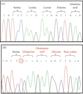
Concerning the SLC37A4 gene, four changes were found, corresponding to a known missense mutation, a novel insertion (frameshift) and two neutral mutations: c.1287G > A (rs8192696) and c.1076-28C > T (rs201063857). The p.G149E (c.654G > A) mutation in exon four was found in one homozygous patient. In another homozygous individual, the insertion of a T base between nucleotides 1338 and 1339 was detected in exon eleven. This change results in a reading frame modification and creates a premature stop codon (Figure 1).
In addition to these mutations, some SNPs were also identified in this sample. In the remaining seven individu-als, no disease causative mutation was found. Clinical and molecular data are summarized in Table 1.
The above results show that GSD1a was more preva-lent than GSDIb in our case series. Considering theG6PC
gene, p.R83C alters the amino acid that contributes to the active center of the enzyme. It is considered one of the most frequent mutations, seen both worldwide (Lei et al., 1995a,b; Parvariet al., 1995, 1997; Stroppianoet al., 1999; Rakeet al., 2000) and in Brazil (Reis et al., 2001). The present results corroborate these findings identify this as the most prevalent one among the mutant alleles. This be-ing the case, the evaluation of this mutation by RFLP may allow the disease diagnosis without the need for automated sequencing, reducing cost and time of examination, since the restriction site mutation alters the enzymeHgaI cleav-age pattern.
The remaining change (p.Q347X), already described in the literature, leads to truncation of the carboxy-terminal 11 amino acid residues in human G6Pase and inactivates its activity (Leiet al., 1994). It is also frequently observed in individuals with impaired G6Pase, being common among Caucasian patients in the United States (Leiet al., 1995a) and Europe (north and south), representing 70% of all mu-tant alleles in these regions (Chevalier-Porstet al., 1996; Rakeet al., 1999). Nevertheless, in the present sample, it was detected only in one allele of one patient, this being compound heterozygous for p.R83C.
In theSLC37A4gene, a missense mutation p.G149E and an insertion c.1338_1339insT were found, each one in an individual, both homozygous. In addition to these, SNP c.1076-28C > T (rs201063857) was recently reported and shown to be present at a low frequency of heterozygosity (0.004%) in 570 participants of European descent (NCBI, 2013). Our sample showed a high frequency, probably rep-resenting difference in ethnic background.
The amino acid glycine at position 149 is located in the transmembrane domain of the enzyme glucose 6-phos-phate translocase, and the substitution of glycine to glutamic acid (p.G149E) changes the conformation of this enzyme. It is a rare change that has been reported only once in a Chinese family (Lamet al., 2000).
Finally, the present study allowed the identification of a new mutation, c.1338_1339insT, in individual 8. This patient is the fourth child of a consanguineous couple (first cousins) of Italian origin, whose third child died at the age of seven months with similar clinical symptoms. Pregnancy was complicated by maternal hypertension, and delivery was at 38 weeks of gestational age, weighting 3,590 g and measuring 51 cm. The child had hyperbilirubinemia after 48 h requiring exchange transfusion, and was released at the 5thday after birth. At 10 days, a single convulsion was documented and at the age of three months hepatomegaly was denoted. During investigation, glycemia after fasting was 2 mg, total cholesterol was 251 mg%, and triglyceride 814 mg% (both with a reference value: < 200), alkaline phosphatase was 477 IU/L (reference value: < 400), and CK 144 IU/L (reference value: < 190). Liver biopsy was
performed for a histological study, which was compatible with GSD. After starting treatment with frequent meals, cornstarch, and dietary restriction of lipids and sugars (fructose, lactose, galactose, and sucrose), he presented normal neuromotor and somatic development, reaching a final height of 189 cm. Nevertheless, biochemical follow-up revealed variable degrees of hypercholesterolemia and hypertriglyceridemia, and he also developed hyperurice-mia at the age of nine years, which was treated with 200 mg allopurinol twice a day. Renal function was always normal and neutropenia was never seen during routine exams, but at the age of 23 he presented an acute episode of sepsis and died after two days in another hospital with no further infor-mation available.
Although functional studies could not be performed to confirm that this mutation causes the disease, some as-pects support the hypothesis. First, the insertion of the T base changed the reading frame, resulting in a different amino acid sequence and a premature stop codon. Second, no other causative mutation was found in this individual. Finally, the presence of parental consanguinity is compati-ble with the finding of this mutation in homozygosity.
In seven patients with clinical symptoms suggestive of GSDla and GSDIb no mutation was found in coding re-gions (exons) or adjacent introns. Mutations in other parts of the gene, including introns, were not studied, but it seems unlikely that they may cause an abnormal phenotype in these individuals. An alternative explanation to the nega-tive results in this molecular study is the possibility of a misdiagnosis, since even with a careful evaluation based on laboratory and clinical findings, overlap with other types of GSD is possible. Thus further molecular studies would be indicated.
The molecular analysis of the two genes enabled di-agnosis confirmation of GSDIa and Ib in four patients, without the need for liver biopsy. In addition, the identifica-tion of the mutaidentifica-tion provided an addiidentifica-tional tool for genetic counseling. Despite the limited number of subjects, our re-sults showing that p.R83C was the most frequent mutation inG6PCin the present sample is compatible with interna-tional (Leiet al., 1995a,b; Parvariet al., 1995, 1997; Strop-pianoet al., 1999; Rakeet al., 2000) and previous Brazilian studies (Reiset al., 2001). Thus, in every patient with GSD Ia or Ib, we propose to start with an investigation of this mutation and then to sequence theG6PCgene. For patients without identifiable genetic mutations, enzymatic essay or a further molecular study of other GSD genes remain diag-nostic tools. Finally, in this study a new mutation was found and described.
References
Burchell A (1990) Molecular pathology of glucose-6-phospha-tase. FASEB J 4:2978-2988.
Chen YT (2001) Glycogen storage disease. In: Scriver CR, Beaudet AL, Sly WS and Valle D (eds) The Metabolic and
Molecular Bases of Inherited Disease. McGraw Hill, New York, pp 1521-1551.
Chevalier-Porst F, Bozon D, Bonardot AM, Bruni N, Mithieux G, Mathieu M and Maire I (1996) Mutation analysis in 24 French patients with glycogen storage disease type 1a. J Med Genet 33:358-360.
Chou JY, Matern D, Mansfield BC and Chen YT (2002) Type I glycogen storage diseases: Disorders of the glucose-6-phos-phatase complex. Curr Mol Med 2:121-143.
Diament A, Schmidt BJ and Ramos JLA (1994) Erros inatos do metabolismo. In: Marcondes E (ed) Pediatria Básica. Sar-vier, São Paulo, pp 722-726.
Fernandes J, Huijing F and Van de Kamer JH (1969) A screening method for liver glycogen diseases. Arch Dis Child 44:311-317.
Froissart R, Piraud M, Boudjemline AM, Vianey-Saban C, Petit F, Hubert-Buron A, Eberchweiler PT, Gajdos V and Labru-ne P (2011) Glucose-6-phosphatase deficiency. OrphaLabru-net J Rare Dis 6:27.
Gerin I, Veiga-da-Cunha M, Achouri Y, Collet J and Schaftingen EV (1997) Sequence of a putative glucose 6-phosphate translocase, mutated in glycogen storage disease type 1b. FEBS Lett 419:235-238.
Gitzelmann R and Bosshard NU (1993) Defective neutrophil and monocyte functions in glycogen storage disease type Ib: A literature review. Eur J Pediatr 152(Suppl 1):S33-S38. Hicks J, Wartchow E and Mierau G. (2011) Glycogen storage
dis-eases: A brief review and update on clinical features, genetic abnormalities, pathologic features, and treatment. Ultrastruct Pathol 35:183-196.
Kure S, Hou DC, Suzuki Y, Yamagishi A, Hiratsuka M, Fukuda T, Sugie H, Kondo N, Matsubara Y and Narisawa K (2000) Glycogen storage disease type Ib without neutropenia. J Pediatr 137:253-256.
Lam WC, Sin SY, Lau ET, Lam YY, Poon P and Tong SF (2000) Prenatal diagnosis of glycogen storage disease type1b using desnaturing high performance liquid chromatography. Prenat Diag 20:765-768.
Lei KJ, Shelly LL, Pan C, Sidbury JB and Chou JY (1993) Muta-tions in the glucose 6-phosphatase gene that cause glycogen storage disease type 1a. Science 262:580.
Lei KJ, Pan CJ, Shelly LL, Liu JL and Chou JY (1994) Identifica-tion of mutaIdentifica-tions in the gene for glucose-6-phosphatase, the enzyme deficient in glycogen storage disease type 1a. J Clin Invest 93:1994-1999.
Lei KJ, Shelly LL, Baochuan L, Sidbury JB, Chen Y and Nordlie RC (1995a) Mutations in the glucose 6-phosphatase gene are associated with glycogen storage disease types 1a and 1aSP but not 1b and 1c. J Clin Invest 95:234-240.
Lei KJ, Chen YT, Chen H, Wong LJ, Liu JL, McConkie-Rosell A, Van Hove JL, Ou HC, Pan LY,et al.(1995b) Genetic basis of glycogen storage disease type Ia: Prevalent mutations at the glucose-6-phosphatase locus. Am J Hum Genet 57:766-771.
Marcolongo P, Barone V, Priori G, Pirola B, Giglio S, Zammarchi E, Parenti G, Burchell A, Benedetti A and Sorrentino V (1998) Structure and mutation analysis of the glycogen stor-age disease type 1b gene. FEBS Lett 436:247-250. Martens DH, Kuijpers TW, Maianski NA, Rake JP, Smit GP and
disease type Ib mutations without neutropenia or neutrophil dysfunction. J Inherit Metab Dis 29:224-225.
Moses SW (1990) Pathophysiology and dietary treatment of the glycogen storage diseases J Pediatr Gastroenterol Nutr 11:155-174.
Parvari R, Lei KJ, Bashan N, Hershkovitz E, Korman SH, Barash V, Lerman-Sagie T, Mandel H, Chou JY and Moses SW (1997) Glycogen storage disease type 1a in Israel. Biochem-ical, clinBiochem-ical, and mutational studies. Am J Med Genet 72:286-290.
Parvari R, Moses S, Hershkovitz E, Carmi R and Bashan N (1995) Characterization of the mutations in the glucose-6-phos-phatase gene in Israeli patients with glycogen storage dis-ease type 1a: R83C in six Jews and a novel V166G mutation in a Muslim Arab. J Inher Metab Dis 18:21-27.
Rake JP, ten Berg AM, Verlind E, Visser G, Niezen-Koning KE, Buys CH, Smit GP and Scheffer H (1999) Glycogen storage disease type1a: Four novel mutations (175delGG, R170X, G266V and V338F) identified. Hum Mutat 13:173-176. Rake JP, Ten Berge AM, Verlind E, Niezen-Koning KE, Buys
CH, Smit GP and Scheffer H (2000) Glycogen storage dis-ease type 1a: Recent experience with mutation analysis, a summary of mutations reported in the literature and a newly developed diagnostic flowchart. Eur J Pediatr 159:322-330. Reis FC, Caldas HC, Norato DY, Schwartz IV, Giugliani R, Burin
MG and Sartorato EL (2001) Glycogen storage disease type Ia: Molecular study in Brazilian patients. J Hum Genet 46:146-149.
Rubin E, Gorstein F, Schwarting R, Rubin R and Strayer D (2006) Patologia - Bases Clinicopatológicas da Medicina. 6th ed. Guanabara Koogan, Rio de Janeiro, pp 1648.
Stroppiano M, Regis S, DiRocco M, Caroli F, Gandullia P and Gatti R (1999) Mutations in the glucose-6-phosphatase gene of 53 Italian patients with glycogen storage disease type Ia. J Inherit Metab Dis 22:43-49.
Tamhankar PM, Boggula V, Girisha KM and Phadke SR (2012) Profile of patients with Von Gierke disease from India. In-dian Pediatr 49:228-230.
van Schaftingen E and Gerin I (2002) The glucose-6-phosphatase system. Biochem J 362:513-532.
Visser G, Rake JP, Fernandes J, Labrune P, Leonard JV, Moses S, Ullrich K and Smit GP (2000) Neutropenia, neutrophil dys-function, and inflammatory bowel disease in glycogen stor-age disease type Ib: Results of the European Study on Gly-cogen Storage Disease type I. J Pediatr 137:187-191.
Internet Resources
National Center for Biotechnology Information (NCBI),
http://www.ncbi.nlm.nih.gov/pro-jects/SNP/snp_ss.cgi?ss=ss491655203 (October 29, 2013).
Associate Editor: Carlos F.M. Menck