Biodegradation of crude oil by individual bacterial strains and a mixed
bacterial consortium
Santina Santisi
1,2, Simone Cappello
1, Maurizio Catalfamo
1, Giuseppe Mancini
3,
Mehdi Hassanshahian
4, Lucrezia Genovese
1, Laura Giuliano
1, Michail M. Yakimov
11
Institute for Coastal Marine Environment, National Counsel of Research, Messina, Italy.
2
School in “Biology and Cellular Biotechnology”, Faculty of Sciences, University of Messina, Messina, Italy.
3
Department of Industrial Engineering, University of Catania, Catania, Italy.
4
Department of Biology, Faculty of Sciences, Shahid Bahonar University of Kerman, Kerman, Iran.
Submitted: December 2, 2013; Approved: June 6, 2014.
Abstract
Three bacterial isolates identified asAlcanivorax borkumensisSK2,Rhodococcus erythropolisHS4 andPseudomonas stutzeriSDM, based on 16S rRNA gene sequences, were isolated from crude oil enrichments of natural seawater. Single strains and four bacterial consortia designed by mixing the single bacterial cultures respectively in the following ratios: (Alcanivorax: Pseudomonas, 1:1), (Alcanivorax:Rhodococcus,1:1), (Pseudomonas:Rhodococcus, 1:1), and (Alcanivorax: Pseudomo-nas:Rhodococcus, 1:1:1), were analyzed in order to evaluate their oil degrading capability. All ex-periments were carried out in microcosms systems containing seawater (with and without addition of inorganic nutrients) and crude oil (unique carbon source). Measures of total and live bacterial abun-dance, Card-FISH and quali-, quantitative analysis of hydrocarbons (GC-FID) were carried out in or-der to elucidate the co-operative action of mixed microbial populations in the process of bio-degradation of crude oil. All data obtained confirmed the fundamental role of bacteria belonging to Alcanivoraxgenus in the degradation of linear hydrocarbons in oil polluted environments.
Key words:Alcanivorax,Pseudomonas,Rhodococcus, bioremediation, bioaugmentation.
Introduction
Petroleum hydrocarbons are the most widespread contaminants within the marine environment. Pollution by hydrocarbons in marine environments may be the conse-quence of various natural (natural seepages) and/or an-thropogenic activities (discharge during tanks and/or ships transportation and/or pipeline failures) as well as the chro-nic pollution (ships, harbours, oil terminals, freshwater run-off, rivers and sewage systems).
The “fate” of petroleum in the sea water largely de-pends on mechanical (wave, wind), physical (temperature, UV) and chemical (pH, dissolved oxygen and nutrient con-centration) factors which may differently influence its natu-ral transformation (oil weathering) and bio-degradation (Nikolopoulou and Kalogeraki, 2010). At an early stage
light fractions of oil are naturally removed; mostly by evap-oration, thence by photo-oxidation and by geo-chemicals reactions. Heavy fractions are instead dispersed or dis-solved and only a small portion may be removed by the pro-cess of biodegradation. Although chemical-physical phe-nomena play an important role in the process of oil detoxification, the ultimate and complete degradation is mainly accomplished by marine microflora, dominant bac-teria in this role (Della Torreet al., 2012).
As reported in different studies, a wide variety of ma-rine bacteria are known to degrade petroleum hydrocar-bons, and those, distributed over several (sub)phyla (a-,b-, andg-Proteobacteria; Bacteroidetes/Chlorobi group) have been described so far (Rooling et al., 2004; Cappello et al., 2007).
DOI: http://dx.doi.org/10.1590/S1517-838246120131276
Send correspondence to S. Cappello. Institute for Coastal Marine Environment, CNR of Messina, Sp. San Raineri 86, 98121 Messina, Italy. E-mail: [email protected].
In the natural environment, biodegradation of crude oil involves a succession of species within the consortia of the present microbes (Alkatibet al., 2011). Indeed, since a single species can metabolize only a limited range of hy-drocarbon substrates, a consortium of many different bacte-rial species, with broad enzymatic capacities, is usually involved in oil degradation (Roolinget al., 2002). Although some bacteria, belonging toPseudomonas(Das and Chan-dar, 2011) andRhodococcusgenera (Hassanshahianet al., 2010 and 2012) have shown able to degrade hydrocarbons (Teramotoet al., 2010), in marine environments the key micro-organisms in the bio-degradation process has been identified as bacteria related toAlcanivoraxgenus (Yaki-movet al., 2007; Cappello and Yakimov 2010).
On the above mentioned basis, bioremediation tech-niques have been developed and improved for cleaning up oil-polluted marine environments as an alternative to che-mical and physical techniques (Alkatibet al., 2011). Bio-remediation can be described as the conversion of pollut-ants (hydrocarbons) by micro-organisms (bacteria) into energy, cell mass and biological waste products (Niko-lopoulou and Kalogeraki, 2010). Nevertheless, the rates of uptake and mineralization of many organic compounds (hydrocarbons) by bacteria in polluted seawater is limited due to the poor availability of nitrogen and phosphorus (Yakimovet al., 1998; Kasaiet al., 2002a, b; Cappello and Guglielmino, 2006; Cefalìet al., 2002). For that reason, in the application of biostimulation techniques the growth of oil-degrading bacteria can be strongly enhanced by
fertil-ization with inorganic nutrients (Nikolopoulou and Kalo-geraki, 2010).
In order to elucidate the cooperative action of mixed microbial populations in the biodegradation of crude oil, we have built up artificial consortia made up of two/three bacteria. By using these consortia, we have been able to in-vestigate the capability of efficient biodegradation of crude oil could be accomplished by the mixed populations. All experiments have been carried out into microcosms sys-tems containing seawater (with and without inorganic nu-trients); oil has been used as the only carbon source.
The knowledge of the efficiency and the activities of bacteria in oil-polluted sites may be helpful for the bio-remediation of oil spills, since human action, by using spe-cific microbial consortia, can be planned in order to clean up oil pollution (Denaroet al., 2005).
Material and Methods
Bacterial strains
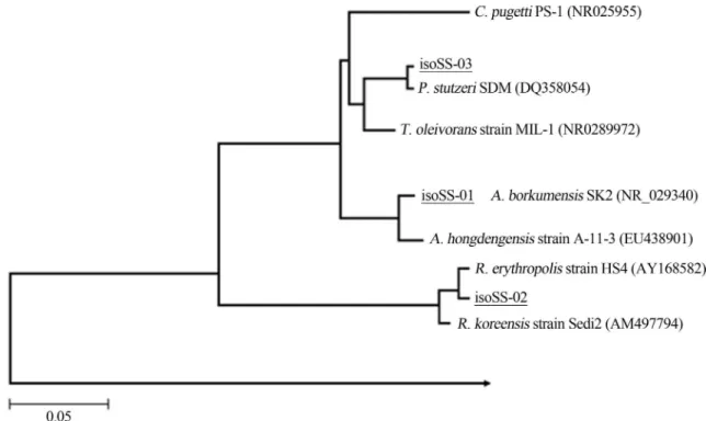
Three bacterial strains named isoSS-01, correspond-ing toAlcanivorax borkumensisstrain SK2T(Genbank ac-cession number Y12579; =DSM 11573T; 99%), isoSS-02 (Rhodococcus erythropolis HS4; Genebank accession number AY168582; 99%) and isoSS-03 (Pseudomonas stutzeri SDM; Genebank accession number DQ358054; 98%) were used in all the experiments (Fig. 1). Strain isoSS-01 belong to a collection of hydrocarbon-degrading bacteria hold at IAMC-Messina, strains isoSS-2 and
SS03 were isolated from natural seawater from crude oil enrichments in previously research. All strains used in this study were isolated from natural seawater from crude oil enrichments.
Analysis of 16S rRNA genes
Total DNA extraction of bacterial strains was per-formed by the MasterPure Complete DNA&RNA Purifica-tion Kit (Epicenter, Biotechnologies, Madison, WI) in ac-cordance with manufacture’s protocol. The 16S rDNA loci
were amplified using 1 primer pair: the 27F
(5’-AGAGTTTGATCCTGGCTCAG-3’, Lane, 1991)
primer and the 1492R (5’
TACGGYTACCTTGTTACGACT-3’, Lane, 1991) uni-versal primer. PCR (polymerase chain reaction) was car-ried out in 50mL of reaction mixture containing 1x reaction buffer, 1x solution Q (both from QIAGEN), 1mM of each primer, 200mM dNTP (Gibco), 1mL of template and 2.5 U of QiagenTaqpolymerase. The PCR reaction was carried out in Mastercycler Gradient (Eppendorf); the PCR condi-tions were as follows: 95 °C for 5 min (1 cycle); 94 °C for 1 min, 50 °C for 1 min and 72 °C for 2 min (35 cycles); with a final extension step at 72 °C for 10 min. PCR products were sequenced using Macrogen Service (Macrocen, Ko-rea). The analysis of the sequences (1400 bp of average length) was performed as previously described by Yaki-movet al.(2005). The sequences similarity of individual inserts was analysed by the FASTA program Nucleotide Database Query available through the EMBL-European Bioinformatics Institute. The phylogenetic affiliation of the sequenced clones, was performed as described by Yakimov et al.(2006).
Growth conditions
Started cultures were prepared by inoculating one loop of microbial cells into 10 mL of ONR7a mineral me-dium based on the composition of seawater was used in this study (Dyksterhouseet al., 1995). Nitrogen was provided in the form of NH4Cl, and was provided in the form of
Na2HPO,. ONR7a contained (per liter of distilled or
deio-nized water) 22.79 g of NaCl, 11.18 g of MgCl2*6H2O,
3.98 g of Na2SO4, 1.46 g of CaCl2, - 2H2O, 1.3 g of TAPSO
{3-[N-tris(hydroxymethyl)
methylamino]-2-hydroxypropanesulfonic acid}, 0.72 g of KCl, 0.27 g of NH4Cl, 89 mg of Na2HPO4* 7H2O, 83 mg of
NaBr, 31 mg of NaHCO3, 27 mg of H3BO3, 24 mg of
SrCI*6H2O, 2.6 mg of NaF, and 2.0 mg of FeCl2*4H20. To
prevent precipitation of ONR7a during autoclaving, three separate solutions were prepared and then mixed together after autoclaving when the solutions had cooled to at least 50 °C; one solution contained NaCI, Na2SO4, KCl, NaBr,
NaHCO3, H2BO3, NaF, NH4Cl, Na2HPO4, and TAPSO (pH
adjusted to 7.6 with NaOH), the second solution contained MgCl2, CaCl2, and SrCI, (divalent cation salts), and the
third solution contained FeCl2; 0.1% (w/v) sterile
tetra-decane (C14H30, Sigma-Aldrich, Milan, Italy) was used as
only energy and carbon source. After growing in a rotary shaker (New Brunswick C24KC, Edison NJ, USA; 150 rpm) at 25 °C for two days, 500mL of the seed culture broth were transferred into a 250 mL Erlenmeyer flask con-taining 100 mL of ONR7a medium supplemented with 1% (w/v) sterile tetradecane. The culture was incubated in a ro-tary shaker (New Brunswick C24KC, Edison NJ, USA; 150 xg) at 25 °C for 5 days.
Consortia
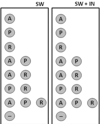
At the beginning (T0) of the experiments selected
mi-croorganisms (isoSS-01,A. borkumensisSK2T; iso-SS-02, R. erythropolisHS4 and iso-SS-03Ps. stutzeriSDM) were added at a final density of 105cell mL-1, in experimental mi-crocosms. Schematic representation of microbial consortia used in this study is indicated below (Fig. 2).
Experimental set-up of microcosms systems
The microcosms systems were performed in 250 mL sterilised Erlenmeyer flasks. Microcosms were incubated at 22±1 °C for 15 days with shaking (100g). All experi-ments were carried out in triplicate.
Two different series of experimentations were carried out. In the first experiment (identified as “SW”) bacterial cultures were carried out in natural seawater sterilized by
filtration through a 0.2-mm syringe filter (Sartorius); in the second experiment (identified “SW+IN”) cultures were carried out in sterile natural seawater with addition of inor-ganic nutrients to reach higher concentrations than those obtained in natural water (final concentrations: KH2PO40.077 g L-1, NH4Cl 0.2 g L-1and NaNO30.1 g L-1).
Microcosms untreated (no bacteria inoculation) were used in each experiment series as negative (abiotic) control. Crude oil was added in all experimentation.
At the beginning (T0) of the experiments, 1000 ppm
of sterile crude oil (Arabian Light Crude Oil; ENI Technol-ogy S.p.A.) were added into SW and SW+IN microcosms. Crude oil was introduced, in microcosm systems, after physical weathering (100 x g, 25 °C for 48 h); crude oil was supplemented with 0.1% (v/v) of squalene (C30H50,
Sigma-Aldrich, Milan) as internal spike for measure of bio-degradation rate.
Sampling strategy and parameters assayed
At the beginning (T0) and at the end (T15) of the
exper-imental period, sub-samples of each bacterial cultures were taken aseptically. Measures of direct bacterial count (DAPI), microbial viability (Live/Dead staining) and mi-crobial activity (Card-FISH) were carried out. Measure of oil degradation was carried out as well. All experiments were carried out twice and all parameters detected were measured three times.
Total bacterial abundance (DAPI count)
After a short-time (30”) ultrasonic treatment ( Ultra-sonic Bath Branson 1200, Branson, USA), the total bacte-rial cell counts were performed by DAPI (4’,6-diamidino-2- phenylindole 2HCl, Sigma-Aldrich S.r.L., Milan, Italy) staining on samples fixed by formaldehyde (2% final con-centration), according to Porter and Feig (1980) and Cap-pelloet al., 2012. Slides were examined by epifluorescence by using Axioplan 2 Imaging (Zeiss; Carl Zeiss Inc., Thornwood, N.Y.) microscope. Results were expressed as number of cells mL-1.
Determination of living and dead bacteria
Living and dead bacteria (L/D) were enumerated after staining with the Live/Dead (BacLight bacterial Viability Kit (Invitrogen Corp; Molecular Probes, Inc Eugene, OR, USA). The above mentioned method allowed discrimina-tion, within the total bacterial community, of the living cells, labelled by SYTO 9 and green-fluorescing, from the
dead ones, labelled by propidium iodide and red-fluo-rescing (Zampinoet al., 2004). Cell counts, performed by an Axioplan epifluorescence microscope (Zeiss; Carl Zeiss Inc., Thornwood, N.Y., USA) equipped with a 100 W Hg lamp using fluorescein (BP 450-490; FT 510; LP 520) and rhodamine (BP 546/12; FT 580; LP 590) filter sets (for live and dead cells, respectively). Data obtained were reported as the mean value of (living and dead) cells mL-1.
Card-FISH
Card-FISH analysis was carried out according to pro-tocol developed by Pernthaler et al. (2002). Aliquot of 1 mL of bacterial culture was filtered on 0.22 mm poly-carbonate membranes (diameter 25 mm) by using a vac-uum filtration device (Millipore, Milan, Italy). Filters for Card-FISH counts were embedded in low-gelling point (0.1% agarose, Sigma-Aldrich, Milan), dried at 37 °C for 20 min, and dehydrated with 95% ethanol. The bacteria on the polycarbonate membrane were then permeabilized by lysozyme (solution (EDTA 0.05 M; 1 M Tris-HCl, pH 8.0; MilliQ water and 10 mg mL-1lysozyme) for 60 min at 37 °C and in some cases a treatment with achromopeptidase (60 U, 0.01 M NaCl, 0.01 M Tris-HCl [pH 8.0]) was per-formed. Filters were incubated at 37 °C for 30 min and hy-bridized with oligonucleotide probes modified at the 5’ end with horseradish peroxidase (HRP). Probes used in this work are listed in the Table 1.
After the hybridization and amplification steps, slides were examined by an Axioplan epifluorescence micro-scope (Zeiss; Carl Zeiss Inc., Thornwood, N.Y., USA) equipped with an appropriate filter sets for Card-FISH. Be-fore counting, the slides were stored at -20 °C for several days without any loss of fluorescence intensity. Cell counts were reported as the mean value of cells mL-1.
Hydrocarbon analysis
The composition of the Total Extracted and Resolved Hydrocarbons and their derivates (TERHCs) were analysed by high-resolution GC-FID (DANI Master GC Fast Gas Chromatograph System, DANI Instruments Sp.A., Milan). After acidification, TERHCs from samples were extracted at room temperature on a shaking table by using dichloro-methane (CH2Cl2, Sigma-Aldrich, Milan; 10% v/v). This
procedure was repeated three times, and the CH2Cl2phase
was combined and treated with sodium sulfate anhydrous (Na2SO4,Sigma-Aldrich, Milan) in order to remove any
re-sidual water (Ehrhardtet al., 1991; Wanget al., 1998; Dutta
Table 1- Oligonucleotide probes used in Card-FISH for this study.
Probe Sequence (5’ to 3’) of probe Specificity Source
NON-Eub338 ACA TCC TAC GGG AGG C Negative Control (Wallneret al. ,1993)
Eub338 GCT GCC TCC CGT AGG AGT Domain Bacteria (Amannet al. ,1990)
and Harayama 2001; Denaroet al., 2005). Extracts were concentrated by rotary evaporation (Rotavapor model R110; Büchi Labortechnik AG, Switzerland) at room tem-perature (< 30 °C), followed by evaporation under a stream of nitrogen and taken up into a solution containing hepta-methyl-nonane as an internal standard (79mg mL-1). Indi-ces selected for this study were:n-C17/Pristane (nC17/Pr), n-C18/Phytane (nC18/Ph) in order to evaluate the relative biodegradation ofn-alkanes.
Biodegradation efficiency (BE) of TERCHs
The degradation of TERCHs was expressed as the percentage of TERCHs degraded in relation to the amount of the remaining fractions in the appropriate abiotic control samples. The biodegradation efficiency (BE), based on the decrease in the total concentration of hydrocarbons, was calculated by using the expression described by Michaudet al., 2004:
100 - (As * 100 / Aac)
where As = total area of peaks in each sample, Aac = total area of peaks in the appropriate abiotic control, BE (%) = Biodegradation efficiency.
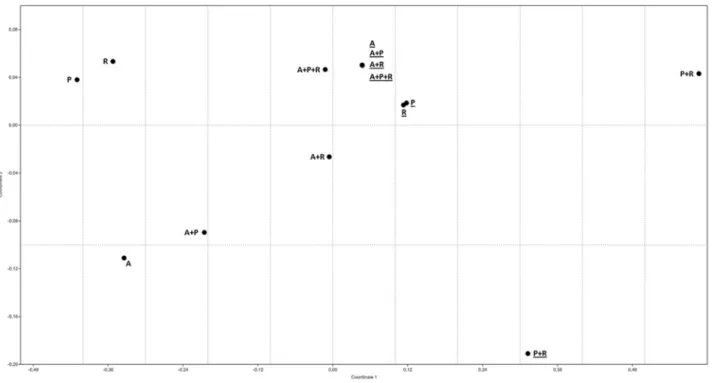
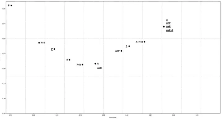
Statistical analysis and nMDS
The experimental data are presented in terms of arith-metic averages of at least three replicates and the standard deviations are indicated by error bars. The non-metric multi-dimensional (nMDS) scaling plot were done using
PAST (PAlaeontological STatistics Software ver. 1.88; Hammeret al., 2001).
Results
Total bacterial abundance (DAPI count)
After 15 days of cultivation, the bacterial abundance was measured by direct DAPI count; and data obtained were compared with the quantity of cells present at the be-ginning of the experimental period (T0). The data obtained
showed, how in seawater added with inorganic nutrients it was possible to observe a general increase of microbial abundance (systems “A”, “A + P”, “A + R,” “P + R”, and “A + P + R ”) with mean values of 108cell mL-1. In cultures performed using seawater (without inorganic nutrients), bacterial abundance present, at the end of experimental pe-riod, mean values of 106 cell mL-1 (systems “P”, “R”, “A + P” and “A + P + R”); in microcosms indicated as “A”, “A + R”, and “P + R”) values of ~105cell mL-1were ob-served (Fig. 3).
Determination of living and dead bacteria
Data of living and dead bacteria (L/D) enumerated us-ing the Live/Dead stainus-ing are showed in Figure 4.
The results obtained after 15 days of cultivation shown as the vital bacterial fraction, present in microcosm performed in seawater with addition of inorganic nutrients, was greater than that observed in the microcosms per-formed in sea water. In particular in microcosms indicated
as SW the percentage of dead cells was about four or six times greater than the initial time.
Card-FISH
The qualitative measure of microbial abundance, into the experimental systems named “A+P” and “A+R”, was carried out by using the card-FISH method. Values of abun-dance of cells hybridized using probes for Eubacteria (EUB338) resulted to be similar to the values obtained from the measure of total bacterial abundance (DAPI count) in the same conditions.
Data obtained put in evidence as almost total cells of experimentations carried out with seawater without inor-ganic nutrients were hybridized by probes for Eubacteria. The same result was not obtained during experimentations carried out with seawater added with inorganic nutrients (in such a case a number of cells of a lower logarithmic order has been obtained). The data obtained showed as the quan-tity of cells of Alcanivorax borkumensis (in “A + P”, SW + IN; “A + R”, SW and “A+ R”, SW + IN systems) present values lower (of a logarithmic order) those obtained in total cells (Fig. 5).
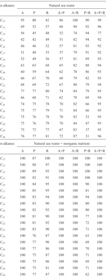
Rate of degradation of n-alkanes
The percentage degradation of n-alkanes (C12-C30)
present in the crude oil was calculated by comparison of the gas chromatograms of the non degraded (abiotic) control
and the degraded sample for each experimental conditions (Table 2 and Fig. 6).
During experimentations performed with natural sea-water the condition identified as “A+P+R” showed a better rate degradation (~ 90%); also in system “A+R” in other conditions is possible to observe a degradation of almost all n-alkanes (rate of degradation > of 60%).
The data obtained show that, during growth in natural seawater added with inorganic nutrients, conditions “A”, “R”, “A+P”, “A+ R” and “A+P+R” n-alkanes present in the crude oil were totally degraded; in contrast, conditions “P” and “P+R” present a low rate of degradation of n-alkanes.
For all strains, n-alkanes with a medium length (C12
-C18) were degraded to a greater extent (rate of degradation
> of ~ 70%) than and long chains (C19-C30) because
long-chain n-alkanes are solid and their low solubility inhibits degradation by bacteria (Figs. 7 and 8).
Biodegradation efficiency (BE) of TERCHs
After 15 days of experimentation, measure of dation of the TERHCs revealed as major rates of oil degra-dation are, in general, observed in systems carried out in natural seawater with inorganic nutrients (Table 3). In SW experiment the maximum rate of total oil degradation is ob-served in “A+P+R” (~ 97%) and “A+R” system (~ 83%). Other conditions present similar values. In system SW+IN the experimentations identified “A”, “A+P”, “A+R” and “A+P+R” the degradation of oil is total; values of ~ 90%,
~ 64% and ~ 30% of total oil degradation were observed for “P”, “R” and “P+R” experiments (Fig. 6).
Discussion
The recovery of petroleum contaminated sites could be achieved by either physicochemical or biological meth-ods. Due to negative consequences of the physicochemical approach, more attention is now given to the exploitation of biological alternatives (Okoh, 2006).
Biological treatments are having more importance, mainly because of the low environmental impact, the costs (in general cheaper than other cleanup technologies), the capability to destroy organic contaminants, and the possi-bility of beneficial use of treated sediments (Rulkens and Bruning, 2005). Different studies have shown better results using bioremediation strategies (Beolchini et al., 2010; Rocchettiet al., 2011, 2012).
In general, bioremediation is often based onin-situ stimulation of the microbial community (biostimulation) or amending the microbial community with an inoculum of hydrocarbon-degrading bacteria (bioaugmentation). In both cases, the successful result of bioremediation depends on appropriate hydrocarbon-degrading consortia and envi-ronmental conditions.
In this study we have analyzed the cooperative action of mixed microbial populations in the biodegradation of
crude oil during different culture conditions. All data obtained confirmed the fundamental role of bacteria be-longing to Alcanivorax genus in degradation of linear hydrocarbons in oil polluted environments. Indeed, all experimentations carried out in seawater (with or without inorganic nutrients) whit presence ofAlcanivoraxshowed maximum rates of oil degradation.
Capability ofAlcanivoraxgenus to use hydrocarbons as the only sources of energy and organic carbon was widely (Yakimovet al., 1998; Scheineret al., 2006). Kasai (2002) and Cappello (2012) explain these characteristics in ability of this strain to produce a lipidic bio-surfactant that increases the bioaviable of contaminant and the ability to use this (Yakimov et al., 1998; Scheiner et al., 2006). Alcanivorax borkumensisSK2 surfactant propose as one of the most efficient of bacterial surfactants; the possible pres-ence of this surfactant can justify an increase in the rates of degradation by both the bacteria that possible microbial consortia. This defines an increment of rates of degradation by both the bacteria and possible microbial consortia (Yakimovet al., 1998; Scheineret al., 2006).
The presence ofAlcanivoraxin natural environment or enrichment by laboratory is generally combined with the presence of other bacterial strains, such asPseudomonas sp. and Rhodococcus sp., that participating in bio-degradation phenomena. However,Pseudomonas sp.and Rhodococcus sp., can not be classified such as
carbonoclastic bacteria (Marine Obligate Hydrocarbono-clastic Bacteria, OMHCB; Yakimovet al., 2007), but these are heterotrophic bacteria that participate in the biode-gradation processes via “syntrophy metabolic”" in which the degradation of pollutant compounds takes place via a metabolic chain, in which the product of the catabolism of a bacterial species is identified as a source of carbon for met-abolic another.
Analysis of microbial abundance in cultures in study showed, however, a divergence of the correlation between microbiological data and those of biodegradation. In exper-imentation carried out withAlcanivoraxandPseudomonas (system “A”, “P” and “A+P”) was possible to observe after 15 days to incubation in seawater with and without inor-ganic nutrients an increase of microbial biomass.
Data obtained during cultivation of Rhodococcus erythropolis(as single strain and/or as consortium) did not show, apparently, increment of microbial abundance. This condition may be due to an underestimation of the direct count (DAPI count) in cultures as result from an ineffi-ciency of methodology used by us for the separation of mi-crobial cells from oil remain (dislodging).
Rhodococcus erythropolisHS4, in presence of linear hydrocarbons, is usually produced trehalose lipids (Rapp, 1979); these molecules are composed of a disaccharide in combination with a long chain of fatty acids. The presence of these molecules define a general reduction of superficial (surface) tension such as increase of cellular hydropho-bicity and consequently increase of bacterial tackiness.
Another important aspect was obtained to qualitative measures of microbial abundance. Card-FISH analysis car-ried out to estimate quantitative abundance of bacteria be-longing toAlcanivoraxgenus in microbial consortia tested in this study. Therefore Card-FISH measures were realized for identified consortia A+P and A+R realized in seawater with and without inorganic nutrients. Hybridization with EUB-338 probe showed values similar to these obtained by direct DAPI count; for against assays carried out with ALK probe (specific probe toAlcanivorax genus) evidence as only the 15% of total cells were hybridized. This result can seem discordant with biodegradation results. However, it is important remember that the sample to Card-FISH was
col-Table 2- Percentage ofn-alkane degradation in crude oil by strains in this study after 15 days of incubation at 22±1 °C with shaking (100g). Table Top, bacterial culture s in natural sea water; Table Lower, Bacterial cul-tures performed in natural seawater with inorganic nutrients. A, Alcanivorax borkumensis SK2; P, Pseudomonas stuzteri SMD; R, Rhodococcus erythropolisHS4.
n-alkanes Natural sea water
A P R A+P A+R P+R A+P+R
C12 95 80 82 86 100 90 99
C13 69 52 57 68 94 92 96
C14 56 45 48 52 74 94 77
C15 42 42 49 51 82 94 92
C16 46 46 52 57 81 93 92
C17 51 48 53 57 79 91 92
C18 52 49 56 57 81 89 93
C19 63 63 68 65 82 88 94
C20 60 59 64 62 78 86 93
C21 66 67 70 68 79 82 93
C22 68 69 72 67 80 79 94
C23 77 77 80 74 84 79 95
C24 66 68 71 62 78 69 94
C25 74 75 78 70 82 66 95
C26 75 77 79 71 84 66 95
C27 75 76 78 70 83 53 95
C28 75 76 79 70 84 47 95
C29 73 73 77 67 83 37 95
C30 76 77 81 72 87 31 96
n-alkanes Natural sea water + inorganic nutrient
A P R A+P A+R P+R A+P+R
C12 100 87 100 100 100 100 100
C13 100 88 97 100 100 100 100
C14 100 89 95 100 100 100 100
C15 100 82 91 100 100 100 100
C16 100 84 95 100 100 90 100
C17 100 85 95 100 100 81 100
C18 100 83 94 100 100 94 100
C19 100 83 90 100 100 80 100
C20 100 82 92 100 100 80 100
C21 100 81 90 100 100 77 100
C22 100 81 92 100 100 72 100
C23 100 83 90 100 100 71 100
C24 100 76 87 100 100 63 100
C25 100 77 90 100 100 68 100
C26 100 77 86 100 100 70 100
C27 100 75 87 100 100 71 100
C28 100 75 86 100 100 69 100
C29 100 75 81 100 100 71 100
C30 100 77 87 100 100 62 100
Table 3- Biodegradation efficiency (BE) of TERCHs. The experimental data are presented in terms of arithmetic averages.
Code Natural sea water Natural sea water+IN
A 64 100
P 48 64
R 64 90
A+R 66 100
A+P 83 100
P+R 73 60
Figure 6- Relative values of major TERHC fractions of Arabian Light Crude Oil detected in SW and SW+IN cultures after 15 days of incubation; data expressed as the percentages compared to negative abiotic control (0). A,Alcanivorax borkumensisSK2; P,Pseudomonas stuzteriSMD; R,Rhodococcus erythropolisHS4. Experimentations carried out in natural seawater in absence (SW) and presence (SW + IN) of inorganic nutrients were indicated, re-spectively, with grey and dark grey bars.
lected after 15 days of incubation, therefore is possible that the cells were collected in advance stationary phase and/or not more active. Supposing that the oil degradation process began early of the end of experiment, Alcanivorax sp. , dominant at the first experimental phase, tended to disap-pear or decrease once hydrocarbons have been degraded, whilePseudomonas sp.andRhodococcus sp. cells could become dominant using metabolic compounds or cellular lysates like nutritional source.
Acknowledgments
This work was supported by grants of National Coun-sel of Research (CNR) of Italy and by: i) EC Project “Un-raveling and exploiting Mediterranean Sea microbial diver-sity and ecology for XEnobiotics’ and pollutants’ clean up” (ULIXES-FP7-KBBE-2010-3.5-03); ii) Italian Project PRIN2010-2011 “La “System Biology” nello studio degli effetti di xenobiotici in organismi marini per la valutazione dello stato di salute dell’ambiente: applicazioni biotec-nologiche per potenziali strategie di ripristino”; iii) Na-tional Operative Project PON R&C 2007-2013 “Sviluppo di Tecnologie Innovative per il trattamento dei rifiuti li-quidi della navigazione finalizzate alla Tutela dell’Ambiente Marino” (STI-TAM); iv) National Opera-tive Project PON R&C 2007-2013 “Sviluppo di tecnologie innovative per la Sostenibilità Energetica ed Ambientale di cantieri nautici ed aree Portuali” (SEA-PORT).
References
Alkatib MA, Alam MDZ, Muyibi SAet al.(2011) An isolated bacterial consortium for crude oil biodegradation. Afr J Biotechnol 10:18763-18767.
Atlas RM (1981) Microbial degradation of petroleum hydrocar-bons: an environmental perspective. Microbiol Rev 45:180-209.
Beolchini F, Rocchetti L, Regoli Let al.(2010) Bioremediation of marine sediments contaminated by hydrocarbons: experi-mental analysis and kinetic modelling. J Hazard Mater 182:403-407.
Cappello S, Caruso G, Zampino Det al.(2007) Microbial com-munity dynamics during assays of harbour oil spill bio-remediation: a microscale simulation study. J Appl Micro-biol 102:184-194.
Cappello S, Denaro R, Genovese Met al.(2007) Predominant growth ofAlcanivoraxduring experiments on “oil spill bio-remediation” in mesocosms. Microbiol Res 162:185-190. Cappello S, Genovese M, Della Torre Cet al. Effect of
bio-emulsificant exopolysaccharide (EPS2003) on microbial
community dynamics during assays of oil spill bioreme-diation: A microcosm study. Mar Pollut Bull 64:2820-2282.
Cappello S, Guglielmino SPP (2006) Effects of growth tempera-ture on polystyrene adhesion ofPseudomonas aeruginosa ATCC27853. Braz J Microbiol 37:205-207.
Cappello S, Yakimov MM (2010)Alcanivorax. In handbook of hydrocarbon and lipid microbiology. In: Timmis KN, McGenity TJ, van der Meeret al.(eds). Springer, Berlin, pp. 1738-1745.
Cefalì E, Patanè S, Arena Aet al.(2002) Morphologic variations in bacteria under stress conditions: Near-field optical stud-ies. Scanning 24:274-283.
Das N, Chandran P (2011) Microbial degradation of petroleum hydrocarbon contaminants: an overview. Biotechnol Res Int doi: 10.4061/2011/941810.
Della Torre C, Tornambè A, Cappello Set al.(2012) Modulation of CYP1A and genotoxic effects in European seabass Dicentrarchus labrax exposed to weathered oil: A mesocosm study. Mar Environ Res 76:48-55.
Denaro R, D’Auria G, Di Marco Get al.(2005) Assessing termi-nal restriction fragment length polymorphism suitability for the description of bacterial community structure and dynam-ics in hydrocarbon-polluted marine environments. Environ Microbiol 7:78-87.
Dutta TK, Harayama S (2001) Analysis of long-side chain alky-laromatics in crude oil for evaluation of their fate in the envi-ronment. Environ Sci Technol 35:102-107.
Dykesterhouse S, Gray J, Herwig RPet al.(1995)Cycloclasticus pugetii, gen. et sp. nov., an aromatic hydrocarbon-degrading bacterium from marine environments. Int J Syst Evol Micro-biol 45:116-123.
Ehrhardt M, Klungsøyr J, Law RJ (1991) Hydrocarbons: review of methods for analysis in sea water, biota, and sediments. ICES Techniques in Marine Environmental Sciences, pp 44. Hammer Ø, Harper DAT, Ryan PD (2001) PAST: Paleontological
statistics software package for education and data analysis. Palaeontol Electronica 4:9-14.
Hassanshahian M, Emtiazi G, Cappello S (2012) Isolation and characterization of crude-oil-degrading bacteria from the Persian Gulf and the Caspian Sea. Mar Pollut Bull 64:7-12. Hassanshahian M, Emtiazi G, Kermanshahi RKet al. (2010) Comparison of oil degrading microbial communities in sedi-ments from Persian Gulf and Caspian Sea. Soil Sediment Contam 19:277-291.
Kasai Y, Kishira H, Harayama S (2002) Bacteria belonging to the genusCycloclasticusplay a primary role in the degradation of aromatic hydrocarbons released in a marine environment. Appl Environ Microbiol 68:5625-5633.
Kasai Y, Kishira H, Sasaki Tet al.(2002) Predominant growth of Alcanivorax strains in oil-contaminated and nutrient-supplemented seawater. Environ Microbiol 4:141-147. Lane DJ (1991) 16/23S rRNA sequencing. In nucleic acid
tech-niques in bacterial systematic. Stackebrandt E, Goodfellow M (ed.) Wiley, New York pp. 115-175.
Michaud L, Lo Giudice A, Saitta M et al. (2004) The biode-gradation efficiency on diesel oil by two psychrotrophic Antarctic marine bacteria during a two-month-long experi-ment. Mar Pollut Bull 49:405-409.
Nikolopoulou M, Kalogerakis N (2010) Biostimulation strategies for enhanced bioremediation of marine oil spills including chronic pollution. In: Timmis KN (ed) Handbook of hydro-carbon and lipid microbiology. Springer-Verlag, Berlin, pp 2521-2529.
Okoh AI (2006) Biodegradation alternative in the cleanup of pe-troleum hydrocarbon pollutants. Biotechnology and Molec-ular Biology Review 1:38-50.
Pernthaler A, Pernthaler J, Amann R (2002) Fluorescence in situ hybridization and catalyzed reporter deposition for the iden-tification of marine bacteria. Appl Environ Microbiol 68:3094-3101.
Rocchetti L, Beolchini F, Ciani Met al.(2011). Improvement of bioremediation performance for the degradation of petro-leum hydrocarbons in contaminated sediments. Appl Envi-ron Soil doi:10.1155/2011/319657.
Rocchetti L, Beolchini F, Hallberg KBet al.(2012) Effects of prokaryotic diversity changes on hydrocarbon degradation rates and metal partitioning during bioremediation of con-taminated anoxic marine sediments. Mar Pollut Bull 64:1688-1698.
Röling WFM, Milner MG, Jones DMet al.(2002). Robust hydro-carbon degradation and dynamics of bacterial communities during nutrient-enhanced oil spill bioremediation. Appl En-viron Microbiol 68:5537-5548.
Röling WFM, Milner MG, Martin Jones Det al.(2004) Bacterial community dynamics and hydrocarbon degradation during a field-scale evaluation of bioremediation on a mudflat beach contaminated with buried oil. Appl Environ Microbiol 70:2603-2613.
Rulkens WH, Bruning H (2005). Cleanup technologies for dredged fine sediments: review and future challenges. Olfenbuttel RF, Withe PJ (eds) Finding Achievable Risk Re-duction Solutions Remediation of Contaminated Sediments. Battelle Press, Columbus, p. C6-01.
Schneiker S, Martins dos Santos VA, Bartels Det al.(2006) Ge-nome sequence of the ubiquitous hydrocarbon-degrading marine bacteriumAlcanivorax borkumensis. Nat Biotechnol 24:997-1004.
Wallner G, Amann R, Beisker W (1993) Optimizing fluorescent in situ hybridization with rRNA-targeted oligonucleotide probes for flow cytometric identification of microorgan-isms. Cytometry 14:136-143.
Wang Z, Fingas M, Blenkinsopp Set al.(1998) Comparison of oil composition changes due to biodegradation and physical weathering in different oils. J Chromatogr A 809:89-107. Yakimov MM, Cappello S, Crisafi Eet al.(2006) Phylogenetic
survey of metabolically active microbial communities asso-ciated with the deep-sea coral Lophelia pertusafrom the Apulian Plateau, Central Mediterran Sea. Deep-Sea Res 53:62-75.
Yakimov MM, Denaro R, Genovese Met al.(2005) Natural mi-crobial diversity in superficial sediments of Milazzo Har-bour (Sicily) and community successions during microcosm enrichment with various hydrocarbons. Environ Microbiol 7:1426-1441.
Yakimov MM, Golyshin PN, Lang Set al.(1998)Alcanivorax borkumensisgen. nov., sp. nov., a new, hydrocarbon-degra-ding and surfactant-producing marine bacterium. Int J Syst Bacteriol 48:339-348.
Yakimov MM, Timmis KN, Golyshin PN (2007) Obligate oil-degrading marine bacteria. Curr Opin Biotechnol 18:257-266.
Zampino D, Zaccone R, La Ferla R (2004) Determination of liv-ing and active bacterioplankton: a comparison of methods. Chemistry and Ecology 20:411-422.
Zulfiqar AM, Safia A (2012). Degradation of petroleum hydrocar-bons by oil field isolated bacterial consortium. Afr J Bio-technol 11:650-658.
Associate Editor: Lara Durães Sette