Genomic characterization of Brazilian
hepatitis C virus genotypes 1a and 1b
Departamento de Virologia, Instituto Oswaldo Cruz, FIOCRUZ, 21040-360 Rio de Janeiro, RJ, Brasil
M.G. Peig Ginabreda, C.F.T. Yoshida and C. Niel
Abstract
Parts of 5’ non-coding (5’ NC) and of E1 envelope regions of the hepatitis C virus (HCV) genome were amplified from sera of 26 Brazilian anti-HCV antibody-positive patients using the reverse tran-scription-polymerase chain reaction (RT-PCR). Fourteen samples were PCR positive with primers from the 5’ NC region and 8 of them were also positive with primers from the E1 region. A genomic segment of 176 bp from the E1 region of 7 isolates was directly sequenced from PCR products. The sequences were compared with those of HCV strains isolated in other countries and the Brazilian isolates were classified by phylogenetic analysis into genotypes 1a and 1b. This could have a clinical importance since it has been shown that individuals infected with type 1 viruses are less likely to respond to treatment with interferon than individuals infected with types 2 and 3 viruses. Two quasispecies isolated from the same patient with an interval of 13 months differed by two base substitutions (1.1%). The sequence of another isolate presented a three-nucleotide deletion at codon 329.
Correspondence C. Niel
Departamento de Virologia Instituto Oswaldo Cruz, FIOCRUZ 21040-360 Rio de Janeiro, RJ Brasil
Fax: 55 (021) 270-6397
Research supported by CNPq and Fundação Banco do Brasil (No. 10/4662-0).
Received July 25, 1996 Accepted January 6, 1997
Key words •Hepatitis C virus •Genotype 1a •Genotype 1b •Nucleotide sequence •Brazilian isolates
Introduction
Hepatitis C virus (HCV), the major caus-ative agent of parenterally transmitted non-A, non-B hepatitis (1), belongs to the Flaviviridae family. The genome of HCV is a single-stranded RNA with positive polarity of about 9,400 bases, which has a long open reading frame encoding structural proteins (core, E1, E2) and nonstructural proteins (NS2, NS3, NS4A, NS4B, NS5A, and NS5B) with untranslated regions at both the 5’ and 3’ ends. Considerable genetic heterogeneity has been reported among isolates (2,3). Com-parison of reported sequences has shown that some parts of the genome are more variable than others (4-6), the most
con-served part being the 5’ non-coding (5’ NC) region. Six major genotypes (1 to 6) have been identified, some of which contain sev-eral subtypes referred to as a, b, c, etc. (7-11). New genotypes 7 to 10 have been re-cently proposed (12,13).
be used such as restriction fragment length polymorphism (14,15), hybridization assays such as the line probe assay (16), DNA enzyme immunoassay (17), and type-specif-ic primer nested PCR (18,19). However, all these methods suffer from the same prob-lems: i) unusual mutations at critical sites may occasionally produce erroneous results and ii) some samples are unclassifiable. Nucleotide sequencing followed by the anal-ysis of the viral genome is the definitive method for identifying various HCV geno-types.
Few data on HCV genotypes circulating in South America are available. In the pres-ent study, we demonstrate the presence of HCV genotypes 1a and 1b in Brazilian pa-tients by direct sequencing of PCR products and show the phylogenetic localization of the corresponding isolates.
Material and Methods
Serology
Samples were obtained from sera referred to the National Reference Center for Viral Hepatitis for serological analysis between 1994 to 1995. Sera were from patients living in the city of Rio de Janeiro, Brazil, and being followed at several health institutions. Twenty-six anti-HCV antibody-positive sera were selected by in-house 2nd-generation enzyme-linked immunosorbent assay
(ELISA) with controlled specificity and sen-sitivity according to available commercial kits, and using recombinant antigens from the core, NS3, and NS5 regions obtained from the Research Foundation for Microbial Disease of Osaka University, Japan. Sera were also tested by ELISA for the presence of hepatitis B surface antigen (HBsAg) using the Hepanostika HBsAg Uni-form II system (Organon Teknika, Boxtel, The Netherlands).
RNA extraction and RT-PCR
HCV RNA was extracted from 100 µl serum by the acid guanidinium isothiocyanate phenol-chloroform method (20) and finally resuspended in diethylpyrocarbonate-treated water. Reverse transcription was carried out with random primers and 200 U of Moloney murine leukemia virus reverse transcriptase (Gibco-BRL) in a final volume of 20 µl at 37oC for 1 h. One third of the cDNA was
amplified by PCR with primers designed from the 5’ NC and E1 regions. In some cases, nested PCR was done with internal primers. The oligonucleotides used as prim-ers are shown in Table 1. PCR assays were carried out in a volume of 50 µl in the presence of 0.2 mM of each dNTP, 3 mM MgCl2, and 1 unit of Taq DNA polymerase
(Gibco-BRL). After an initial denaturation for 2 min at 94oC, DNA was amplified for 35
cycles at 94oC for 15 s, 50oC for 45 s, and
72oC for 1 min followed by a final
elonga-tion of 7 min at 72oC. When required, 1 µl of
the product was subjected to nested PCR for 30 cycles under the same conditions (only increasing the MgCl2 concentration to 5 mM).
To avoid contamination, RNA extraction and reverse transcription, pre-PCR reagent prepa-ration, DNA amplification, and gel electro-phoresis of PCR products were performed in four separate rooms. In each series of experi-ments, five samples together with a negative and a positive control were tested. None of the 7 nucleotide sequences determined were identical to each other, which permits us to
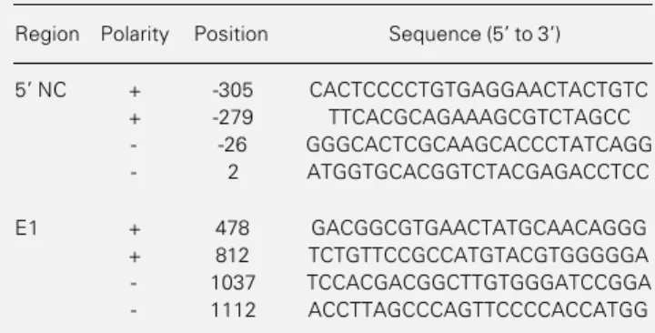
Table 1 - Sequence and genomic location of the oligonucleotides used as PCR primers.
Region Polarity Position Sequence (5' to 3')
5' NC + -305 CACTCCCCTGTGAGGAACTACTGTC
+ -279 TTCACGCAGAAAGCGTCTAGCC
- -26 GGGCACTCGCAAGCACCCTATCAGG
- 2 ATGGTGCACGGTCTACGAGACCTCC
E1 + 478 GACGGCGTGAACTATGCAACAGGG
+ 812 TCTGTTCCGCCATGTACGTGGGGGA
- 1037 TCCACGACGGCTTGTGGGATCCGGA
eliminate the possibility of cross-contamina-tion.
Nucleotide sequencing
Nested PCR products were made single-stranded and sequenced directly by the dideoxynucleotide chain termination method (21). Briefly, one of the internal primers used in the nested PCR was phosphorylated at the 5’ end: 150 pmol of primer was incu-bated in the presence of 10 mM ATP and 15 units of T4 polynucleotide kinase at 37oC for
30 min and the reaction was stopped by heating at 65oC for 10 min. After PCR, DNA
fragments were purified and the 5’ phospho-rylated strand was degraded by lambda exo-nuclease digestion (PCR template prep for SS DNA sequencing, Pharmacia), leaving a single-stranded DNA to be sequenced (T7 sequencing kit, Pharmacia). Primers used for sequencing were the internal PCR prim-ers of the E1 region.
Phylogenetic analysis
Alignment of multiple nucleic acid se-quences was performed with the University of Wisconsin Genetic Computer Group (GCG) PileUp program. This program uses the unweighted pair-group method with arith-metic average (UPGMA) procedure result-ing in a clustered order of the sequences based on the degree of similarity that is represented as a dendrogram (22). For easier identification, DNA databank sequences were handled using their accession number.
Results and Discussion
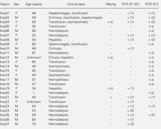
PCR is becoming a universal detection method for a number of DNA and RNA viruses present in the serum of patients. HCV is one of the viruses most extensively inves-tigated by PCR analysis. Two segments of the HCV genome were amplified by RT-PCR from the sera of 26 anti-HCV
antibody-positive patients. Table 2 shows sex, age, and clinical data of the patients. Only one patient was HBsAg positive. Fourteen (54%) samples were PCR positive when using prim-ers of the 5’ NC region (12 after the first round of PCR and 2 after nested PCR). Of these 14 positive samples, 8 were also posi-tive with primers of the E1 envelope region (4 after one-round PCR and 4 after nested PCR). The fact that a larger number of samples were positive when using primers derived from the 5’ NC region may be due to the elevated conservation of this region. The primers from this region showed one or no mismatch when compared with published sequences of the most common genotypes 1, 2, and 3, while sequences of the selected E1 primers were from a much more variable region.
isolates. The E1 region has been shown to be one of the most appropriate regions for this purpose (30) while the 5’ NC and core re-gions, also frequently used, are less effective in distinguishing between genotypes, sub-types, and isolates (23).
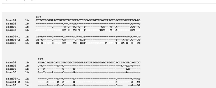
The nucleotide (nt) sequence of a 176-bp DNA fragment of the E1 region from nt 837 to nt 1012 was determined for 7 samples and the sequences are shown in Figure 1. Se-quences of our samples were compared by computer analysis with those of 15 HCV isolates from different parts of the world representing all the six major genotypes cur-rently known. A phylogenetic analysis was carried out and is presented as a dendrogram in Figure 2. Samples Braz04-1, Braz04-2, and Braz08 were assigned to genotype 1a whereas samples Braz01, Braz02, Braz07,
and Braz35 were assigned to genotype 1b. Our results demonstrating the infection of South American patients with types 1a and 1b viruses corroborate previous sequence data showing the presence of types 1b in Peru (7) and 1a (31) and 1b (32) in Argen-tina. The data also agree with the character-ization of Brazilian HCV isolates from geno-type 1 by hybridization line probe assay (16). Furthermore, genotype 1 has been shown to be the predominant type among the blood donors of São Paulo State (33).
Two sequences (Braz04-1 and Braz04-2) were derived from viruses from the same asymptomatic patient, whose blood was col-lected two times with an interval of 13 months. These two sequences differed by two base substitutions: in sample Braz04-2, T was replaced by C at position 858 and C
Table 2 - Clinical and serological data of the patients and detection of HCV in the serum by RT-PCR.
The HCV genome was detected after one-round PCR (1) or only after nested PCR (2). n.d., Not determined. HBsAg, Hepatitis B surface antigen.
Patient Sex Age (years) Clinical data HBsAg PCR (5 NC) PCR (E1)
Braz01 F 46 Hepatomegaly, transfusion - + (1) + (1)
Braz02 M 49 Cirrhosis, transfusion, hepatomegaly - + (1) + (2)
Braz04 F 56 Transfusion, asymptomatic n.d. + (1) + (2)
Braz05 F 30 Transfusion - - n.d.
Braz06 M 56 Hemodialysis - - n.d.
Braz07 F 33 Hemodialysis - + (1) + (1)
Braz08 M 54 Hepatitis - + (1) + (2)
Braz09 F 62 Splenomegaly, transfusion - - n.d.
Braz10 M 46 Cirrhosis - + (1)
-Braz11 M 67 Hemodialysis - - n.d.
Braz12 M Unknown Chronic hepatitis n.d. - n.d.
Braz13 F 65 Transfusion - - n.d.
Braz14 M 45 Asymptomatic - - n.d.
Braz15 F 50 Transfusion - - n.d.
Braz16 F 40 Asymptomatic - - n.d.
Braz17 M 57 Hemophiliacs - - n.d.
Braz18 M 47 Transfusion - - n.d.
Braz19 F 34 Hepatitis n.d. + (1)
-Braz20 F 1 Hemodialysis - - n.d.
Braz21 M 40 Transfusion + + (1) + (1)
Braz22 F Unknown Transfusion - + (1)
-Braz23 M 43 Hemodialysis - + (1) + (1)
Braz24 M 53 Hemodialysis - + (2)
-Braz35 M 59 Hemodialysis - + (1) + (2)
Braz36 M 80 Hemodialysis - + (1)
-was replaced by A at position 886. These changes did not modify the amino acid se-quence and therefore may not have origi-nated from random misincorporation during PCR. The mutation rate was about 1 x 10⁻²
base substitutions per genome site per year, a value similar to that found for the NS5-coding region (34) and higher than that of 1-2 x 10⁻³ reported for the entire genome (2,35).
Isolate Braz08 presented a three-nucle-otide deletion, from nt 985 to nt 987, which would result in the lack of one amino acid residue, located at position 329 of the polyprotein and at the C-terminal side of the E1 envelope protein. We examined 278 HCV E1 region sequences currently available in DNA databanks. Since we found 8 inser-tions and 3 deleinser-tions, all varying from 1 to 4 codons, we may conclude that small inser-tions and deleinser-tions are not rare events in the E1 region. However, no deletion has been reported at codon 329. Further studies are necessary to understand the biological sig-nificance of these genome modifications.
Acknowledgments
We are grateful to Dr. A. Takamizawa of the Research Foundation for Microbial
837 Braz01 1b TCTCTGCGGATCTGTTCTTCTCTTCTCCCAGCTGTTCACCTTCTCGCCTCGCCATCATCA Braz02 1b ---C--C--TA---GG---G-Braz07 1b ---C---T-C--TG-G--T---GT---T--A---GGT--G-Braz35 1b
---CT-C--TG-T--T---TGT---T--A---GGT----Braz04-1 1a CT-G---G---CT----TG--GGT---T---G-GC--CTG Braz04-2 1a CT-G---G---CT---G--GGT---T---A-G-GC--CTG Braz08 1a CT-G---G---CT----TG--GGT---T---T--CA-G--C--CTG
937 Braz01 1b ATAACAGGTCATCGTATGGCTTGGGATATGATGATGAACTGGTCACCTACGACAGCCCT Braz02 1b G-G---C--C---A--AG-T---Braz07 1b G--T---C---G---AG---Braz35 1b
G--T----A---C---G---A---Braz04-1 1a Braz04-2 1a ---G---C--C---G---C---G--AT-Braz08 1a ---G---C--C---G---C---
--G--GG-Figure 1 - Partial nucleotide sequence of the E1 region from nt 837 to nt 1012. Samples Braz01, Braz02, Braz07, and Braz35 were assigned to genotype 1b, and the others to genotype 1a. Samples Braz04-1 and Braz04-2 were from the same patient, whose blood was collected two times with an interval of 13 months. The sequence of patient Braz08 presented a one-codon deletion (nt 985-987).
Figure 2 - Phylogenetic analysis of partial E1 sequences of the HCV genome. HCV genotypes are indicated by numerals. The Brazilian isolates studied here are underlined, and the other isolates are reported as their Genbank accession number and country of origin. Den, Denmark; Dom, Dominican Republic; Ger, Germany; Hon, Hong-Kong; Ind, Indonesia; Sou, South Africa.
References
1. Choo Q-L, Kuo G, Weiner AJ, Overby LR, Bradley DW & Houghton M (1989). Isola-tion of a cDNA clone derived from a blood-borne non-A, non-B viral hepatitis ge-nome. Science, 244: 359-362.
2. Ogata N, Alter HJ, Miller RH & Purcell RH (1991). Nucleotide sequence and muta-tion rate of the H strain of hepatitis C virus. Proceedings of the National Acade-my of Sciences, USA, 88: 3392-3396. 3. Okamoto H & Mishiro S (1994). Genetic
heterogeneity of hepatitis C virus. Intervi-rology, 37: 68-76.
4. Hijikata M, Kato N, Ootsuyama Y, Nakagawa M, Ohkoshi S & Shimotohno K (1991). Hypervariable regions in the puta-tive glycoprotein of hepatitis C virus. Bio-chemical and Biophysical Research Com-munications, 175: 220-228.
5. Okamoto H, Okada S, Sugiyama Y, Kurai K, Iizuka H, Machida A, Miyakawa Y & Mayumi M (1991). Nucleotide sequence of the genomic RNA of hepatitis C virus isolated from a human carrier: compari-son with reported isolates for conserved and divergent regions. Journal of General Virology, 72: 2697-2704.
6. Weiner AJ, Brauer MJ, Rosenblatt J, Richman KH, Tung J, Crawford K, Bonino F, Saracco G, Choo Q-L, Houghton M & Han JH (1991). Variable and hypervariable domains are found in the regions of HCV corresponding to the flavivirus envelope and NS1 proteins and the pestivirus enve-lope glycoproteins. Virology, 180: 842-848.
7. Bukh J, Purcell RH & Miller RH (1993). At least 12 genotypes of hepatitis C virus predicted by sequence analysis of the pu-tative E1 gene of isolates collected world-wide. Proceedings of the National Acade-my of Sciences, USA, 90: 8234-8238. 8. Bukh J, Purcell RH & Miller RH (1994).
Sequence analysis of the core gene of 14 hepatitis C virus genotypes. Proceedings of the National Academy of Sciences, USA, 91: 8239-8243.
9. McOmish F, Yap PL, Dow BC, Follett EAC, Seed C, Keller AJ, Cobain TJ, Krusius T, Kolho E, Naukkarinen R, Lin C, Lai C, Leong S, Medgyesi GA, Hejjas M, Kiyokawa H, Fukuda K, Cuypers T, Saeed AA, Al-Rasheed AM, Lin M & Simmonds P (1994). Geographical distribution of hepatitis C virus genotypes in blood do-nors: an international collaborative survey.
Journal of Clinical Microbiology, 32: 884-892.
10. Simmonds P, Holmes EC, Cha T-A, Chan S-W, McOmish F, Irvine B, Beall E, Yap PL, Kolberg J & Urdea MS (1993). Classifi-cation of hepatitis C virus into six major genotypes and a series of subtypes by phylogenetic analysis of the NS-5 region.
Journal of General Virology, 74: 2391-2399.
11. Simmonds P, Smith DB, McOmish F, Yap PL, Kolberg J, Urdea MS & Holmes EC (1994). Identification of genotypes of hepatitis C virus by sequence compari-sons in the core, E1, and NS-5 regions.
Journal of General Virology, 75: 1053-1061.
12. Stuyver L, Wyseur A, van Arhem W, Lunel F, Laurent-Puig P, Pawlotsky J-M, Kleter B, Bassit L, Nkengasong J, van Doorn L-J & Maertens G (1995). Hepatitis C virus genotyping by means of 5-UR/core line probe assays and molecular analysis of untypeable samples. Virus Research, 38: 137-157.
13. Tokita H, Okamoto H, Tsuda F, Song P, Nakata S, Chosa T, Iizuka H, Mishiro S, Miyakawa Y & Mayumi M (1994). Hepati-tis C virus variants from Vietnam are clas-sifiable into the seventh, eighth and ninth major genetic groups. Proceedings of the National Academy of Sciences, USA, 91: 11022-11026.
14. Nakao T, Enomoto N, Takada N, Takada A & Date T (1991). Typing of hepatitis C virus genomes by restriction fragment length polymorphism. Journal of General Virology, 72: 2105-2112.
15. McOmish F, Chan S-W, Dow BC, Gillon J, Frame WD, Crawford RJ, Yap P-L, Follett EAC & Simmonds P (1993). Detection of three types of hepatitis C virus in blood donors: investigation of type-specific dif-ferences in serological reactivity and rate of alanine aminotransferase abnormali-ties. Transfusion,33: 7-14.
16. Stuyver L, Rossau R, Wyseur A, Duhamel M, Vanderborght B, Van Heuverswyn H & Maertens G (1993). Typing of hepatitis C virus isolates and characterization of new subtypes using a line probe assay. Jour-nal of General Virology, 74: 1093-1102. 17. Viazov S, Zibert A, Widell A, Calvicchini A,
Schreier E & Roggendorf M (1994). Typ-ing of hepatitis C virus isolates by DNA enzyme immunoassay. Journal of Virologi-cal Methods, 48: 81-92.
18. Okamoto H, Sugiyama Y, Okada S, Kurai K, Akahane Y, Sugai Y, Tanaka T, Sato K, Tsuda F, Miyakawa Y & Mayumi M (1992). Typing hepatitis C virus by polymerase chain reaction with type-specific primers: application to clinical surveys and tracing infection sources. Journal of General Vi-rology, 73: 673-679.
19. Widell A, Shev S, Månsson S, Zhang Y-Y, Foberg U, Norkrans G, Frydén A, Weiland O, Kurkus J & Nordenfelt E (1994). Genotyping of hepatitis C virus isolates by a modified polymerase chain reaction assay using type specific primers: Epide-miological applications. Journal of Medi-cal Virology, 44: 272-279.
20. Chomczynski P & Sacchi N (1987). Single step method of RNA isolation by acid guanidinium thiocyanate-phenol-chloro-form extraction. Analytical Biochemistry, 162: 156-159.
21. Sanger F, Nicklen S & Coulson AR (1977). DNA sequencing with chain-terminating inhibitors. Proceedings of the National Academy of Sciences, USA, 74: 5463-5467.
22. Sneath PHA & Sokal RR (1973). Numeral Taxonomy. Freeman and Co., San Fran-cisco.
23. Shukla DD, Hoyne PA & Ward CW (1995). Evaluation of complete genome se-quences and sese-quences of individual gene products for the classification of hepatitis C viruses. Archives of Virology, 140: 1747-1761.
24. Holland JJ, De La Torre JC & Steinhauer DA (1992). RNA virus populations as quasispecies. Current Topics in Microbiol-ogy and ImmunolMicrobiol-ogy, 176: 1-20. 25. Pozzato G, Kaneko S, Moretti M, Croce
LS, Franzin F, Unoura M, Bercich L, Tiribelli C, Crovatto M, Santini G & Kobayashi K (1994). Different genotypes of hepatitis C virus are associated with different severity of chronic liver disease.
Journal of Medical Virology, 43: 291-296. 26. Yoshioka K, Kakumu S, Wakita T, Ishikawa T, Itoh Y, Takayanagi M, Higashi Y, Shibata M & Morishima T (1992). Detection of hepatitis C virus by polymerase chain re-action and response to interferon-alpha therapy: relationship to genotypes of hepatitis C virus. Hepatology, 16: 293-299.
28. Dusheiko G, Schmilovitzweiss H, Brown D, McOmish F, Yap PL, Sherlock S, McIntyre N & Simmonds P (1994). Hepa-titis C virus genotypes - an investigation of type-specific differences in geographic origin and disease. Hepatology, 19: 13-18.
29. Pontisso P, Gerotto M, Chemello L, Casarin C, Tisminetzky S, Baralle F & Alberti A (1995). Hepatitis C virus geno-types HCV-1a and HCV-1b: The clinical point of view. Journal of Infectious Dis-eases,171: 760.
30. Mellor J, Holmes EC, Jarvis LM, Yap PL, Simmonds P & The International HCV Col-laborative Study Group (1995). Investiga-tion of the pattern of hepatitis C virus sequence diversity in different geographi-cal regions: implications for virus classifi-cation. Journal of General Virology, 76: 2493-2507.
31. Oubiña JR, Quarleri JR, Rudzinski M, Parks C, Badia I & González Cappa SM (1995). Genomic characterization of hepa-titis C virus isolates from Argentina. Jour-nal of Medical Virology, 47: 97-104. 32. Cha T-A, Beall E, Irvine B, Kolberg J, Chien
D, Kuo G & Urdea MS (1992). At least five related, but distinct, hepatitis C viral geno-types exist. Proceedings of the National Academy of Sciences, USA, 89: 7144-7148.
33. Bassit L, Vanderborght B, Dorlhiac-Llacer PE, Chamone DAF & Alquezar AS (1994). Anti-HCV cPCR positivity and HCV sub-types among screening positive blood do-nors from São Paulo. Revista da Socieda-de Brasileira Socieda-de Medicina Tropical, 27 (Suppl 1): 98.
34. Murakawa K, Esumi M, Kato T, Kambara H & Shikata T (1992). Heterogeneity within the nonstructural protein 5-encod-ing region of hepatitis C viruses from a single patient. Gene,117: 229-232. 35. Okamoto H, Kojima M, Okada S,