Evaluation of chromogenic media and seminested PCR
in the identification of
Candida
species
Enas Daef
1, Ahmed Moharram
2, Salwa Seif Eldin
1, Nahla Elsherbiny
1,
Mona Mohammed
11Medical Microbiology and Immunology Department, Faculty of Medicine, Assiut University,
Assiut, Egypt.
2Botany Department, Faculty of Science, Assiut University, Assiut, Egypt.
Submitted: June 25, 2012; Approved: April 1, 2013.
Abstract
Identification ofCandidacultured from various clinical specimens to the species level is increasingly necessary for clinical laboratories. Although sn PCR identifies the species within hours but its cost-effectiveness is to be considered. So there is always a need for media which help in the isolation and identification at the species level. The study aimed to evaluate the performance of different chromogenic media and to compare the effectiveness of the traditional phenotypic methods vs.
seminested polymerase chain reaction (sn PCR) for identification ofCandidaspecies. One hundred and twenty sevenCandidastrains isolated from various clinical specimens were identified by con-ventional methods, four different chromogenic media and sn PCR. HiCrome Candida Differential and CHROMagar Candida media showed comparably high sensitivities and specificities in the iden-tification ofC. albicans,C. tropicalis,C. glabrataandC. krusei. CHROMagar Candida had an extra advantage of identifying all C. parapsilosis isolates. CHROMagar-Pal’s medium identified C. albicans,C. tropicalisandC. kruseiwith high sensitivities and specificities, but couldn’t identifyC. glabrataorC. parapsilosis. It was the only medium that identifiedC. dubliniensiswith a sensitivity and specificity of 100%. Biggy agar showed the least sensitivities and specificities. The overall con-cordance of the snPCR compared to the conventional tests including CHROMAgar Candida in the identification ofCandidaspecies was 97.5%. The use of CHROMAgar Candida medium is an easy and accurate method for presumptive identification of the most commonly encounteredCandidaspp.
Key words:chromogenic media, sn PCR,Candida.
Introduction
Invasive candidiasis is recognized as a leading cause of morbidity and mortality in both immunocompetent and immunocompromised critically ill patients. In intensive care unit (ICU) patients, the most common types of
Candidainfections are bloodstream, intra-abdominal, uri-nary tract and catheter-related infections. Until recently,
Candida albicans was by far the predominant species, causing up to two-thirds of all cases of invasive candidiasis. However, a shift toward non-albicans Candidaspp. (NAC) with reduced susceptibility to commonly used antifungal agents, was recently observed (Nguyen et al., 1996).
Changing candidal epidemiology and the availability of newer antifungal drugs with different antifungal spectrums means that physicians can no longer make therapeutic deci-sions based on the broad identification of fungi as either yeasts or moulds (Hospenthalet al., 2004). Prompt diagno-sis of infection is of utmost importance as rapid initiation of appropriate antifungal therapy is crucial for reducing mor-tality (Fluitet al., 2000).
Identification to the species level of yeasts cultured from various clinical specimens is increasingly necessary for clinical laboratories. Nowadays, a large variety of
Candida spp. identification methods are commercially available, and they differ in principles, discrimination
Send correspondence to N. Elsherbiny. Medical Microbiology and Immunology Department Faculty of Medicine, Assiut University, 71515 Assiut, Egypt. E-mail: [email protected].
power and cost. Generally yeast identification procedures start with a germ tube test in clinical laboratories. When the yeast cannot be identified with this method, further tests such as culturing on cornmeal agar, carbohydrate fermenta-tion and carbohydrate assimilafermenta-tion tests are performed. De-tection of growth patterns on cornmeal agar takes 24-72 h and sugar assimilation tests may take 72 h to two weeks. These procedures are labor intensive and take longer to de-termine the diagnosis and judge the proper antifungal agent (Baradkaret al., 2010).
In order to facilitate rapid identification, several chro-mogenic substrate containing culture media have been de-veloped. These special media yield microbial colonies with varying colors secondary to chromogenic substrates that re-act with enzymes secreted by microorganisms (Bernalet al., 1996). Chromogenic agar carries the potential of im-proving identification of yeast, especially in mixed cultures (Murrayet al., 2005).
In order to overcome the limitations of conventional diagnostic tests, DNA-based methods have been developed for the detection ofCandidaspecies and offer a potentially more sensitive means of identification of the yeasts and they appear reliable and easy to use (Liguoriet al., 2007). DNA amplification with universal fungal primers followed by detection using species-specific probes greatly im-proved the sensitivity ofCandidadetection (Wahyuningsih
et al., 2000). This study aimed to evaluate the performance of four different chromogenic media and to compare the ef-fectiveness and practical applicability of the traditional phenotypic methods vs. sn PCR for identification of
Candidato the species level.
Material and Method
Clinical isolates
One hundred and twenty sevenCandidastrains were isolated from different clinical specimens from patients with nosocomial infections admitted to different ICUs of Assiut University Hospitals (in southern Egypt) during the period from October 2009 to May 2011. TheCandida iso-lates were from urine (55 isoiso-lates), endotracheal aspirates (42), sputum (17), blood (5), bed sores (4) , oropharyngeal specimens (2) and wounds (2).
Conventional methods
Clinical specimens were processed for the isolation of
Candida species according to standard procedures and were identified by conventional means (McGinnis, 1994). First cultures were obtained on Sabouraud-dextrose agar (SDA) with chloramphenicol after 48 h at 37 °C. All of the yeast isolates were examined by wet mount , Gram staining and germ tube (Bhavan et al., 2010), morphology and chlamydospore production on cornmeal tween 80 agar and sugar assimilation tests were done using auxanograms (Barnettet al., 2000) and KB006 HiCandida Identification Kit (HiMedia, Mumbai, India) which is a combination of
12 tests for identification ofCandidaspecies. The plastic strip has twelve wells with sterile medium for different bio-chemical tests as Medium for Urease detection test , and Carbohydrate Utilization Test (with eleven different sugars in respective wells as Melibiose, Lactose, Maltos, Sucrose, Galactose, Cellobiose, Inositol, Xylose, Dulcitol, Raffi-nose, Trehalose).
Candidaspecies identification on chromogenic media
Separate colonies from Candida isolates on SDA were subcultured on each of the following chromogenic media: HiCrome Candida Differential agar (HiMedia, Mumbai, India), CHROMagar Candida (Paris, France), CHROMagar Candida supplemented with Pal’s medium and on BIGGY Agar (HiMedia, Mumbai, India). Plates were incubated at 37 °C for 24-48 h. Preparation and identi-fication of species were performed according to the manu-facturer’s instructions.
Seminested PCR
Extraction of Candida DNA from culture
DNA was extracted from liquid culture ofCandida
grown for 20 - 24 h at 30 °C in YPD (1% yeast extract, 2% peptone, 2% dextrose) according to Harjuet al.(2004). Re-peated freezing-thawing of cells in a lysis buffer was used to disrupt the cell wall and release genomic DNA. Cell lysis was followed by extraction with chloroform and ethanol precipitation.
PCR primers
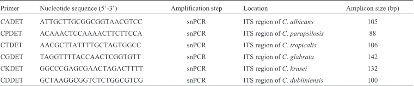
A 22-bp forward primer, CTSF (5-CGCATCGAT GAAGAACGCAGC-3), and a 25-bp reverse primer, CTSR (5-TCTTTTCCTCCGCTTATTGATATGC-3), ca-pable of amplifying the 3 end of 5.8S rDNA and the 5 end of 28S rDNA, including the intervening internally tran-scribed spacer 2 (ITS2) region, were synthesized by metabion international AG, Germany. Species-specific oligonucleotide primers for seminested PCR (snPCR) were derived from the ITS2 regions of C. albicans, C. parapsilosis, C. tropicalis,C. glabrata,C. krusei andC. dubliniensis and are described in Table 1 (Fujita et al., 1994; Ahmadet al., 2002; Khanet al., 2009).
DNA amplification and detection
reac-tion mixture consisted of 1mL AmpliTaq PCR buffer I; 1 U of AmpliTaq DNA polymerase; 5 pmol of CTSR together with 5 pmol of CADET, CPDET, CGDET, CTDET, CKDET, or CDDET; 1mL of the first PCR product; and 0.1 mM each deoxynucleoside triphosphate. PCR cycling was carried out under the following conditions:
An initial denaturation step at 94 °C for 3 min fol-lowed by 30 cycles of denaturation at 94 °C for 1 min, an-nealing at 60 °C for 30 s, and extension at 72 °C for 1 min. with a final extension step at 72 °C for 10 min. Optimum amplification was determined to be obtained with 30 cycles of the first PCR followed by 20 cycles of the snPCR accord-ing to Ahmadet al.(2002). Appropriate negative controls were included in each test run, including controls omitting the DNA template during PCR assays.
Amplified DNA fragments were separated by agarose gel electrophoresis at 80 V for 1 to 2 h on gels composed of 1% (wt/vol) agarose (Sigma-Aldrich, Germany).The gels were exposed to UV light and photographed. The sizes of amplified DNA fragments were identified by comparison with molecular size marker DNA (100-bp DNA ladder).
Statistical analysis
Statistical analysis was performed using SPSS soft-ware version 16 (SPSS Inc., Chicago, USA). Values are presented as percentages of the group from which they were derived. Sensitivities and specificities of chromo-genic media for the species were calculated as follows:
Sensitivity true positive
(true positive false nega =
+ tive)
´100
Sensitivity true negative
(true negative false posi =
+ tive)
´100
Percent agreement was determined by the number of isolates positive by conventional methods /number of PCR-positive isolates x 100.
Results
Candidaidentification by conventional methods
Among the 127Candidaisolates, the majority were non- albicans(95/127, 75%).C. tropicalis was the most frequent (46.5% or 59 strains) followed by C. glabrata
(17.4% or 22 stains). On the other hand,C. albicans consti-tuted 25% of the isolates (32/127). The remaining species constituted much lower percentages as shown in Figure 1.
Candidaidentification on four chromogenic media
By culture on four chromogenic media, different iso-lated Candida spp. showed different colony colors and morphology which are summarized in Table 2.
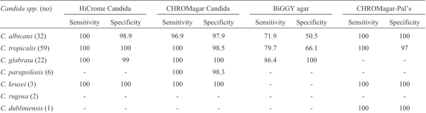
The sensitivities and specificities of the chromogenic media in comparison to the conventional methods are shown in Table 3. HiCrome Candida and CHROMagar Candida media had comparably high sensitivities and specificities in identification ofC. albicans,C. tropicalis , C. glabrataandC. krusei. CHROMagar Candida was the only medium that identifiedC. parapsilosis with a high sensitivity and specificity.BiGGY agar showed the lowest sensitivity and specificity in the identification of C. albicans, C. tropicalis and C. glabrata. CHROMagar Candida medium supplemented by Pal’s medium was the only chromogenic medium that detected C. dubliniensis
with 100% sensitivity and specificity.
PCR results
Out of the 127 Candidaisolates, only 123 isolates were tested by PCR. C. rugosa (2 isolates) and C. haemulonii(1 isolate) were not tested. Regarding PCR
am-Figure 1- Frequency of differentCandidaspecies identified by conven-tional methods.
Table 1- Primers for species identification by snPCR.
Primer Nucleotide sequence (5’-3’) Amplification step Location Amplicon size (bp)
CADET ATTGCTTGCGGCGGTAACGTCC snPCR ITS region ofC. albicans 105
CPDET ACAAACTCCAAAACTTCTTCCA snPCR ITS region ofC. parapsilosis 88
CTDET AACGCTTATTTTGCTAGTGGCC snPCR ITS region ofC. tropicalis 106
CGDET TAGGTTTTACCAACTCGGTGTT snPCR ITS region ofC. glabrata 142
CKDET GGCCCGAGCGAACTAGACTTTT snPCR ITS region ofC. krusei 132
plification ofCandidarDNA with universal fungal prim-ers, the first round of PCR amplification of rDNA was per-formed with genomic DNA from different Candidaspp. and non-fungal organisms with CTSF and CTSR primers and the amplified products were detected by agarose gel electrophoresis (AGE). The PCR amplification with these two primers and genomic DNA from C. albicans, C. tropicalis, C. glabrata, C.krusei, C. dubliniensis, and C. parapsilosisresulted in amplification of DNA fragments of 344, 334, 423, 335, 337 and 317 bp respectively (Figure 2).
In sn PCR, reamplification of the product of the first PCR with CTSR and the species-specific primers corre-sponding to the ITS2 sequences from C. albicans, C. tropicalis,C. glabrata,C. krusei,C. dubliniensis, andC. parapsilosis resulted in specific amplification of single DNA products of the expected sizes (105, 106, 142, 132, 100, 88 bp respectively) (Figure 3) .
Figure 2- PCR amplification of genomic DNAs of 6Candidaspp. with universal fungal primers. Lane M: 100-bp molecular size marker,Lane 1: C. tropicalis,Lane 2:C. albicans, Lane 3:C. glabrata, Lane 4:C. krusei, Lane 5:C. dubliniensis,and Lane 6:C. parapsilosis.
Table 2 -Colony colors of different isolated Candida spp. on four different chromogenic media. Candida spp. (no.) Colony colors on different chromogenic media (no.) HiCrome Candida CHROMagar Candida BiGGY agar CHROMagar-Pal’s C. albicans (32) Light green (32) Light Green (31) Dark green (1) Dark brown (23) Light brown (9) Light green (32) C. tropicalis (59) Bluish green a(49) Blue purple a(10) Blue (59) Dark brown (47) Light brown (12) Dark blue (39) Light blue (19) Light green (1) C. glabrata (22) Cream white (22) Dark pink (22) Creamy (19) Light brown (3) Cream white (22) C. parapsilosis (6) Dark pink (5) Cream white (1) White pale pink (6) Light brown (6) Cream white (4) Pale pink (2) C. krusei (3) Purple fuzzy bc (3) Pink fuzzy bc (3) Light brown (3) Pink fuzzy bc (3) C. rugosa (2) Light blue c (2) Light green c (1) Light blue c (1) Light brown (2) Light blue c (2) C. dubliniensis (1) Light green (1) Light green (1) Dark brown (1) Dark green d(1) C. kefyr (1) Greenish brown (1) White pale pink (1) Light brown (1) Pale pink (1) C. haemulonii (1) Greenish brown (1) White pale pink (1) Light brown (1) Pale pink (1) a: Halo which diffused into surrounding agar, b: Large rough, c: flat pale edges, d: rough with fringe.
The results of snPCR in species identification were concordant with those of the conventional phenotypic methods for all isolates of C. albicans, C. tropicalis, C. glabrata,C. krusei, andC. dubliniensisspecies. But for the 3 isolates of C. parapsilosis, there was discrepancy be-tween snPCR and the phenotypic tests. Thus, the overall concordance of the snPCR compared to the conventional phenotypic tests in the identification of Candidaspecies was found to be 97.5% (120 of 123) as shown in Table 4.
Discussion
Rapid presumptive identification of yeast species is quite difficult in resource- limited countries due to the lack of training, proper reagents and equipments. In order to reduce the financial burden of the poor patients, these laboratories do not go beyond the germ tube test and limit their diagnosis toC. albicansor NAC. The biochemical assimilation and fermentation tests are not used in these laboratories due to lack of resources, expertise and time required for these tests which increase the cost of mycol-ogy cultures (Pfalleret al., 1996). As a result, direct selec-tion of appropriate agents for antifungal therapy or prophylaxis becomes almost impossible. That’s why there is always a need for media which help not only in the iso-lation but also in the identification at the species level (Nadeemet al., 2010).
Chromogenic media have the advantage of rapid identification ofCandidaspecies, technically simple prep-aration (by boiling), rapid and cost effective compared to technically demanding time consuming and expensive con-ventional method (Vijayaet al., 2011). In this study, the colony appearance of 127 Candida isolates on four chromogenic media was compared in order to identify the most easy and reliable one for rapid identification of differ-entCandidaspecies. Compared to the results of the con-ventional methods, the sensitivity and specificity of HiCrome Candida Differential agar (HiMedia, Mumbai, India) were determined as 100% and 98.9% forC. albicans, 100% and 100% forC. tropicalis, 100% and 99% forC. glabrata, and 100% and 100% forC. krusei.
RegardingC albicans, our results are comparable to many previous studies. Willinger et al. (2001) reported 98.8% sensitivity and 100% specificity. Baradkar et al.
(2010) reported a sensitivity and specificity of HiChrom Candida agar to be 96.55 and 96.42% respectively. Yuce-soy and Marol (2003)reported the sensitivity and specific-ity to be 99.4% and 100% respectively.
ForC. tropicalis, similar results were previously re-ported by Yucesoy and Marol (2003). Baradkar et al.
(2010) found the sensitivity and specificity to be 100%. On the other hand, Penget al.(2007) showed 100% sensitivity and 78.8% specificity.
We couldn’t distinguish C. dubliniensis from C. albicansisolates as we had only one isolate that gave light green colored growth so it is difficult to evaluate HiChrom Candida agar for the identification of this species.
For C. parapsilosis, 83% of the isolates produced pink colonies while 17% produced cream colonies similar to those of C. glabrata. AsC. glabrata doesn’t produce pseudohyphae on corn meal/Tween 80 agar, combination of the two media can distinguish the two species within 48 h as compared to 48 h to 2 weeks required for sugar assimila-tion tests (Willingeret al., 2001). These results were similar to those reported by Girgiset al.(2009) and Vijayaet al.
(2011). We reported a sensitivity and specificity for identi-fication ofC. glabratato be 100% and 99% respectively comparable to Willingeret al.(2001) but higher than other studies (Yucesoy and Marol, 2003; Peng et al., 2007;
Table 3- Sensitivity and specificity of the chromogenic media in relation to the conventional phenotypic methods.
Candida spp.(no) HiCrome Candida CHROMagar Candida BiGGY agar CHROMagar-Pal’s
Sensitivity Specificity Sensitivity Specificity Sensitivity Specificity Sensitivity Specificity
C. albicans(32) 100 98.9 96.9 97.9 71.9 50.5 100 100
C. tropicalis(59) 100 100 100 98.5 79.7 66.1 100 97
C. glabrata(22) 100 99 100 100 86.4 100 -
-C. parapsilosis(6) - - 100 98.3 - - -
-C. krusei(3) 100 100 100 100 - - 100 100
C. rugosa(2) - - -
-C. dubliniensis(1) - - - 100 100
Table 4- Agreement between the conventional phenotypic methods and sn PCR in the identification ofCandidaspecies.
Candida spp. No. of isolates identified by: % agreement Conventional
phenotypic methods
snPCR
C. albicans 32 32 100
C. tropicalis 59 59 100
C. glabrata 22 22 100
C. parapsilosis 6 3 50
C. krusei 3 3 100
C. dubliniensis 1 1 100
Baradkaret al., 2010). The results in this study concerning
C. parapsilosisdiffered from Baradkaret al. (2010) who described that the majority of growth gave cream colored colonies (8/10) while the minority (2/10) produced pink colonies.C. rugosaproduced characteristic easily identifi-able colony color and morphology on HiCrome Candida Differential agar. OtherCandidaspecies (C. kefyrandC. haemulonii) could not be differentiated from each other on this medium as both produced greenish brown colonies. Unfortunately, we couldn’t find any other reports evaluat-ing HiCrome Candida Differential agar (HiMedia, Mumbai, India) for identification of these less common
Candidaspecies.
The sensitivity and specificity of CHROMagar Candida (Paris, France) were determined as 96.9% and 97.9% forC. albicans, 100% and 98.5% forC. tropicalis, 100% and 100% forC. glabrata, 100% and 100% forC. krusei, and 100% and 98.3% for C. parapsilosis respec-tively. C. rugosa appears to typically produce a readily identifiable and unique color/colony type on CHROMAgar Candida as previously reported (Hospenthalet al., 2006). In agreement, Nadeem et al. (2010) reported that CHROMAgar Candida correctly identified the majority of
C. albicans,C. tropicalis,C. glabrata, andC. kruseiwith comparable high sensitivities and specificities.
RegardingC. glabrata, we didn’t agree with Hospen-thalet al. (2006), who reported that they couldn’t distin-guishC. glabratafrom others that gave pink to lavender coloured colonies.C. kefyrandC. haemuloniicould not be differentiated from C. parapsilosis on CHROMagar Candida. Also,C. dubliniensiscould not be distinguished fromC. albicansgreen colonies. These results were consis-tent with those of Hospenthalet al.(2006) and Nadeemet al.(2010).
The addition of Pal’s agar to CROMagar Candida medium facilitated the differentiation of C. dubliniensis
fromC. albicanswith 100% sensitivity and specificity for both species, whereC. albicansisolates appeared as light green smooth colonies whileC. dubliniensisshowed distin-guishable dark green rough colonies which were accompa-nied by the production of chlamydospores. These results were in accordance with those of Sahand et al. (2005). Other Candida species that were assayed in this study showed rough or smooth colonies in CHROMagar Candida supplemented with Pal’s agar; however, they never devel-oped green colonies nor produced chlamydospores, making their misidentification as C. albicans or C. dubliniensis
highly unlikely. Interestingly,C. tropicalis andC. krusei
retained a distinguishable blue and pink-and-rough colony aspect, respectively, that allowed their easy identification on CHROMagar Candida medium supplemented with Pal’s agar with sensitivity and specificity detected as 100% and 97% forC. tropicalisand 100% forC. kruseirespectively. On the other hand,C. glabrata,C. parapsilosis,C. kefyr
andC. haemuloniiproduced indistinguishable pale pink to
cream white smooth colonies making it difficult to identify them on CHROMagar Candida supplemented with Pal’s medium. These results were in harmony with Sahandet al.
(2005).
The sensitivity and specificity of BiGGY agar were detected as 71.9% and 50.5% forC. albicans, 79.7% and 66.1% for C. tropicalis, and 86.4% and 100% for C. glabratarespectively. It was difficult to differentiate the colors ofC. albicansandC. tropicaliscolonies on this me-dium. In the present study, mostC. glabratastrains (86.4%, 19/22) showed distinctive appearance on BiGGY agar after 48 h. Brown color was only observed at the first streaks of the cultures, where the colonies were very crowded, while other areas especially single colonies were very small and creamy colored. These data agreed with those of Yücesoy and Marol (2003) with the exception ofC. krusei which they reported to show typical distinctive appearance on BiGGY agar with 100% sensitivity and specificity. The dis-crepancy may be due to the small number ofC. krusei(3 isolates) tested in our study compared to 12 isolates in the study of Yücesoy and Marol (2003). The lower sensitivity and specificity of BiGGY agar to identify commonly iso-latedCandidaspecies potentially limits the clinical useful-ness of this agar despite its low cost compared to other chromogenic media.
As Culture-based phenotypic identification tech-niques are slow and especially prone to misidentification of fungal pathogens. Numerous DNA-based methods have developed to diagnoseCandidainfections. In the present study, snPCR assays targeting species-specific sequences in the rDNA have been used for the specific detection of six clinically importantCandidaspecies. We used the sn PCR amplification method according to many previous studies (Ahmadet al., 2002, 2004; Çerikçiogluet al., 2010). The reamplification step introduced has resulted in maximum sensitivity of the assay with a specificity of 100% (Ahmad
et al., 2004; Çerikçiogluet al., 2010).
The target for snPCR amplification was rDNA which is considered to be most suitable for sensitive detection of
Candidabecause it is present in multiple copies (50 to 100
copies) perCandida genome (Reisset al., 1998).
More-over, between the highly conserved rDNA subunits are the internally transcribed spacers, which contain sequences unique to eachCandidaspecies, and thus the use of primers
corresponding to these regions facilitates species identifi-cation (Ahmedet al., 2002).
The results presented in this study showed a 97.5% overall concordance of the snPCR with the conventional methods including the use of CHROMagar Candida me-dium in the identification ofCandidaspecies which were comparable to those of Ahmadet al.(2002) and Girgiset al.
The discrepancy we found was among 3 isolates which were phenotypically identified asC. parapsilosisbut they showed negative results when tested with all spe-cies-specific primers used in the study (50%). Although the precise reason for this discrepancy remains unclear, it could be related to inadequacy of the presently available phenotypicCandidaidentification methods. Further con-firmation by DNA sequencing may be required to correctly identify these isolates. Ahmad et al. (2004) reported a closely related finding. They found that no amplification was detected in snPCR fromC. krusei,C. lusitaniaeandC. dubliniensis.
The snPCR used in this study has an average process-ing time of 9 to 10 h, does not require the use of hybridiza-tion probes and radioactive substances, and is specific. The snPCR appears to be at least 100 times more sensitive than the multiplex PCRs (Ahmedet al., 2002).
In conclusion, CHROMAgar Candida medium (CHROMagar Company, Paris, France) is an easy and ac-curate method for presumptive identification of the most commonly encountered Candida spp. (C. albicans, C. tropicalis, C. glabrata, C. parapsilosis and C. krusei) within 48 h. Although sn PCR identified the species within 8-10 h but its cost-effectiveness should also be considered. So these phenotypic methods should be considered in the identification ofCandidaspecies in areas with limited re-sources.
References
Ahmad S, Khan Z, Mustafa AS, Khan ZU (2002) Seminested PCR for diagnosis of candidemia: comparison with culture, antigen detection, and biochemical methods for species identification..J Clin. Microbiol. 40(7):2483-2490. Ahmad S, Mustafa AS, Khan Z, Al-Rifaiy AI, Khan ZU (2004)
PCR-enzyme immunoassay of rDNA in the diagnosis of candidemia and comparison with amplicon detection by agarose gel electrophoresis. Int. J Med Microbiol 294:45-51. Baradkar VP, Mathur M, Kumar S (2010) Hichrom Candida agar for identification of Candida species. Indian. J. Pathol. Microbiol 53(1):93-94.
Barnett JA, Pane RW, Yarrow D (2000) Yeasts: Characteristics and Identification. Cambridge University Press: Cambridge, 3rd edition, pp 26-27.
Bernal S, Mazuelos ME, Garcia M, Aller AI, Martinez MA, Gutierrez MJ (1996) Evaluation of CHROMagarCandida
medium for the isolation and presumptive identification of species of Candida of clinical importance Diagn Microbiol Infect Dis 24(4):201-204.
Bhavan PS, Rajkumar R, Radhakrishnan S, Seenivasan C, Kan-nan S (2010) Culture and Identification ofCandida albicans
from Vaginal Ulcer and Separation of Enolase on SDS-PAGE. Interna J Biol 2:84-94.
Çerikçioglu N, Aksu B, Dal, TD, Deniz U, Bilgen HS, Özek E, Söyletir G (2010) Seminested PCR for detection and identi-fication of Candida species directly from blood culture bot-tles. New. Microbiologica. 33:57-62.
Fluit AC, Jones ME, Schmitz FJ, Acar J, Gupta R, Verhoef J (2000) Antimicrobial susceptibility and frequency of occur-rence of clinical blood isolates in Europe from the SENTRY antimicrobial surveillance program, 1997 and 1998. Clin In-fect Dis 30(3):454-460.
Fujita S, Lasker BA, Lott TJ, Reiss E, Morrison CJ (1995) Micro-titration plate enzyme immunoassay to detect PCR-ampli-fied DNA fromCandidaspecies in blood. J Clin Microbiol 33:962-967.
Girgis SA, El-Mehalawy AA, Rady LM (2009) Comparison be-tween culture and non-culture based methods for detection of Nosocomial fungal infections ofCandidaspp. in inten-sive care unit patients. Egypt. Acad. J. Biolog. Sci. 1:37-47. Harju S, Fedosyuk H, Peterson KR (2004) Rapid isolation of yeast
genomic DNA: Bust n’ Grab. BMC Biotechnol 4:8. Hospenthal DR, Beckius ML, Floyd KL, Horvath LL, Murray CK
(2006) Presumptive identification ofCandidaspecies other
than C. albicans, C. krusei, and C. tropicalis with the
chromogenic medium CHROMagar Candida. Ann Clin Microbiol Antimicrobiol 3:5-1.
Hospenthal DR, Murray CK, Rinaldi MG (2004) The role of antifungal susceptibility testing in the therapy of candi-diasis. Diagn Microbiol Infect Dis 48(3):153-160.
Khan Z, Mustafa AS, Alam FF (2009) Real-time LightCycler polymerase chain reaction and melting temperature analysis for identification of clinically important Candida spp. J Microbiol Immunol Infect 42:290-295.
Liguori G, Lucariello A, Colella G, De Luca A, Marinelli P (2007) Rapid identification of Candida species in oral rinse solu-tions by PCR. J Clin Pathol 60:1035-1039.
McGinnis MR (1994) Mycology.InIsenberg H. D. (ed.). Clinical microbiology procedure handbook. American Society for Microbiology, Washington D.C., pp 6.1-6.12.
Murray CK, Beckius ML, Green JA, Hospenthal DR (2005) Use of chromogenic medium for the isolation of yeasts from clinical specimens. J Med Microbiol 54:981-985.
Nadeem, S.G.; Hakim, S.T.; Kazmi, S.U. (2010). Use of CHROMagar Candida for the presumptive identification of
Candida species directly from clinical specimens in
re-source-limited settings. Libyan J Med.5:2144-2150. Nguyen MH, Peacock JE, Morris A J, Tanner DC, Nguyen ML,
Snydman DR, Waqener MM, Rinaldi MG, Yu VL (1996) The changing face of candidemia: emergence of
non-Candida albicansspecies and antifungal resistance. Am. J
Med 100(6):617-623.
Peng, C.F.; Lee, K.M.; Lee, S.H. (2007). Characterization of two chromogenic media of Candida ID2 and CHROMagar
Candida for preliminary identification of yeasts.J Biomed Lab Sci 19:63-68.
Pfaller MA, Houston A, Coffmann S (1996) Application of CHROMagar Candida for rapid screening of clinical speci-mens for Candida albicans, Candida tropicalis, Candida
krusei, andCandida(Torulopsis)glabrata. J Clin Microbiol
34(1):58-61.
Sahand IH, Moragues MD, Eraso E, Villar-Vidal M, Quindos G, Ponton J (2005). Supplementation of CHROMagarCandida
medium with Pal’s medium for rapid identification of
Candida dubliniensis. J Clin Microbiol 43 (11):5768-5770.
Sahiner F, Ergünay K, Ozyurt M, Ardyç N, Hosbul T, Hazne-daroglu T (2011) Phenotypic and genotypic identification of
Candida strains isolated as nosocomial pathogens.
Mikrobiyol Bul 45:478-488.
Vijaya D, Harsha TR, Nagaratnamma T (2011) Candida
Speciation Using Chromagar. J Clin Diagn Res 5:755-757. Wahyuningsih R, Freisleben HJ, Sonntag HG, Schnitzler P (2000)
Simple and rapid detection ofCandida albicansDNA in
se-rum by PCR for diagnosis of invasive candidiasis.J Clin
Microbiol38:3016-3021.
Willinger B, Hillowoth C, Selitsch B, Manafi M (2001) Perfor-mance of Candida ID, a new chromogenic medium for pre-sumptive identification of Candida species, in comparison to CHROMagar Candida. J Clin Microbiol 39(10):3793-3795. Yucesoy M, Marol S (2003) Performance of CHROMagar
Candida and BIGGY agar for identification of yeast species. Ann Clin Microbiol Antimicrobiol 2:1-8.