ABSTRACT
Candida albicans and
non-Candida albicans
Candida species on acrylic
substrate
123
1- MSc. PhD, Sr. Lecturer, Department of Microbiology, Modern Dental College & Research Centre, Indore (M.P.), India.
2- BDS, MDS, Department of Prosthodontics with Crown, Bridge & Implantology, Modern Dental College & Research Centre, Indore (M.P.), India. 3- BDS, MDS, Department of Oral Medicine, Diagnosis & Radiology, Modern Dental College & Research Centre, Indore (M.P.), India.
Dr. Apurva K Pathak - Department of Microbiology, Modern Dental College & Research Centre - Gandhinagar - Indore (M.P.) - India - 453112 - Phone: +91 731 2882695 - Ext. 341 - Fax: +91 731 2882697 - e-mail: [email protected]
!"#$%&''()*+,#$-&'$')+4$(&'$'
O
Candida species,but the interaction among the different species of the Candida is yet to be completely
of four species of Candida in both single-species and multi-species combinations on the surface of dental acrylic resin strips was evaluated. Material and Methods: The species of
Candida, isolated from multiple species oral candidiasis of the neutropenic patients, were used for the experiment. Organisms were cultured on Sabouraud dextrose broth with 8% !"#$&'&
violet assay. Student’s t-test and ANOVA were used to compare in vitro
for the individual species of Candida and its different multi-species combinations. Results: ! of individual species (C. glabrata>C. krusei>C. tropicalis>C. albicans) and in its multi-species’ combinations (the highest for C. albicans with C. glabrata and the lowest for all the ' *+! !!Candida albicans Candida species (NCAC) than for C. albicans species with intra-species variation. Presence of C. albicans in
/C. tropicalis
with all other NCAC species.
Key words:0&*1
INTRODUCTION
Candida is the most frequently isolated fungal !!2 ranging from superficial mucosal infections to systemic mycoses. Oral fungal infections develop frequently in immunocompromised patients, particularly in patients with prolonged, severe neutropenic episodes2. In vivo studies indicate
that microbial contamination of denture acrylic resin occurs quite rapidly and implanted devices like denture prostheses provide refuge to candidal organisms as either single-species or multi-species 5,13. Consequently, the immunocompromised
denture wearers are more prone to the fungal
infections.
One of the major factors contributing to the virulence of Candida is its ability in acclimatize to a variety of different habitats for growth and formation of surface-attached microbial 7 9; & matrix of extracellular polymeric substance (EPS), which display phenotypic features that differ from
72! 7,16,19.
In vitro studies indicate that microbial contamination of denture acrylic resin occurs quite rapidly and the yeast cells adhere strongly to denture materials4,13,18.As bacteria and non-Candida
Candida albicans in vivo, it is likely that extensive interspecies interactions take place in these adherent populations16,17,19.
A large number of in vitro model systems have been used to investigate characteristics of
!C.
albicans and bacteria1,3,10,11,12,17. Some studies have
also been done on the interaction between different species of Candida on in vitro
formation20,25. The study on the interaction between
different species of Candida on in vitro multi-species
27.
forming ability of clinical isolates of four species of Candida in single-species and multi-species combination on the surface of dental acrylic resin strips, commonly used for dental appliances, was evaluated.
MATERIAL AND METHODS
Organisms
The test organisms included 24 isolates of 4 different species of Candida i.e. C. albicans, C. kruzi, C. glabrata and C. tropicalis (six isolates of each), isolated from the oral lesions of multiple species "BDCandida species) Oropharyngeal Candidiasis (OPC) of the neutropenic patients (absolute neutrophil count <1.5×10⁹ cells/L)28. Pure culturing
$ Microbiology, Modern Dental College and Research *+Candida species was conducted by culture characteristics on HiChrome
Candida agar medium (HiMedia, Mumbai, India), assessing germ tube, chlamydospore formation and sugar assimilation patterns6,22.
Simulated acrylic resins plates fabrication
T h i r t y - s i x s q u a r e a c r y l i c r e s i n s (polymethylmethacrylate) strips of 10x10x3 mm were fabricated. The acrylic resins strips were prepared as described by Samaranayake and MacFarlane21 (1980) with some modifications.
Q 27 boiled out, packed with the denture base resins, and heat polymerized according to manufacturer instructions at a temperature 73°C for 6 h. Strips 27!0 surface of strips was polished on buff wheel with pumice slurry. Other surface was left untouched to simulate intaglio surface. The resultant acrylic resins strips were immersed in distilled water for 1 week to leach excess monomer. Following this strips were disinfected by dipping in 70 % alcohol for 1 min, washed with sterile distilled water, dried and used for the experiment after checking their sterility.
'HWHUPLQDWLRQRIELR¿OPSURGXFWLRQ
Sabouraud dextrose broth (SDB) prepared from powdered Sabouraud broth (HiMedia, Mumbai, India) supplemented with 60 g of glucose per liter " ! WX !Y WZ' (Qualigens, Navi Mumbai, India), was according to manufacturer’s instructions. Fresh pure cultures of testing organism were prepared on SDA medium by subculturing clinical isolate. A loop full of organisms from each SDA plate was inoculated # "WZ glucose concentration) for 24 h at 35±2°C. The turbidity of each suspension was adjusted to the equivalent of 1x10⁷ CFU/mL with SDB as determined
by comparative plate counts.
Next, 1 mL of suspension of isolated species and testing combination of different species was
Figure 1- Optical Density of de-staining solution #
prepared by mixing equal volume of tested species, inoculated into a test tube with a screw cap (HiMedia, Mumbai, India) containing 9 mL of SDB, 7 1x10⁶ CFU/mL. Strips were placed in SDB, and then
incubated at 35±2°C for 24 h without agitation. After 24 h of incubation, the culture broth in the tube was aspirated gently, and then acrylic strips were taken out for further investigation.
The acrylic resins strips, on which biofilms developed, were washed once with distilled water,
and then incubated in a crystal violet (HiMedia, Mumbai, India) staining solution (0.1% in distilled water) for 15 min. These were then washed three times with distilled water. The stain was then dissolved in de-staining solution (95% ethanol) and absorbance in terms of optical density (OD) was measured at 570 nm as previously described15.
Untreated acrylic strips were used as a control for the amount of the crystal violet stain in the de-staining solution. The absorbance values of controls were subtracted from the test values to minimize
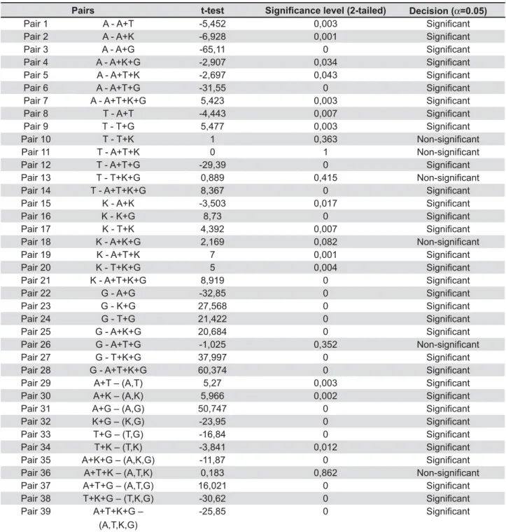
t-test *8&)+9 :8α;'<'=9
Pair 1 A - A+T -5,452 0,003
Pair 2 A - A+K -6,928 0,001
Pair 3 A - A+G -65,11 0
Pair 4 A - A+K+G -2,907 0,034
Pair 5 A - A+T+K -2,697 0,043
Pair 6 A - A+T+G -31,55 0
Pair 7 A - A+T+K+G 5,423 0,003
Pair 8 T - A+T -4,443 0,007
Pair 9 T - T+G 5,477 0,003
Pair 10 T - T+K 1 0,363
Pair 11 T - A+T+K 0 1
Pair 12 T - A+T+G -29,39 0
Pair 13 T - T+K+G 0,889 0,415
Pair 14 T - A+T+K+G 8,367 0
Pair 15 K - A+K -3,503 0,017
Pair 16 K - K+G 8,73 0
Pair 17 K - T+K 4,392 0,007
Pair 18 K - A+K+G 2,169 0,082
Pair 19 K - A+T+K 7 0,001
Pair 20 K - T+K+G 5 0,004
Pair 21 K - A+T+K+G 8,919 0
Pair 22 G - A+G -32,85 0
Pair 23 G - K+G 27,568 0
Pair 24 G - T+G 21,422 0
Pair 25 G - A+K+G 20,684 0
Pair 26 G - A+T+G -1,025 0,352
Pair 27 G - T+K+G 37,997 0
Pair 28 G - A+T+K+G 60,374 0
Pair 29 A+T – (A,T) 5,27 0,003
Pair 30 A+K – (A,K) 5,966 0,002
Pair 31 A+G – (A,G) 50,747 0
Pair 32 K+G – (K,G) -23,95 0
Pair 33 T+G – (T,G) -16,84 0
Pair 34 T+K – (T,K) -3,841 0,012
Pair 35 A+K+G – (A,K,G) -11,87 0
Pair 36 A+T+K – (A,T,K) 0,183 0,862
Pair 37 A+T+G – (A,T,G) 16,021 0
Pair 38 T+K+G – (T,K,G) -30,62 0
Pair 39 A+T+K+G – (A,T,K,G)
-25,85 0
Table 1- Paired samples t-test of Candida species combinations$
$C. albicans; T., C. tropicalis; K., C. krusei; G., C. glabrata;
background interference.
Statistical analysis
The ODs of the amount of the crystal violet in the de-staining solution, measured for different Candida
species, were compared by the paired Student’s t-test by using the SPSS Win 12.0 program (SPSS Inc, Chicago, IL, USA). Differences between the isolated species and its multi-species combinations ! P of 0.05. The null hypothesis (HO) rejected in favor of the "^_'!` (0.05) if; T>tn-1, į/2 (value of the Student table
with n -1 degrees of freedom). The null hypothesis ^X }~X " formation ability among the tested species) and "'^_}~X" ! the tested species).
RESULTS
Twenty-four Candida species isolated from clinical samples and were used for this study. Amongst the 24 isolates, 6 each of C. albicans, C. glabrata, C. krusei, and C. tropicalis were used to ! species and in multi-species combination. All the isolates produce moderate to high degree of "! _'+!C. glabrata were reported to be the highest on acrylic substances (OD - 0.3833±0.02066), whereas; of C. albicans
to be the lowest (OD - 0.1467±0.01366). In case of multiple species conditions, it was observed that the C. albicans had a positive impact on highest degree of slim production occurred when
C. albicans were inoculated with C. glabrata
(OD- 0.7850±0.03209); whereas C. krusei had a negative impact in combination. Under multiple species condition when all the four species were ! severely hampered (OD – 0.1133±0.01033).
To test the hypothesis of no difference or no relationship between biofilm-forming ability of isolated species with that of in-combination of other species, paired t-test was performed (Table 1). For all the four species, 7 different multi-species combinations were prepared, which made a total of ! null hypothesis in favor of alternate hypothesis. Only 15% of pairs showed no difference between ! single-species and in its multi-species combination.
DISCUSSION
mucosal infections can lead to severe oral and esophageal candidiasis, resulting in disseminated candidiasis and sometimes early death. One of the most important virulence factors of Candida
an important clinical consequence, as it confers resistance to antifungal therapy and capacity for immune defenses3,5,6,12,19. Changes in the oral
environment effected by tooth loss or denture ! ! 2 The carriage rates of single and multiple Candida
! ! in denture wearers11. In the light of above fact
Candida, isolated from multiple species candidimia ability on acrylic surface in vitro, in single-species and multi-species combination.
Candida
on polymethylmethacrylate strips which occur essentially in three overlapping phases: early (0-11 h), intermediate (12-30 h), and maturation (38-72 h) phases. The early stage is characterized by adherence and development of blastospores into distinct microcolonies. By 18 to 24 h, the Candida
structure comprising a mixture of yeasts, germ tubes, and young hyphae; this intermediate phase is distinguished by the production of extracellular polymeric substance (EPS). During maturation, the 7# network of yeasts, pseudohyphae, and hyphae are embedded4,18.
+!
C. albicans, C. krusei, C. tropicalis and C. glabrata
recovered from multiple species candidimia lesions, was evaluated by measuring absorbance of the de-staining solution containing crystal violet dye (crystal violet assay). The present showed C. glabrata forming thickest slim layer on acrylic resins strip followed by C. krusei, C. tropicalis
and C. albicans, ! ! previous researchers9,24. However Silva, et al.23
(2009) reported that C. glabrata was high in both protein and carbohydrates, which that probably enabled it to adsorbed more amount of crystal violet contents.
Shin, et al.22 (2002) observed that biofilm
formation was most frequent for isolates of C. tropicalis (80%), followed by C. parapsilosis (73%),
C. glabrata (28%), and C. albicans (8%). This ! probably due to the sources of isolates. Different strains of the same Candida species were found
also reported in this study, indicating “strong” and 97;!! each Candida species26. Shin, et al.22 (2002) also
!! for NCAC than for albicans species using similar protocol.
Presence of one species of microorganism on a surface can promote the adhesion of another14.
Thein, et al.25 (2007) reported competitive
C. albicans
and C. krusei; whereas Pereira-Cenci, et al.20
(2008) did not report competitive interaction C. albicans and C. glabrata. In the present study, the highest degree of slim production was seen with the combination of C. albicans and C. glabrata, followed by the combination of C. albicans, C. tropicalis and C. glabrata, and the lowest slim production was seen of Candida. Presence of C. albicans in multi-species inoculated with all other NCAC species; whereas
C. tropicalis retarded the production of slim under multi-species condition except with C. albicans. In this way, C. albicans might be able to successfully provide a substratum to the NCAC species on the acrylic prosthesis.
0! agents. Studies have demonstrated drug resistance
when Candida !
like denture acrylic. The possible mechanisms of ! !! matrix; phenotypic switching, surface-induced expression of resistance genes and a small number of “persistent” cells8. Presence of two or more
!!! Synergistic effects of these factors can pose major problems to the clinicians.
CONCLUSION
& forming ability was found greater for NCAC than for
albicans species, isolated from multi-species oral candidiasis of the neutropenic patients. Presence of C. albicans the slim production with all other NCAC species, whereas C. tropicalis impeded the production of slim under multi-species condition except for C. albicans.
ACKNOWLEDGEMENTS
Authors are greatly thankful to Dr. P.V. Wanjari, Honorable Dean, Modern Dental College and Research Centre, Indore, for providing academic and materialistic supports, without which it was
hard to perform this work. We also acknowledge the helpful comments and suggestions of the reviewers of JAOS.
REFERENCES
_ 1 & & # $! Candida albicans and Staphylococcus epidermidis. J Med Microbiol. 2002;51:344-9.
D1&#! #!Candida infections in children with cancer. Path Onco Res. 2006;12:237-41.
&#$!Candida to antifungal agents. Methods Enzymol. 1999;310:644-56. 4- Chandra J, Kuhn DM, Mukherjee PK, Hoyer LL, McCormick T,
1&! !Candida
albicans: development, architecture, and drug resistance. J Bacteriol. 2001;183:5385-94.
5- Coogan MM, Fidel PL Jr, Komesu MC, Maeda N , Samaranayake LP. Candida and mycotic Infections. Adv Dent Res. 2006;19:130-8. 6- Costa CR, Cohen AJ, Fernandes OF, Miranda KC, Passos XS, Souza LK, et al. Asymptomatic oral carriage of Candida species in HIV-infected patients in the highly active antiretroviral therapy era. Rev Inst Med Trop Sao Paulo. 2006;48:257-61.
*Q0 1995;15:137-40.
W$! Candida infections.
Rev Iberoam Micol. 2002;19:139-43.
^#$!&Candida species on the surface of catheter materials in vitro. Infect Immun. 2002;62:915-21.
10- Henriques M, Azeredo J, Oliveira R. Candida albicans and Candida dubliniensis biomass and activity. Br J Biomed Sci. 2006;63:5-11.
11- Jenkinson HF, Douglas LJ. Interactions between Candida species and bacteria in mixed infections. In: Brogden KA, Q!$* ASM Press; 2002. p. 357-73.
12- Jin YY, Yip HK, Samaranayake YH, Yau JY, Samaranayake &! Candida albicans is unlikely to contribute to high levels of oral yeast carriage in cases of human *DXX/_D_ 7.
_ $ * 7 1 Comparison of biofilms formed by Candida albicans and Candida parapsilosis on bioprosthetic surfaces. Infect Immun. 2002;70:878-88.
14- Leung JW, Liu YL, Desta T, Libby E, Inciardi JF, Lam K. Is there ! _W/WDX _ 0+ 1 ! ! necessary for Pseudomonas aeruginosa Microbiol. 1998;30:295-304.
16- Peeters E, Nelis HJ, Coenye T. Comparison of multiple methods ! J Microbiol Methods. 2008;72:157-65.
_##&#* enzymatic activity (proteinase and phospholipase) of Candida albicans from edentulous patients, with and without denture stomatitis. Pesq Odontol Bras. 2000;14:119-122.
_W*+*11*#!*In vitro Candida colonization on acrylic resins and denture liners: 2!!! bacteria. Int J Prosthodont. 2007;20:308-10.
20- Pereira-Cenci T, Deng DM, Kraneveld EA, Manders EM, Del Bel Cury AA, Ten Cate JM, Crielaard W. The effect of Streptococcus mutans and Candida glabrata on Candida albicans on different surfaces. Arch Oral Biol. 2008;53:755-64.
21- Samaranayake LP, MacFarlane TW. An in vitro study of the adherence of Candida albicans to acrylic surfaces. Arch Oral Biol. 1980;25:603-9.
DD#^###^#$^# & Candida species recovered from nonneutropenic patients: comparison of bloodstream isolates with isolates from other sources. J Clin Microbiol. 2002;40:1244-8. 23- Silva S, Henriques M, Martins A, Oliveira R, Williams D, 1 & Candida albicans Candida species: 2009;47:681-9.
D**+*Q PWJ. Molecular and cellular mechanisms that lead to Candida $DXX/WW_X_
+)#*"6Candida albicans and non-Candida albicansCandida#++
D+#7^#7* of dual species Candida1 Oral Biol. 2007;52:1200-8.
D + #7 ^ #7 In vitro Candida albicans and non-albicans Candida species under dynamic and anaerobic conditions. Arch Oral Biol. 2007;52:761-7.
D+#*#7^#7 LP. Community lifestyle of Candida Mycoses. 2009;52:467-75.