ISSN 0100-879X
CLINICAL INVESTIGATION
www.bjournal.com.br
www.bjournal.com.br
Volume 42 (8) 692-775 August 2009
Institutional Sponsors
The Brazilian Journal of Medical and Biological Research is partially financed by
Braz J Med Biol Res, August 2009, Volume 42(8) 707-711
Flow rate, pH and calcium concentration of saliva of children
and adolescents with type 1 diabetes mellitus
Flow rate, pH and calcium concentration of
saliva of children and adolescents with type
1 diabetes mellitus
A.R. Moreira
1, I.A. Passos
2, F.C. Sampaio
1, M.S.M. Soares
1and R.J. Oliveira
31
Programa de Pós-graduação em Odontologia,
2Departamento de Odontologia Restauradora,
3Departamento de Medicina, Centro de Ciências da Saúde, Universidade Federal da Paraíba,
João Pessoa, PB, Brasil
Correspondence to: I.A. Passos, Departamento de Odontologia Restauradora, Universidade Federal da
Paraíba, Campus I, 58051-900 João Pessoa, PB, Brasil
E-mail: isabelaapassos@yahoo.com.br
Alterations in salivary parameters may increase the caries risk in diabetic children, but, contradictory data on this issue have been reported.The aims of this study were to compare salivary parameters (flow rate, pH and calcium concentration) between healthy and type 1 diabetes mellitus (T1DM) individuals. The sample consisted of 7- to 18-year-old individuals divided into two groups: 30 subjects with T1DM (group A) and 30 healthy control subjects (group B). Fasting glucose levels were determined. Unstimulated and stimulated saliva was collected. The pH of unstimulated saliva was measured with paper strips and an electrode. Calcium concentrations in stimulated saliva were determined with a selective electrode. Group A individuals had inadequate blood glucose control (HbA1C >9%), with means ± SD unstimulated salivary flow rate of 0.15 ± 0.1 mL/min compared
to 0.36 ± 0.2 mL/min for group B (P < 0.01). Stimulated salivary flow rate was similar by both groups and above 2.0 mL/min. Saliva pH was 6.0 ± 0.8 for group A and significantly different from 7.0 ± 0.6 for group B (P < 0.01). Salivary calcium was 14.7 ± 8.1 mg/ L for group A and significantly higher than 9.9 ± 6.4 mg/L for group B (P < 0.01). Except for elevated calcium concentrations in saliva, salivary parameters favoring caries such as low saliva pH and unstimulated salivary flow rate were observed in T1DM individuals.
Key words: Type 1 diabetes mellitus; Saliva pH; Salivary flow rate
Received September 22, 2008. Accepted April 28, 2009
Introduction
Diabetes mellitus (DM) is a group of metabolic dis-eases characterized by high blood glucose levels due to problems in insulin secretion or insulin action, or both. As a result, chronic hyperglycemia with frequent disorders of carbohydrate metabolism can be associated with obesity, protein and electrolyte disorders, and other diseases (1).
The number of children with type 1 and 2 diabetes is increasing in many countries. Gillespie et al. (2) pointed out that the proportion of high-risk children increased in the last 50 years, supporting the hypothesis that the rise of type 1 diabetes is due to a major environmental effect. A decade ago, the prevalence of type 1 diabetes among children under 15 years of age in the Americas was
esti-mated at 88,000, with 40% of the cases in Latin America and the Caribbean (3). In spite of the lack of recent epide-miological surveys of DM, the Brazilian estimate is 5 million diabetic patients. Even though only 10% of these cases are type 1, this is the most common type affecting children (4,5).
708 A.R. Moreira et al.
flow rates between diabetic and non-diabetic individuals. Mata et al. (11) demonstrated higher salivary calcium concentrations in diabetic individuals than in healthy sub-jects. Conversely, Lopez et al. (8) observed lower salivary calcium concentrations in diabetic patients. Reuterving et al. (12) and Edblad et al. (10) did not find any difference between normal and diabetic children regarding calcium concentrations in saliva. There are reports indicating that diabetic patients have more acidic saliva (8,11,13), whereas others have not reported this difference (10,12). It is still not clear whether caries risk can be higher in diabetic pediatric patients due to affected salivary factors as com-pared to controls (9,14). In addition to the contradictory report, salivary parameters should be measured in dia-betic children because most clinical trials have been car-ried out on adults with periodontal disease (15). Thus, the aim of this study was to compare salivary parameters (flow rate, pH and calcium concentration) of healthy and type 1 diabetes mellitus (T1DM) Brazilian children and adoles-cents.
Material and Methods
Sixty patients aged 7 to 18 years comprised the sample and were divided into two groups: A) 30 T1DM patients selected at a public health pediatric endocrinology service in João Pessoa, Brazil, and B) 30 (gender-matched, male/ female 15/15) healthy individuals selected from a public school. Children of both groups were from the same neigh-borhood reflecting the same social strata. The parents of all subjects who participated in the study gave written informed consent. The research protocol was approved by the Human Research Ethics Committee of the Federal University of Paraíba (Protocol #209-B).
Glucose salivary concentration was determined with a glucosimeter (Accu-Chek Advantage, Roche Diagnostica Brasil, Brazil). The upper limit of fasting glucose concen-tration considered to be normal was 100 mg/dL. For capil-lary glucose, normal values were considered to be those below 140 mg/dL (5). For group A, glycosylated hemoglo-bin (HbA1C) data were also obtained from the patient’s
medical records.
The subjective experience of xerostomia was diag-nosed by means of the question: “Do you feel that your mouth is dry frequently?”
Saliva was always collected 2 h after the last meal and at the same time of day (2:00 pm). This was done to standardize the collections according to the circadian rhythm (16). The subjects were also requested to refrain from any oral hygiene prior to saliva collection. The un-stimulated and un-stimulated salivary flow rates and saliva pH
were determined immediately after collection. Unstimu-lated saliva samples were obtained by having the volun-teers spit into plastic vials following the procedures pro-posed by Navazesh et al. (17,18). The stimulated salivary flow was determined with the aid of 2% citric acid accord-ing to the technique described by Fox et al. (19). Low unstimulated (≤0.1 mL/min) and stimulated salivary (≤0.5 mL/min) flow was classified according to the standards reported by Bolwig and Rafaelsen (20) and Sreebny and Valdini (16). Aliquots of stimulated and unstimulated saliva were collected into plastic vials and frozen (-20°C) until analysis for calcium level determination.
Saliva pH was determined immediately after collection of the unstimulated saliva samples in order to avoid any time-related pH changes or loss of CO2. The analyses
were performed using a pH electrode connected to a potentiometer (Orion 710A, Orion Research Inc., USA) after calibration at pH standards of 4.0 and 7.0. For 10 samples with very low volume (<100 μL), pH was deter-mined with the aid of a “narrow range” pH paper strip (Merck, Germany). Calcium concentrations in stimulated saliva were determined with a calcium selective electrode (Orion 93-20, Orion Research Inc.) together with a poten-tiometer (Orion 710A, Orion Research Inc.) calibrated at calcium standards of 1.25, 2.50, 5.00, 12.50, and 25.00 mg/L in an ionic strength adjuster following manufacturer recommendations. Taking into account all standards, only calibration curves with 5% less than variation and r ≥0.99 were accepted. The reproducibility of sample analysis was good since, by applying the Nernstian slope of 28 mV/ decade at 25°C, a potential drift of less than 0.2 mV/h was observed between duplicate samples. Prior to the analy-sis, saliva samples were thawed and centrifuged for 3 min at 7000 g.
Differences between means were determined by the Student t-test using the Statistical Package for Social Sciences version 11.0 (SPSS Inc., USA). The association of salivary parameters and glucose levels was analyzed by the Pearson product-moment correlation coefficient. An alpha value of 0.05 was selected a priori as an indicator of statistical significance. The minimum sample size required was previously estimated at 26 subjects in each group based on the salivary calcium levels observed in a pilot study (N = 6).
Results
136.8 mg/dL and mean HbA1C was 9.34 ± 1.9%. Half of the
individuals in group A showed high glucose levels (HbA1C
>9%) indicating poor metabolic control of diabetes melli-tus.
The mean value of unstimulated salivary flow for group B was more than 2-fold higher than group A, whereas there was no significant difference between groups regarding the mean values of stimulated salivary flow (Table 1). Nineteen T1DM individuals (63%, group A) complained of dry mouth, whereas none of the healthy control subjects (group B) complained. Two patients in group B showed sign of hyposalivation (<0.2 mL/min) but did not complain of dry mouth. Fourteen T1DM patients were diagnosed with hyposalivation and 3 of them had lower stimulated salivary flow (>0.5 mL/min). Mean saliva pH was 6.0 ± 0.8 and 7.0 ± 0.6 for groups A and B, respectively. Mean calcium concentration in saliva was significantly higher for group A than for group B (Table 1).
Salivary parameters and capillary glucose concentra-tions were not associated. A positive association was detected between capillary glucose concentrations and calcium concentrations in saliva (N = 60, Pearson’s r = 0.27, P < 0.05).
Discussion
In the present study, T1DM patients presented a sig-nificant decrease in unstimulated salivary flow. Moreover, dry mouth was a major complaint of many T1DM patients. These observations are similar to those reported by Siudikiene et al. (7) and Lopez et al. (8). A reduction of unstimulated salivary flow rate may reflect the presence of outlying neuropathy in T1DM patients, with an implication in the control of saliva secretion (4,14,16,21). Dehydration may cause irreversible structural changes in the salivary glands of T1DM patients (22). However, in the present study, dehydration and peripheral neuropathy were not observed. Even though a detailed evaluation was not
carried out, the very low unstimulated saliva rates (<0.1 mL/min) observed suggest salivary gland hypofunction (16,18). The low unstimulated salivary flow in young T1DM patients is particularly relevant since this condition is usu-ally more frequent among old adults and is an indication of a high caries risk in T1DM children (7,23,24).
No differences in unstimulated salivary flow rates be-tween T1DM and healthy control subjects have been ob-served in some clinical studies (7-9,19). However, differ-ences in saliva collection techniques, glucose metabolic control methods, degrees of patient dehydration, and the wide age span in some studies can limit direct compari-sons. In our study, saliva was collected in the afternoon due to operational reasons. Lower salivary flow values are more likely to occur early in the morning compared with late morning hours and afternoon collections (25). Thus, our values may have underestimated the number of pa-tients with hyposalivation even though the influence of a circadian rhythm on the saliva flow of individuals with hyposalivation is still a matter of debate (26).
The low saliva pH of T1DM patients is strong evidence of reduced buffer salivary capacity and increased caries risk. Certainly, the low unstimulated salivary flow contrib-uted to the low pH values observed, but factors not ex-plored in this report, such as plaque accumulation and microbiological shift to a more cariogenic biofilm, are also significant variables (7). The stimulated salivary measure-ments may reflect the saliva produced during meals (26). Our data on stimulated salivary flow showed no specific trend, though lower values were found among T1DM pa-tients (Table 1).
Lopez et al. (8) found low calcium concentrations in the saliva of T1DM patients compared to controls. A recent study showed no significant differences in calcium electro-lyte concentrations (median values close to 10.0 mg/L) between healthy, controlled diabetic patients and uncon-trolled diabetic patients (21). Unfortunately, the wide range of concentrations observed (from 0 to 34.5 mg/L) and the
Table 1. Table 1.Table 1. Table 1.
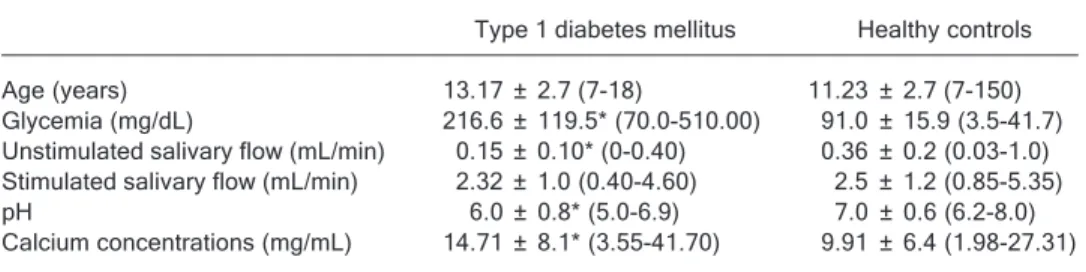
Table 1. Salivary parameters in children with type 1 diabetes mellitus and healthy controls.
Type 1 diabetes mellitus Healthy controls
Age (years) 13.17 ± 2.7 (7-18) 11.23 ± 2.7 (7-150) Glycemia (mg/dL) 216.6 ± 119.5* (70.0-510.00) 91.0 ± 15.9 (3.5-41.7) Unstimulated salivary flow (mL/min) 0.15 ± 0.10* (0-0.40) 0.36 ± 0.2 (0.03-1.0) Stimulated salivary flow (mL/min) 2.32 ± 1.0 (0.40-4.60) 2.5 ± 1.2 (0.85-5.35) pH 6.0 ± 0.8* (5.0-6.9) 7.0 ± 0.6 (6.2-8.0) Calcium concentrations (mg/mL) 14.71 ± 8.1* (3.55-41.70) 9.91 ± 6.4 (1.98-27.31)
710 A.R. Moreira et al.
small number of subjects in the subgroups (less than 12) do not permit a conclusion to be made. In a study with 60 diabetic patients, Mata et al. (11) observed higher calcium concentrations (>50 mg/L) in the saliva of DM patients than in matched healthy controls. The high sensitivity of the atomic absorbance spectrophotometer technique used to measure calcium electrolytes in saliva cannot be disre-garded. This technique can measure strong protein-cal-cium binding complexes and it is certainly much more sensitive than the specific ion electrode and probably more sensitive than the TLC photometer used by Reznick et al. (21). On the other hand, the ion-specific electrode directly measures free ionic calcium in saliva, which is of clinical relevance for the development of caries (27).
Calcium concentrations in blood can be lower in dia-betic patients compared to normal controls. This effect may be related to renal problems such as impaired absorp-tion and altered calcium excreabsorp-tion into urine. As a result, salivary calcium can also be altered. Therefore, high urine and salivary calcium concentrations are expected to occur in patients with endocrine complications that induce trace element disturbances (28). This may explain the weak but positive correlation between capillary glucose concentra-tion and calcium concentraconcentra-tion in saliva reported in the present study.
A positive relationship between salivary flow rate and calcium concentrations is expected to occur (29). Calcium concentration is about 45% higher in the submandibular glands than in the parotid glands. However, high calcium concentrations in saliva may be related to the parotid glands more than to other salivary glands (29). In fact,
Mata et al. (11) observed higher calcium concentrations in stimulated saliva than in unstimulated saliva of diabetic patients. Thus, for the T1DM patients of our study, daily intra-oral calcium concentrations might be lower than the values measured for stimulated saliva. Unfortunately, due to the small volume of the unstimulated saliva samples, the calcium analysis was not performed.
Our cross-sectional study design does not allow a detailed analysis of the caries risk in these patients. Never-theless, the low unstimulated salivary flow rate values compared to the healthy controls can be regarded as a negative factor to be considered when assisting diabetic children. Elevated calcium concentrations in saliva can be regarded as a favorable indicator for the oral health of these patients. In addition, T1DM children can be careful about their diet, which can be a positive aspect. On the other hand, these favorable variables may not be sufficient to cope with a constant low salivary pH in patients with insulin-dependent diabetes mellitus. How much calcium is of clinical relevance for these patients? Certainly, other variables such as dental plaque accumulation associated with poor salivary parameters can counteract the benefi-cial calcium effect.
Finally, our data support the view that an appropriate evaluation of salivary clinical parameters, such as salivary flow rate and buffer capacity, is recommended when as-sisting diabetic children. These data can be obtained by simple techniques (17). An adequate oral hygiene pro-gram with exposure to fluoride and frequent use of artificial saliva can be of the utmost importance for controlling development of caries in diabetic patients.
References
1. Sicree R, Shaw JE, Zimmet PZ. The global burden of diabe-tes. In: Gan D (Editor), Diabetes atlas. 2nd edn. Brussels: International Diabetes Federation; 2003. p 15-71.
2. Gillespie KM, Bain SC, Barnett AH, Bingley PJ, Christie MR, Gill GV, et al. The rising incidence of childhood type 1 diabetes and reduced contribution of high-risk HLA haplo-types. Lancet 2004; 364: 1699-1700.
3. Barcelo A. Diabetes in the Americas. Epidemiol Bull 2001; 22: 1-3.
4. Alemzadeh R, Wyatt DT. Diabetes mellitus. In: Kliegman RM, Behrman RE, Jensen HB, Stanton BF (Editors), Nelson textbook of pediatrics. 17th edn. Chapter 583. Philadelphia: W.B. Saunders; 2003. p 1947-1972.
5. Brasil, Ministério da Saúde, Secretaria de Políticas de Saú-de. Plano de reorganização da atenção à hipertensão arte-rial e ao diabetes mellitus: manual de hipertensão artearte-rial e diabetes mellitus. In: Projetos PeR (Editor), Programáticas
Estratégicas. Brasília: Ministério da Saúde, No. 59; 2001. p 102.
6. Belazi MA, Galli-Tsinopoulou A, Drakoulakos D, Fleva A, Papanayiotou PH. Salivary alterations in insulin-dependent diabetes mellitus. Int J Paediatr Dent 1998; 8: 29-33. 7. Siudikiene J, Machiulskiene V, Nyvad B, Tenovuo J,
Nedzelskiene I. Dental caries and salivary status in children with type 1 diabetes mellitus, related to the metabolic con-trol of the disease. Eur J Oral Sci 2006; 114: 8-14. 8. Lopez ME, Colloca ME, Paez RG, Schallmach JN, Koss
MA, Chervonagura A. Salivary characteristics of diabetic children. Braz Dent J 2003; 14: 26-31.
9. Swanljung O, Meurman JH, Torkko H, Sandholm L, Kaprio E, Maenpaa J. Caries and saliva in 12-18-year-old diabetics and controls. Scand J Dent Res 1992; 100: 310-313. 10. Edblad E, Lundin SA, Sjodin B, Aman J. Caries and salivary
2001; 25: 53-60.
11. Mata AD, Marques D, Rocha S, Francisco H, Santos C, Mesquita MF, et al. Effects of diabetes mellitus on salivary secretion and its composition in the human. Mol Cell Bio-chem 2004; 261: 137-142.
12. Reuterving CO, Reuterving G, Hagg E, Ericson T. Salivary flow rate and salivary glucose concentration in patients with diabetes mellitus influence of severity of diabetes. Diab Metab 1987; 13: 457-462.
13. Dawes C. What is the critical pH and why does a tooth dissolve in acid? J Can Dent Assoc 2003; 69: 722-724. 14. Tenovuo J, Alanen P, Larjava H, Viikari J, Lehtonen OP.
Oral health of patients with insulin-dependent diabetes mel-litus. Scand J Dent Res 1986; 94: 338-346.
15. Yalda B, Offenbacher S, Collins JG. Diabetes as a modifier of periodontal disease expression. Periodontol 2000 1994; 6: 37-49.
16. Sreebny LM, Valdini A. Xerostomia. Part I: Relationship to other oral symptoms and salivary gland hypofunction. Oral Surg Oral Med Oral Pathol 1988; 66: 451-458.
17. Navazesh M, Kumar SK. Measuring salivary flow: chal-lenges and opportunities. J Am Dent Assoc 2008; 139 (Suppl): 35S-40S.
18. Navazesh M, Christensen C, Brightman V. Clinical criteria for the diagnosis of salivary gland hypofunction. J Dent Res 1992; 71: 1363-1369.
19. Fox PC, Busch KA, Baum BJ. Subjective reports of xerosto-mia and objective measures of salivary gland performance. J Am Dent Assoc 1987; 115: 581-584.
20. Bolwig TG, Rafaelsen OJ. Salivation in affective disorders. Psychol Med 1972; 2: 232-238.
21. Reznick AZ, Shehadeh N, Shafir Y, Nagler RM. Free radi-cals related effects and antioxidants in saliva and serum of adolescents with type 1 diabetes mellitus. Arch Oral Biol 2006; 51: 640-648.
22. Karjalainen KM, Knuuttila ML, Kaar ML. Salivary factors in children and adolescents with insulin-dependent diabetes mellitus. Pediatr Dent 1996; 18: 306-311.
23. Moore PA, Guggenheimer J, Etzel KR, Weyant RJ, Orchard T. Type 1 diabetes mellitus, xerostomia, and salivary flow rates. Oral Surg Oral Med Oral Pathol Oral Radiol Endod 2001; 92: 281-291.
24. Bardow A, ten Cate JM, Nauntofte B, Nyvad B. Effect of unstimulated saliva flow rate on experimental root caries. Caries Res 2003; 37: 232-236.
25. Flink H, Tegelberg A, Lagerlof F. Influence of the time of measurement of unstimulated human whole saliva on the diagnosis of hyposalivation. Arch Oral Biol 2005; 50: 553-559.
26. Dawes C. Salivary flow patterns and the health of hard and soft oral tissues. J Am Dent Assoc 2008; 139 (Suppl): 18S-24S.
27. Carey C, Vogel GL. Measurement of calcium activity in oral fluids by ion selective electrode: method evaluation and simplified calculation of ion activity products. J Res Natl Inst Stand Technol 2000; 105: 267-273.
28. Abou-Seif MA, Youssef AA. Evaluation of some biochemi-cal changes in diabetic patients. Clin Chim Acta 2004; 346: 161-170.