alpha-amylase and unstimulated whole saliva
flow rate in pregnant and non-pregnant
Níveis de cortisol salivar e sérico, alfa-amilase e fluxo
de saliva total não estimulada em gestantes e não gestantes
Abstract
PURPOSE: To compare salivary and serum cortisol levels, salivary alpha-amylase (sAA), and unstimulated whole saliva (UWS) low rate in pregnant and non-pregnant women. METHOD: A longitudinal study was conducted at a health promotion center of a university hospital. Nine pregnant and 12 non-pregnant women participated in the study. Serum and UWS were collected and analyzed every trimester and twice a month during the menstrual cycle. The salivary and serum cortisol levels were determined by chemiluminescence assay and the sAA was processed in an automated biochemistry analyzer. RESULTS: Signiicant differences between the pregnant and non-pregnant groups were found in median [interquartile range] levels of serum cortisol (23.8 µL/dL [19.4–29.4] versus 12.3 [9.6–16.8], p<0.001) and sAA (56.7 U/L [30.9–82.2] versus 31.8 [18.1–53.2], p<0.001). Differences in salivary and serum cortisol (µL/dL) and sAA levels in the follicular versus luteal phase were observed (p<0.001). Median UWS low rates were similar in pregnant (0.26 [0.15–0.30] mL/min) and non-pregnant subjects (0.23 [0.20–0.32] mL/min). Signiicant correlations were found between salivary and serum cortisol (p=0.02) and between salivary cortisol and sAA (p=0.01).
CONCLUSIONS: Serum cortisol and sAA levels are increased during pregnancy. During the luteal phase of the ovarian cycle, salivary cortisol levels increase, whereas serum cortisol and sAA levels decline.
Resumo
OBJETIVO: Comparar os níveis de cortisol sérico e salivar, alfa-amilase salivar (sAA) e luxo de saliva não estimulada (UWS) em gestantes e não gestantes. MÉTODOS: Trata-se de um estudo longitudinal realizado no centro de promoção da saúde de um hospital universitário. Nove gestantes e 12 não gestantes participaram do estudo. Foram coletados e analisados soro e UWS nos três trimestres gestacionais e duas vezes por mês durante o ciclo menstrual. A análise do cortisol salivar e sérico foi realizada com o uso de quimiluminescência e a atividade da sAA foi determinada por meio de analisador automático para bioquímica. RESULTADOS: Foi veriicado que a mediana (intervalo interquartil) dos níveis de cortisol sérico no grupo de gestantes foi maior que 23,8 µL/dL (19,4–29,4) quando comparado ao grupo de não gestantes, que teve média de 12,3 (9,6–16,8; p<0,001). Os níveis de sAA seguiram o mesmo padrão, com médias de 56,7 U/L (30,9–82,2) e 31,8 (18,1–53,2; p<0,001), respectivamente. Foram observadas diferenças dos níveis de cortisol sérico e salivar (µL/dL) e de sAA entre a fase folicular versus a fase lútea (p<0,001). As medianas dos luxos salivares (UWS) foram semelhantes em gestantes (0,26 [0,15–0,30] mL/min) e não gestantes (0,23 [0,20–0,32] mL/min). Foram encontradas correlações signiicativas entre o cortisol salivar e o sérico (p=0,02) e entre o cortisol salivar e a sAA (p=0,01). CONCLUSÕES:
Os níveis de cortisol sérico de sAA durante a gestação elevam-se. Na fase lútea do ciclo ovariano, os níveis de cortisol salivar aumentam ao passo que os níveis de cortisol sérico e sAA diminuem.
Serviço de Odontologia da Universidade de Brasília – UnB – Brasília (DF), Brasil.
1Programa de Pós-graduação em Ciências da Saúde, Universidade de Brasília – UnB – Brasília (DF), Brasil. 2Programa de Pós-graduação da Faculdade de Medicina, Universidade de Brasília – UnB – Brasília (DF), Brasil.
Conlict of interests: none.
Keywords Saliva Cortisol Alpha-amylase Pregnancy Menstrual cycle
Palavras-chave Saliva Cortisol Alfa-amilase Gestação Ciclo menstrual
Correspondence
Aline Lauria Pires Abrão Faculdade de Ciências da Saúde – Universidade de Brasília Campus Darcy Ribeiro, Universidade de Brasília, Asa Norte CEP: 70910-900 Brasília (DF), Brasil
Received
12/22/2013
Accepted with modiications
Introduction
Laboratory analysis of saliva has become an important technique for the assessment of physiological and patho-logical conditions, mostly due to the origin, composition, and functions of saliva, as well as its interactions with other body systems and structures. Other favorable aspects of saliva testing include painless sampling, ease of storage, and low cost of analysis as compared with blood. These factors have driven extensive research into this testing modality1-3,
including validation studies of quantitation of a variety of organic and inorganic compounds in saliva4.
Cortisol is a hormone secreted by the adrenal glands that can be detected in urine, serum, and saliva. Measurement of cortisol levels in saliva is gaining in-creasingly widespread acceptance as a diagnostic method because they correspond only to the unbound, bioactive fraction of cortisol, whereas most serum cortisol is bound to proteins such as corticosteroid-binding globulin (CBG)5,6. Salivary cortisol testing has been used to
as-sess hypothalamic-pituitary-adrenal (HPA) axis function under various cognitive conditions and in the presence of stress and anxiety6,7. During pregnancy, baseline salivary
cortisol concentrations exhibit a constant increase start-ing around gestational week 25; by term, levels are over twice as high as those detected in non-pregnant women8.
Within one week after delivery, salivary cortisol levels returns to baseline8. The physiology of cortisol can be
as-sessed under baseline conditions and in response to speciic stressors9,10. Measurement of changes in baseline cortisol
levels and in cortisol reactivity to stress during pregnancy is important, as high concentrations of cortisol affect fetal development7,10,11 and may lead to low birth weight12.
The enzyme salivary alpha-amylase (sAA) is one of the key protein constituents of saliva and accounts for 10–20% of all proteins produced by the parotid gland13. Its function
includes, but is not restricted to, initiation of digestion in the oral cavity. It also plays a major role in modulation of bacterial adhesion and growth on intraoral surfaces14. Recent
studies have highlighted the utility of sAA as a marker of physical, psychological, or psychosocial stress induced by activation of the autonomic nervous system, which controls the salivary glands13,15-17. Furthermore, increased levels of
sAA have been shown to reduce the likelihood of concep-tion during the fertile window in women18.
Pregnancy-related changes in sAA secretion have rarely been described in the literature. Studies suggest that salivary low and sAA levels remain unchanged during gestation19,20. However, a research has also shown
that pregnant women exposed to stressor agents exhibit increased sAA concentrations17; conversely, other study
have demonstrated less marked changes in sAA levels in response to stress in pregnant versus non-pregnant subjects5.
Salivary cortisol and sAA have been used in medical and psychological research as physiological and psycho-logical markers of psychosocial stress15,21-23. However, data
on baseline sAA and cortisol levels during the menstrual cycle in humans are scarce, conlicting, and inconclusive in relation to changes during pregnancy5,19,20,24.
The aim of the present study was to measure serum and salivary cortisol levels, sAA and unstimu-lated whole saliva (UWS) flow rate in pregnant and non-pregnant women and compare these levels during each trimester of pregnancy (in the pregnant group) and during the follicular and luteal phases of the ovar-ian cycle (in the non-pregnant group). A secondary objective was to ascertain whether correlations exist between these variables.
Methods
Pregnant and non-pregnant women seen at Hospital Universitário da Universidade de Brasília, in Brasília, Brazil, were invited to take part in this longitudinal study. The criteria for inclusion common to both groups were good overall health, age >18 years, no history of miscarriage during the last 2 years, no current systemic pharmacotherapy, and no smoking. Pregnant subjects were required to be in the irst trimester, and non-pregnant participants were required to refrain from hormonal con-traceptive use. All participants underwent an intraoral examination, interview and history-taking, and blood and UWS collection in each trimester of pregnancy (for pregnant participants) and during the follicular and luteal phases of the menstrual cycle (for non-pregnant participants). The study was conducted according to the Declaration of Helsinki and all participants provided written informed consent approval by the Universidade de Brasília School of Medicine Research Ethics Committee (#040/07).
Case series and sample collection
In the pregnant group, irst-trimester samples were collected between gestational weeks 11 and 16; second-trimester samples, between gestational weeks 18 and 22; and third-trimester samples, between weeks 32 and 36. All non-pregnant participants had regular cycles, and menstrual cycle phases were estimated on the basis of information provided during the interview. The self-reported date of onset of menses was used to calculate the follicular phase (6 to 8 days later) and luteal phase (23 to 25 days later).
dental chair for 2 minutes to relax and then instructed to spit, so as to discard any detritus-containing saliva present in the oral cavity. This was deined as time point zero for collection. Participants remained seated, with eyes open and the neck and head lexed forward to facilitate “passive” low of saliva, and were instructed to refrain from moving the tongue, cheeks, or lips. UWS was collected into a 50-mL Falcon® polypropylene
tube. Overall collection time was 6 minutes. Samples exhibiting reddish discoloration (suggesting presence of blood) or cloudiness or turbidity (suggesting exces-sive epithelial cell shedding) were discarded to prevent excessive variation in cortisol and sAA levels. The col-lected samples were immediately sent for analysis. The UWS low rate was expressed as volume of saliva/unit of time (mL/min).
Laboratory analysis of cortisol and sAA levels
For measurement of cortisol levels, saliva samples were centrifuged and the supernatant set aside for analysis. Cortisol levels in serum and saliva (µg/dL) were determined by chemiluminescence assay (Immulite 2000®, Siemens
Medical Solutions Diagnostics, Los Angeles, CA, USA), using reagents and calibration materials provided by the manufacturer. A 10 µl aliquot of serum/saliva was used and the calibration curve ranged from 1–50 µg/dL for cortisol. For measurement of sAA (U/L), saliva samples were diluted in distilled water to a concentration of 1:100 (1%) and processed in an Architect c8000®
auto-mated chemistry analyzer (Abbott Clinical Chemistry, Wiesbaden, Germany), using reagents and calibration materials provided by the manufacturer.
Statistical methods
The sample size was calculated for a two-sided test to have 80% power to detect a clinically signiicant mean (standard deviation) difference of 4.4 (3.5) µg/dL in sali-vary cortisol between groups, based on previous study25,26.
Alpha was set at 0.05. The total number of subjects to be recruited was 20. Nevertheless, it was assumed an attri-tion rate of 30%, which is to be expected in a long-term study once pregnant women are susceptible to intercur-rent conditions. Furthermore, it was also considered that non-pregnant subjects were asked to attend study visits repeatedly without any health reason, nor monetary in-centive to do so. Hence, it was enrolled 26 participants. Data were analyzed in the Statistical Package for the Social Sciences (SPSS® 20.0 for Windows, SPSS Inc.,
IBM Group, Chicago, USA). All tests were two-sided and the signiicance level was set at p<0.05. Initially, the Mann-Whitney U test was used to assess between-group differences (pregnant versus non-pregnant). Afterwards,
within-group differences were assessed. Serum cortisol,
sAA and UWS low rate were compared among pregnancy trimesters by means of analysis of variance (ANOVA), while the Kruskal-Wallis test was used for comparison of salivary cortisol levels. Within-group differences between the follicular and luteal phases of the ovarian cycle in non-pregnant subjects were assessed with the Wilcoxon test (salivary cortisol, sAA, and UWS low rate) or a dependent t-test (serum cortisol). Spearman correlation coeficients were calculated for sAA, UWS low rate, and serum and salivary cortisol.
Results
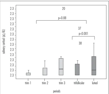
A total of 13 pregnant (primigravida) and 13 non-pregnant women were enrolled. Three miscarriages and one dropout occurred in the pregnant group, and one control was lost to follow-up. Thus, 9 primigravidas with median age (interquartile range) of 28 years (25–31), and 12 non-pregnant women aged 29 (27–32) took part in the study. A total of 27 and 24 samples were collected for each variable per group, respectively. There were no signiicant between-group differences in salivary cortisol levels. However, signiicant within-group differences in median (interquartile range) levels were found among the non-pregnant subjects, with values of 1.0 (1.0–1.05) µg/dL in the follicular phase of the ovarian cycle versus 1.1 (1.0–1.48) µg/dL in the luteal
phase (p<0.001) (Figure 1).
Median serum cortisol levels were signiicantly dif-ferent in the pregnant and non-pregnant groups (23.8 [19.4–29.4] versus 12.3 [9.6–16.8] µg/dL, and also between
follicular versus luteal phase (12.7 [10.2–18.7] versus 12.2
[9.1–15.1] µg/dL (p<0.001) (Figure 2).
periods 20
p=0.88
37
38 p<0.001
trim 1 trim 2 trim 3 trifollicular luteal
salivar
y cor
tisol (μL/dL)
2.3 2.3 2.3 2.3 2.3 2.3 2.3
2.3 2.3 2.3 2.3 2.3 2.3 2.3
There were no differences in median UWS low rate values between pregnant and non-pregnant subjects (0.26 [0.15–0.30] versus 0.23 [0.20–0.32] mL/min), and no
within-group differences among trimesters of pregnancy or between the follicular and luteal phases of the ovarian cycle (data not shown).
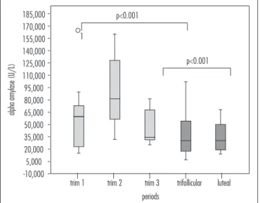
Levels of sAA were signiicantly different between the pregnant and non-pregnant groups (56.7 [30.9–82.2]
versus 31.8 [18.1–53.2] U/L, p<0.001) and between the
follicular and luteal phases in the non-pregnant group (p<0.001) (Figure 3).
Signiicant Spearman correlations were found be-tween salivary and serum cortisol levels (p=0.02) and between salivary cortisol and sAA (p=0.01) (Table 1).
Discussion
Measurement of salivary cortisol levels has been widely used as an alternative to quantitation of this hormone in plasma or serum. Saliva samples are readily obtained and can be collected several times a day, allowing dynamic assessment of free cortisol secretion5,6. Circulating
un-bound cortisol is quickly transported to saliva by passive diffusion, to the extent that some studies report strong correlations between salivary cortisol levels and free (un-bound) cortisol concentrations in plasma and serum6,26,27.
However, if salivary cortisol levels are to be used for diagnostic purposes in clinical practice, analytical meth-ods must be standardized and cutoff values deined on the basis of normal population-wide control samples to serve as a reference range for testing. Research and retail laboratories should validate their salivary cortisol assays before making them available to clinicians2,28.
The interrelatedness between serum and salivary cortisol levels in non-pregnant women and men appears to be the same. However, women in the third trimester of pregnancy and those on oral contraceptives exhibit markedly increased serum cortisol levels, but near-normal salivary cortisol27.
The linear correlation between serum free cortisol and salivary cortisol is usually very strong, independent of changes in CBG concentrations, and similar across all groups: men, pregnant women, non-pregnant women, and oral contraceptive users27.
Conversely, some authors believe these parameters should be interpreted cautiously29,30, as salivary cortisol
concentrations do not correlate linearly with serum levels in some cases. This nonlinearity in association between total salivary and serum cortisol may be attributable to the presence of CBG in plasma. CBG concentrations may be increased during oral contraceptive use and in certain physiological conditions, such as pregnancy27,30.
Figure 3. Between-group and within-group sAA.
periods 1
p<0.001 p<0.001
trim 1 trim 2 trim 3 trifollicular luteal
alpha amylase (U/L)
185,000 170,000 155,000 140,000 125,000 110,000 95,000 80,000 65,000 50,000 35,000 20,000 5,000 -10,000
periods
p<0.001 p<0.001
trim 1 trim 2 trim 3 trifollicular luteal 45
42 39 36 33 30 27 24 21 18 15 12 9 6 3 0
ser
um cor
tisol (μL/dL)
Figure 2. Between-group and within-group serum cortisol.
Table 1. Spearman correlation coeficients
Salivary cortisol
Serum cortisol
Alpha- amylase
UWS low rate Spearman’s rho
Salivary cortisol
Correlation coeficient 1.000 0.324* -0.351* -0.080
Sig. (2-tailed) – 0.020 0.011 0.575
Serum cortisol
Correlation coeficient 1.000 0.135 -0.155
Sig. (2-tailed) – 0.343 0.277
Alpha-amylase
Correlation coeficient 1.000 0.010
Sig. (2-tailed) – 0.943
This might explain the fact that, in the present study, salivary and serum cortisol levels were signiicantly corre-lated from a statistical but not from a clinical standpoint. It bears stressing that blood and saliva sampling took place in a short period of time (30 minutes at most) so as to prevent circadian inluences on measurement of the parameters of interest.
When both CBG and cortisol concentrations are in-creased simultaneously, as in pregnancy, salivary cortisol levels appear to remain within normal limits, although above the baseline levels found in non-pregnant women31,32.
Studies have shown that salivary cortisol levels increase gradually, eventually exceeding the upper limit of normal, during the second half of pregnancy8,26,33. In the present
study, there were no signiicant between-group differences in salivary cortisol levels. Serum cortisol levels, however, were higher in pregnant versus non-pregnant women.
These indings are consistent with the literature9,26,31,32.
In the present study, signiicant differences were found between salivary cortisol levels in the follicular and luteal phases of the ovarian cycle, with higher val-ues found in the latter. A previous study also found that salivary cortisol response patterns were more evident in the luteal phase, although the subjects were exposed to psychosocial stress, unlike the participants of the present study, who were not subjected to any external stressors23.
A review of the literature did not yield any evidence that would explain this difference.
It has been suggested that salivary free cortisol levels are independent of salivary low2. Accordingly, salivary
low rate and salivary cortisol concentration were unre-lated in the present sample. This makes salivary cortisol a good biomarker, as it is unaffected by salivation itself. Although measurement of stimulated saliva is useful for assessment of the functional capacity of the salivary glands, unstimulated saliva is the predominant form during most of the day as well as during sleep, and plays an important role in the maintenance of oral health24,34. Furthermore,
research suggests that if analysis of the biochemical com-ponents of saliva is to be feasible in outpatient clinical practice, whole27 and unstimulated28 saliva must be used;
therefore, UWS was used in the present study.
Although salivary free cortisol has been proposed as a useful parameter for assessment of pituitary-adrenal function, an appropriate biomarker that relects sym-pathoadrenal medullary activity has yet to be found. In this context, sAA has been suggested as a potential parameter for determination of autonomic activity and, thus, a reliable and noninvasively quantiiable indicator of stress-related changes in the human body15. The salivary
glands are innervated by sympathetic and parasympa-thetic nerve ibers alike, so that salivary secretion occurs in response to neurotransmitter-mediated stimulation35.
As this biomarker is produced locally in the oral cavity, it is found in high concentrations in saliva as compared with other markers, such as cortisol, which is a compo-nent of blood serum, produced by the adrenal gland, and transported to the saliva via ultrailtration in the salivary glands36.
There is no established correlation between sAA and salivary or blood levels of cortisol in the literature13,15. In the
present study, salivary cortisol levels correlated with sAA. However, there was no correlation between sAA and serum cortisol. This may be attributable to production in differ-ent sites18,22. Nevertheless, the potential interrelatedness
between these two parameters warrants further investigation. Prior studies diverge as to the sAA response to preg-nancy. Comparison between UWS collected from pregnant and non-pregnant women showed increased sAA levels in the irst and second trimesters of pregnancy as compared to near-term and non-pregnant women24. Other research,
however, failed to ind any signiicant differences in sAA concentrations during pregnancy19. Supporting this
ind-ing, no signiicant changes in sAA levels during gesta-tion were observed in the present study. However, sAA activity was increased in pregnant versus non-pregnant
subjects. We also observed greater sAA activity in the luteal phase of the ovarian cycle as compared with the follicular phase. To the best of our knowledge, no other studies have assessed sAA levels during the distinct phases of the menstrual cycle. One previous study found high levels of sAA, but not of salivary cortisol, to be associated with a reduction in female fertility18. The mechanism
whereby sAA might reduce fertility remains unknown18.
Measurement of salivary low rate plays an important role in the interpretation of changes in salivary protein. However, a previous study showed that no association what-soever exists between sAA activity and salivary low rate37.
In addition, authors have conirmed that sAA measurement can be performed without assessment of salivary low, as the latter does not interfere with sAA activation22,38.
The pres-ent study found no correlation between sAA and salivary low rate and no correlations between cortisol and sali-vary low rates measured in pregnant and non-pregnant women, which conirms that neither cortisol nor sAA levels are signiicantly altered as a function of salivary low rate.
Nevertheless, some limitations of this study should be noted. The small sample size precludes generalization of indings to other populations. Furthermore, in the non-pregnant group, the timing of ovarian cycle phases was estimated solely on the basis of self-provided information, and not evaluated by any tests.
1. Al Kawas S, Rahim ZH, Ferguson DB. Potential uses of human salivary protein and peptide analysis in the diagnosis of disease. Arch Oral Biol. 2012;57(1):1-9.
2. Chiappin S, Antonelli G, Gatti R, De Palo EF. Saliva specimen: a new laboratory tool for diagnostic and basic investigation. Clin Chim Acta. 2007;383(1-2):30-40.
3. Llena-Puy C. The role of saliva in maintaining oral health and as an aid to diagnosis. Med Oral Patol Oral Cir Bucal. 2006;11(5):E449-55.
4. Lima DP, Diniz DG, Moimaz SA, Sumida DH, Okamoto AC. Saliva: relection of the body. Int J Infect Dis. 2010;14(3):e184-8. 5. Nierop A, Bratsikas A, Zimmermann R, Ehlert U. Are stress-induced
cortisol changes during pregnancy associated with postpartum depressive symptoms? Psychosom Med. 2006;68(6):931-7. 6. Gozansky WS, Lynn JS, Laudenslager ML, Kohrt WM. Salivary
cortisol determined by enzyme immunoassay is preferable to serum total cortisol for assessment of dynamic hypothalamic–pituitary– adrenal axis activity. Clin Endocrinol (Oxf). 2005;63(3):336-41. 7. Egliston KA, McMahon C, Austin MP. Stress in pregnancy and
infant HPA axis function: conceptual and methodological issues relating to the use of salivary cortisol as an outcome measure. Psychoneuroendocrinology. 2007;32(1):1-13.
8. Allolio B, Hoffmann J, Linton EA, Winkelmann W, Kusche M, Schulte HM. Diurnal salivary cortisol patterns during pregnancy and after delivery: relationship to plasma corticotrophin-releasing-hormone. Clin Endocrinol (Oxf). 1990;33(2): 279-89.
9. de Weerth C, Buitelaar JK. Physiological stress reactivity in human pregnancy- a review. Neurosci Biobehav Rev. 2005;29(2): 295-312. 10. Li J, Wang ZN, Chen YP, Dong YP, Shuai HL, Xiao XM, et al. Late
gestational maternal serum cortisol is inversely associated with fetal brain growth. Neurosci Biobehav Rev. 2012;36(3):1085-92. 11. Van den Bergh BR, Mulder EJ, Mennes M, Glover V. Antenatal maternal anxiety and stress and the neurobehavioural development of the fetus and child: links and possible mechanisms. A review. Neurosci Biobehav Rev. 2005;29(2):237-58.
12. Bolten MI, Wurmser H, Buske-Kirschbaum A, Papoušek M, Pirke KM, Hellhammer D. Cortisol levels in pregnancy as a psychobiological predictor for birth weight. Arch Womens Ment Health. 2011;14(1):33-41. 13. Nater UM, Rohleder N, Gaab J, Berger S, Jud A, Kirschbaum C,
et al. Human salivary alpha-amylase reactivity in a psychosocial stress paradigm. Int J Psychol. 2005;55(3):333-42.
14. Rohleder N, Nater UM. Determinants of salivary alpha-amylase in humans and methodological considerations. Psychoneuroendocrinology. 2009;34(4):469-85.
15. Nater UM, La Marca R, Florin L, Moses A, Langhans W, Koller MM, et al. Stress-induced changes in human salivary alpha-amylase activity -- associations with adrenergic activity. Psychoneuroendocrinology. 2006;31(1):49-58.
16. Nater UM, Rohleder N. Salivary alpha-amylase as a non-invasive biomarker for the sympathetic nervous system: current state of research. Psychoneuroendocrinology. 2009;34(4):486-96. 17. Guglielminotti J, Dehoux M, Mentré F, Bedairia E, Montravers
P, Desmonts JM, et al. Assessment of salivary amylase as a stress biomarker in pregnant patients. Int J Obstet Anesth. 2012;21(1):35-9.
18. Louis GM, Lum KJ, Sundaram R, Chen Z, Kim S, Lynch CD, et al. Stress reduces conception probabilities across the fertile window: evidence in support of relaxation. Fertil Steril. 2011;95(7):2184-89.
19. Laine M, Tenovuo J, Lehtonen OP, Ojanotko-Harri A, Vilja P, Tuohimaa P. Pregnancy-related changes in human whole saliva. Arch Oral Biol. 1988;33(12): 913-7.
20. D’Alessandro S, Curbelo HM, Tumilasci OR, Tessler JA, Houssay AB. Changes in human parotid salivary protein and sialic acid levels during pregnancy. Arch Oral Biol. 1989;34(10):829-31. 21. Gordis EB, Granger DA, Susman EJ, Trickett PK. Salivary alpha
amylase-cortisol asymmetry in maltreated youth. Horm Behav. 2008;53(1):96-103.
22. van Stegeren AH, Wolf OT, Kindt M. Salivary alpha amylase and cortisol responses to different stress tasks: impact of sex. Int J Psychophysiol. 2008;69(1): 33-40.
23. Kirschbaum C, Hellhammer DH. Salivary cortisol in psychoneuroendocrine research: recent developments and applications. Psychoneuroendocrinology. 1994;19(4):313-33. 24. Salvolini E, Di Giorgio R, Curatola A, Mazzanti L, Fratto G.
Biochemical modiications of human whole saliva induced by pregnancy. Br J Obstet Gynaecol. 1998;105(6):656-60. 25. Backhaus J, Junghanns K, Hohagen F. Sleep disturbances are
correlated with decreased morning awakening salivary cortisol. Psychoneuroendocrinology. 2004;29(9):1184-91.
26. Meulenberg PM, Hofman JA. Differences between concentrations of salivary cortisol and cortisone and of free cortisol and cortisone in plasma during pregnancy and postpartum. Clin Chem. 1990;36(1):70-5.
27. Vining RF, McGinley RA. The measurement of hormones in saliva: possibilities and pitfalls. J Steroid Biochem. 1987;27(1-3):81-94. 28. Lewis JG. Steroid analysis in saliva: an overview. Clin Biochem
Rev. 2006;27(3): 139-46.
29. Levine A, Zagoory-Sharon O, Feldman R, Lewis JG, Weller A. Measuring cortisol in human psychobiological studies. Physiol Behav. 2007;90(1):43-53.
30. Hellhammer DH, Wüst S, Kudielka BM. Salivary cortisol as a biomarker in stress research. Psychoneuroendocrinology. 2009;34(2):163-71.
References
diagnostics still require further research for standardization of analytical methods, validation of results, and deini-tion of analyte reference ranges in a series of populadeini-tions before they can be made available to clinical practice.
31. Greaves MS, West HF. Cortisol and cortisone in saliva of pregnancy. J Endocrinol. 1963;26(2):189-95.
32. Bustamante B, Crabbé J. Parotid saliva cortisol in normal subjects: increase during pregnancy. J Steroid Biochem. 1984;20(6A):133-6. 33. Stahl F, Dörner G. Responses of salivary cortisol levels to
stress-situations. Endokrinologie. 1982;80(2):158-62.
34. Rockenbach MI, Marinho SA, Veeck EB, Lindemann L, Shinkai RS. Salivary low rate, pH, and concentrations of calcium, phosphate, and sIgA in Brazilian pregnant and non-pregnant women. Head Face Med. 2006;2:44.
35. Humphrey SP, Williamson RT. A review of saliva: normal composition, low, and function. J Prosthet Dent. 2001;85(2):162-9.
36. Harmon AG, Towe-Goodman NR, Fortunato CK, Granger DA. Differences in saliva collection location and disparities in baseline and diurnal rhythms of alpha-amylase: a preliminary note of caution. Horm Behav. 2008;54(5):592-6.
37. Bosch JA, Brand HS, Ligtenberg TJ, Bermond B, Hoogstraten J, Amerongen AVN. Psychological stress as a determinant of protein levels and salivary-induced aggregation of Streptococcus gordonii in human whole saliva. Psychosom Med. 1996;58(4):374-82. 38. Rohleder N, Wolf JM, Maldonado EF, Kirschbaum C.