ISSN 0100-879X
BIOMEDICAL SCIENCES
AND
CLINICAL INVESTIGATION
www.bjournal.com.br
www.bjournal.com.br
Volume 43 (3) 182-267 March 2011
Braz J Med Biol Res, March 2011, Volume 44(3) 182-185
doi: 10.1590/S0100-879X2011007500004
Evidence for the endophytic colonization of
Phaseolus vulgaris
(common bean) roots by the diazotroph Herbaspirillum seropedicae
M.A. Schmidt, E.M. Souza, V. Baura, R. Wassem, M.G. Yates, F.O. Pedrosa and R.A. Monteiro
Faculdade de Medicina de Ribeirão Preto Campus
Ribeirão Preto
Institutional Sponsors
The Brazilian Journal of Medical and Biological Research is partially financed by
analiticaweb.com.br S C I E N T I F I C
Evidence for the endophytic colonization
of
Phaseolus vulgaris
(common bean)
roots
by the diazotroph
Herbaspirillum seropedicae
M.A. Schmidt
1, E.M. Souza
1, V. Baura
1, R. Wassem
2, M.G. Yates
1,
F.O. Pedrosa
1and R.A. Monteiro
11Departamento de Bioquímica e Biologia Molecular, 2Departamento de Genética,
Universidade Federal do Paraná, Curitiba, PR, Brasil
Abstract
Herbaspirillum seropedicae is an endophytic diazotrophic bacterium, which associates with important agricultural plants. In the present study, we have investigated the attachment to and internal colonization of Phaseolus vulgaris roots by the H. seropedicae
wild-type strain SMR1 and by a strain of H. seropedicae expressing a red fluorescent protein (DsRed) to track the bacterium in
the plant tissues. Two-day-old P. vulgaris roots were incubated at 30°C for 15 min with 6 x 10⁸ CFU/mL H. seropedicae SMR1
or RAM4. Three days after inoculation, 4 x 10⁴ cells of endophytic H. seropedicae SMR1 were recovered per gram of fresh
root, and 9 days after inoculation the number of endophytes increased to 4 x 10⁶ CFU/g. The identity of the recovered bacteria
was confirmed by amplification and sequencing of the 16SrRNA gene. Furthermore, confocal microscopy of P. vulgaris roots inoculated with H. seropedicae RAM4 showed that the bacterial cells were attached to the root surface 15 min after inoculation;
fluorescent bacteria were visible in the internal tissues after 24 h and were found in the central cylinder after 72 h, showing that H. seropedicae RAM4 is capable of colonizing the roots of the dicotyledon P. vulgaris. Determination of dry weight of common bean inoculated with H. seropedicae SMR1 suggested that this bacterium has a negative effect on the growth of P. vulgaris.
Key words: Herbaspirillum seropedicae; Phaseolus vulgaris; Confocal microscopy
Introduction
Correspondence: R.A. Monteiro, Departamento de Bioquímica e Biologia Molecular, Universidade Federal do Paraná, 81531-990 Curitiba, PR, Brasil. E-mail: [email protected]
Received June 16, 2010. Accepted December 15, 2010. Available online January 14, 2011. Published March 7, 2011. Herbaspirillum seropedicae is a diazotrophic endophyte
of the β-proteobacteria (1), which colonizes internal tissues of
maize, rice, wheat, sorghum, and sugar cane (2,3). This organ-ism has also been isolated from plants other than Poaceae such as banana and pineapple (4,5). H. seropedicae may stimulate
plant growth by supplying fixed nitrogen to the plant, producing
and secreting phytohormones or protecting the host against pathogenic microorganisms (6,7). The bacterial association withpoaceous crops apparently initiates with attachment to root surfaces followed by proliferation at the emergence points of secondary roots and penetration through discontinuities in the epidermis. Rapid occupation of root intercellular spaces then occurs, and the bacteria spread to aerenchyma, xylem vessels and aerial portions (8-10).
Olivares et al. (11) described the isolation of H. seropedicae
from gramineae and also from roots of a legume species ( Ca-janus cajan). However, the authors suspected that fragments of maize roots might have contaminated the legume sample.
Later, Valverde et al. (12) isolated a new species of this genus from the nodules of Phaseolus vulgaris. Based on genotypic and phenotypic characterization, the new isolates were
clas-sified as a novel species for which the authors proposed the
name H. lusitanum sp nov.
The aim of the present study was to determine if H. se-ropedicae is able to establish an endophytic relationship with
P. vulgaris.
Material and Methods
H. seropedicae SMR1 is a spontaneous streptomycin-resistant derivative of the wild-type strain Z78 (13). RAM4 is a strain tagged with the dsred gene, which expresses the red
fluorescent protein, to allow the monitoring of single bacterial
cells in P. vulgaris roots (10). Seeds of P. vulgaris (cv. Uirapuru)
H. seropedicae colonizes P. vulgaris roots 183
Carneiro, Brazil) containing 0.02% Tween-20 (United States Biochemical, USA) solution for 20 min at 30°C. Seeds were
then washed five times with sterile distilled water by shaking for 15 min each time and germinated at 25°C in the dark for
48 h. P. vulgaris roots were then incubated at 30°C for 15 min with 6 x 10⁸ colony forming units (CFU)/mL SMR1 (wild-type) or RAM4 (dsred) grown in NFbHPN medium (14). After incubation,
the plantlets were placed in wells of a 96-well block containing filter paper soaked with plant medium (15) and incubated for
a 14-h light period at 25°C.
The roots of 3-day-old plantlets of P. vulgaris were ana-lyzed at 0, 3, 7, and 9 days after inoculation with 6 x 10⁸
CFU/mL H. seropedicae strain SMR1. After these intervals, approximately 0.05 g fresh roots was surface-sterilized by sequentially washing with 70% ethanol (1 min), 1% sodium hypochlorite containing 0.01% Tween-20 (1
min) and three times with sterile water (5 min). The roots were homogenized using a sterile pestle and mortar, and the extracts diluted in 1 mL sterile saline (0.9% NaCl). The diluted extracts were plated onto solid NFbHPN me-dium in the absence or presence of 80 µg/ mL streptomycin (Sigma, USA). The number of CFU was determined after 24-48 h of incu-bation at 30°C. The identity of the recovered
bacteria was determined by amplification and
sequencing of the 16SrRNA gene (16). The colonization pattern of P. vulgaris
roots by H. seropedicae was observed by inoculating P. vulgaris roots with the RAM4 strain followed by confocal microscopy. Root samples were collected 1, 2 or 3 days after inoculation, washed with water, hand cut, mounted on a microscope slide and imme-diately examined under a BioRad Confocal
Radiance 2001-Eclipse E800 Nikon Micro -scope (USA) equipped with an HeNe laser
(DsRed: excitation, 543 nm; emission filter
LP, 560 nm).
We measured the dry weight of inoculated plants grown in medium containing 0, 0.2, and
4 mM ammonium nitrate (Merck, Germany)
and compared it to that of non-inoculated plants grown in the same medium. The bean seeds were sterilized, germinated and in-oculated as described above. Ten days after inoculation the roots were dried and the weight was determined.
Results
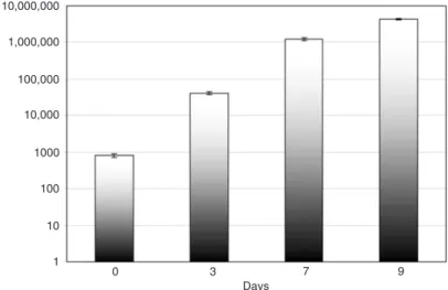
The results indicated that H. seropedicae
colonized P. vulgaris roots progressively from
the first to the 9th day after inoculation. The
number of bacteria recovered from surface
sterilized roots increased from 4 x 10⁴ per gram fresh root 3 days after inoculation to 4 x 10⁶ bacteria 9 days after
inoculation(Figure 1).To confirm that the recovered bac -teria were indeed H. seropedicae, the 16SrRNA gene of
randomly selected colonies was amplified and sequenced.
All sequences were 100% identical to those of H. serope-dicae SMR1.
Under the confocal microscope H. seropedicae
express-ing the DsRed protein showed bright red fluorescence, easily distinguishable from the diffuse fluorescence background
of the contour of the plant cells. After 15 min of incubation with the RAM4 strain, cell clusters were found on the root hairs (Figure 2). These are probable entry sites used by the bacteria, possibly due to a higher concentration of carbon sources at these points (17).
Figure 1. Internal colonization of Phaseolus vulgaris roots by Herbaspirillum seropedicae. The extent of internal colonization was determined 15 min after inoculation (day 0) and on days 3, 7, and 9. Data are reported as CFU/g (mean ± SD) fresh roots from 3 plants per treatment.
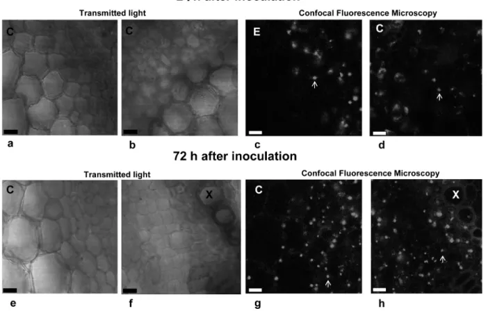
Confocal microscopy images of different regions of a cross-section of P. vulgaris roots inoculated with RAM4 showed that the bacteria had started to invade the root internal tissues 24 h after inoculation. After 72 h the number of bacterial cells increased progressively from the epidermis to the central cylinder (Figure 3).
To investigate the effect of H. seropedicae on P. vulgaris
growth, plantlets were incubated for 15 min with 1 mL of a fresh culture containing 108 CFU, and transplanted to vermiculite
in the presence of different NH4NO3 concentrations. After 10
days, the dry weight of the inoculated plants was significantly
lower than that of the non-inoculated plants under all conditions (Table 1), suggesting that H. seropedicae has a negative effect on the growth of P. vulgaris (cv. Uirapuru).
Discussion
The results showed that H. seropedicae is able to adhere to and colonize P. vulgaris roots internally. H.
lusitanum was also recovered from bean roots 2 weeks
after inoculation (12).
The H. seropedicae colonization process on and in P. vulgaris roots appears to occur in a pattern similar to that
Figure 3. Transmitted light and confocal fluorescence microscopy of a cross-section of Phaseolus vulgaris root 24 (a-d) or 72 h (e-h) after inoculation with Herbaspirillum seropedicae strain RAM4. The images were recorded in two different regions of the cross-section,
i.e., the epidermis (a, c, e, and g) and the central cylinder (b, d, f, and h). Transmitted light microscopy (a, b, e, and f); confocal fluores
-cence microscopy (c, d, g, and h). C = cortex; E = epidermis; X = xylem vessels. Arrows indicate the fluorescent bacteria. Magnification bars = 50 μm.
Table 1. Dry weight of Phaseolus vulgaris roots inoculated or not with Herbaspirillum seropedicae SMR1.
NH4NO3 (mM) Dry weight (g)
Inoculated Uninoculated
0.0 11.8 ± 2.5 28.2 ± 7.7* 0.2 31.8 ± 1.2 41.6 ± 4.7* 4.0 35.6 ± 9.5 58.2 ± 2.6*
Data are reported as means ± SD for 3 plants per treatment. Plants were grown without nitrogen or in the presence of 0.2 or 4 mM NH4NO3 and analyzed 10 days after inoculation. The total
dry weight of bean roots inoculated or not with H. seropedicae
H. seropedicae colonizes P. vulgaris roots 185
of Poaceae (9,10): the bacteria invade the intercellular spaces and disperse in the cortex, eventually reaching the xylem vessels. A notable difference was a lower number of bacterial cells visualized at the time tested, when compared with that of maize (10).
We also evaluated the effect of H. seropedicae inocula-tion on the growth of common bean seedlings (Table 1). The result showed that H. seropedicae has a negative effect on Phaseolus growth, since the root dry weights of plants inoculated with H. seropedicae were lower than those of uni-noculated plants. This is in contrast to the growth-promoting effect of H. seropedicae on rice plants (9), indicating that
H. seropedicae has different effects on different plants. A strong interaction between the plant genotype and the rhizobacteria inoculated has been documented, with ef-fects ranging from variable growth promotion (18,19) to
a slight reduction in yield (20,21). The negative effect of
H. seropedicae on the growth of Phaseolus may be due to interaction between the plant and bacterial molecular factors such as those secreted by the type three secretion system (T3SS) of H. seropedicae. In Rhizobium NGR234, the inactivation of T3SS leads to an increase in the number of root nodules in P. vulgaris cv. BAT93, suggesting that the secretion of certain effector proteins may negatively affect the interaction with Rhizobium NGR234 (Lariguet P, unpublished results).
The present results show that H. seropedicae is not an exclusive endophyte of gramineous plants, but is capable of colonizing other plant types such as the common bean, indi-cating that H. seropedicae is a broad host-range endophyte.
Whether this association benefits the development of common beans under specific conditions is yet to be established.
References
1. Baldani JI, Baldani VLD, Seldin L, Döbereiner J. Characteriza-tion of Herbaspirillum seropedicae gen. nov., sp. nov., a new
root-associated nitrogen-fixing bacterium. Int J Syst Bacteriol
1986; 36: 86-93.
2. Baldani JI, Baldani VLD, Sampaio MJAM, Döbereiner J. A fourth
Azospirillum species from cereal roots. An Acad Bras Cienc
1984; 56: 365.
3. Boddey RM, Oliveira OC, Urquiaga S, Reis VM, Olivares FL,
Baldani VLD, et al. Biological nitrogen fixation associated with
sugar cane and rice: contributions and prospects for improve-ment. Plant Soil 1995; 174: 195-209.
4. Cruz LM, Souza EM, Weber OB, Baldani JI, Döbereiner J, Pedrosa FO. 16S ribosomal DNA characterization of
nitrogen-fixing bacteria isolated from banana (Musa spp) and pineapple (Ananas comosus (L.) Merril). Appl Environ Microb 2001; 67: 2375-2379.
5. Weber OB, Baldani VLD, Teixeira KRS, Kirchhof G, Baldani JI, Döbereiner J. Isolation and characterization of diazotrophic bacteria in banana and pineapple plants. Plant Soil 1999; 210: 103-113.
6. Bashan Y, Holguin G, de-Bashan LE. Azospirillum-plant relation-ships: physiological, molecular, agricultural, and environmental advances (1997-2003). Can J Microbiol 2004; 50: 521-577. 7. Baldani JI, Pot B, Kirchhof G, Falsen E, Baldani VL, Olivares
FL, et al. Emended description of Herbaspirillum; inclusion of [Pseudomonas] rubrisubalbicans, a milk plant pathogen, as Herbaspirillum rubrisubalbicans comb. nov.; and classification
of a group of clinical isolates (EF group 1) as Herbaspirillum
species 3. Int J Syst Bacteriol 1996; 46: 802-810.
8. James EK, Gyaneshwar P, Mathan N, Barraquio WL, Reddy PM, Iannetta PP, et al. Infection and colonization of rice seed-lings by the plant growth-promoting bacterium Herbaspirillum seropedicae Z67. Mol Plant Microbe Interact 2002; 15: 894-906.
9. Roncato-Maccari LD, Ramos HJ, Pedrosa FO, Alquini Y, Chu-batsu LS, Yates MG, et al. Endophytic Herbaspirillum seropedi-cae expresses nif genes in gramineous plants. FEMS Microbiol Ecol 2003; 45: 39-47.
10. Monteiro RA, Schmidt MA, Baura VA, Balsanelli E, Wassem R, Yates MG, et al. Early colonization pattern of maize (Zea mays L. Poales, Poaceae) roots by Herbaspirillum seropedicae
(Burkholderiales, Oxalobacteraceae). Genet Mol Biol 2008; 31: 932-937.
11. Olivares FL, Baldani VLD, Reis VM, Baldani JI, Döbereiner J. Occurrence of endophytic diazotrophs Herbaspirillum spp. in roots, stems and leaves predominantly of Gramineae. Biol Fert Soils 1996; 21: 197-200.
12. Valverde A, Velazquez E, Gutierrez C, Cervantes E, Ventosa A, Igual JM. Herbaspirillum lusitanum sp. nov., a novel
nitrogen-fixing bacterium associated with root nodules of Phaseolus vulgaris. Int J Syst Evol Microbiol 2003; 53: 1979-1983. 13. Pedrosa FO, Teixeira KRS, Machado IMP, Steffens MBR,
Klas-sen G, Benelli EM, et al. Structural organization and regulation of the nif genes of Herbaspirillum seropedicae. Soil Biol Bio-chem 1997; 9: 843-846.
14. Klassen G, Pedrosa FO, Souza EM, Funayama S, Rigo LU. Effect of nitrogen compounds on nitrogenase activity in
Herbaspirillum seropedicae SMR1. Can J Microbiol 1997; 43: 887-891.
15. Broughton WJ, Dilworth MJ. Control of leghaemoglobin
synthe-sis in snake beans. Biochem J 1971; 125: 1075-1080. 16. Magnani GS, Didonet CM, Cruz LM, Picheth CS, Pedrosa FO,
Souza EM. Diversity of endophytic bacteria in Brazilian sugar-cane. Genet Mol Res 2010; 9: 250-258.
17. Bennett RA, Lynch JM. Bacterial growth and development in the rhizosphere of gnotobiotic cereal plants. J Gen Microbiol 1981; 125: 95-102.
18. Roesch LFW, Camargo FAO, Selbach PA, Sá ELS, Passaglia
LMP. Identificação de cultivares de milho eficientes na absorção de nitrogênio e na associação com bactérias diazotróficas. Cienc Rural 2005; 35: 924-927.
19. Baldani VLD, Baldani JI, Döbereiner J. Inoculation of rice plants with endophytic diazotrophs Herbaspirillum seropedicae and
Burkholderia spp. Biol Fertil Soil 2000; 30: 485-491.
20. Didonet AD, Lima OS, Candaten AA, Rodrigues O. Realocação de nitrogênio e de biomassa para os grãos, em trigo submetido a inoculação de Azospirillum. Pesq Agropec Bras 2000; 35: 401-411.