ISSN 0100-879X
BIOMEDICAL SCIENCES
AND
CLINICAL INVESTIGATION
www.bjournal.com.br
www.bjournal.com.br
Volume 43 (5) 381-496 May 2011
Braz J Med Biol Res, May 2011, Volume 44(5) 381-387
doi:
10.1590/S0100-879X2011007500035
Leptin and adiponectin in the female life course
S.B. Lecke, D.M. Morsch and P.M. Spritzer
Faculdade de Medicina de Ribeirão Preto Campus
Ribeirão Preto
Institutional Sponsors
The Brazilian Journal of Medical and Biological Research is partially financed by
analiticaweb.com.br S C I E N T I F I C Hotsite of proteomics metabolomics
Leptin and adiponectin in the female life course
S.B. Lecke, D.M. Morsch and P.M. Spritzer
Unidade de Endocrinologia Ginecológica, Serviço de Endocrinologia, Hospital de Clínicas de Porto Alegre, Laboratório de Endocrinologia Molecular, Departamento de Fisiologia, Universidade Federal do Rio Grande do Sul,
and Instituto Nacional de Hormônios e Saúde da Mulher - CNPq, Porto Alegre, RS, Brasil
Abstract
Adipose tissue secretes a variety of adipokines, including leptin and adiponectin, which are involved in endocrine processes regulating glucose and fatty metabolism, energy expenditure, inflammatory response, immunity, cardiovascular function, and reproduction. The present article describes the fluctuations in circulating leptin and adiponectin as well as their patterns of secretion in women from birth to menopause. During pregnancy, leptin and adiponectin seem to act in an autocrine/paracrine fashion in the placenta and adipose tissue, playing a role in the maternal-fetal interface and contributing to glucose metabo-lism and fetal development. In newborns, adiponectin levels are two to three times higher than in adults. Full-term newborns have significantly higher leptin and adiponectin levels than preterms, whereas small-for-gestational-age infants have lower levels of these adipokines than adequate-for-gestational-age newborns. However, with weight gain, leptin concentrations increase significantly. Children between 5 and 8 years of age experience an increase in leptin and a decrease in adiponectin regardless of body mass index, with a reversal of the newborn pattern for adiponectin: plasma adiponectin levels at age five are inversely correlated with percentage of body fat. In puberty, leptin plays a role in the regulation of menstrual cycles. In adults, it has been suggested that obese individuals exhibit both leptin resistance and decreased serum adiponectin levels. In conclusion, a progressive increase in adiposity throughout life seems to influence the relationship between leptin and adiponectin in women.
Key words: Female; Adiposity; Leptin; Adiponectin
Introduction
Correspondence: P.M. Spritzer, Serviço de Endocrinologia, Hospital de Clínicas de Porto Alegre, Rua Ramiro Barcelos, 2350, 90035-003 Porto Alegre, RS, Brasil. Fax: +55-51-3359-8027/8777. E-mail: [email protected]
Received December 9, 2010. Accepted March 4, 2011. Available online March 25, 2011. Published May 16, 2011.
Adipose tissue is currently recognized as a dynamic organ because of its capacity to secrete hormones and cytokines. White adipose tissue, the predominant form found in adults (brown fat is mainly found in neonates), consists of preadipocytes and adipocytes, macrophages, endothelial cells, fibroblasts, and leukocytes (1), and secretes a variety of adipokines. These adipokines are involved in a range of endocrine processes that regulate glucose and fatty metabolism, energy expenditure, inflam-matory response, immunity, cardiovascular function, and reproduction, among other functions (2,3).
Several aspects related to leptin and adiponectin have been extensively studied, including their metabolic, clinical, and pharmacological characteristics. However, the behavior of these adipokines over the course of life has received less attention. In the present article, fluc-tuations in circulating leptin and adiponectin as well as their patterns of secretion from birth to menopause are reviewed in order to trace a physiological profile of these
adipokines in the female life course.
Leptin and adiponectin
382 S.B. Lecke et al.
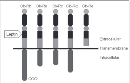
proteins, forming large, high-molecular weight complexes (7), and acts by interacting with the leptin receptor (Ob-R). The Ob-R family is a large single-transmembrane-domain protein belonging to the class I cytokine receptor family (8,9). The Ob-R gene encodes at least five alternatively spliced variants. The long form of Ob-R is considered to be the active leptin receptor, while the shorter forms lack the membrane-spanning region and are believed to be involved in leptin transport (10) (Figure 1). Ob-R is found in regions of the hypothalamus, the gonadotropic cells of the anterior pituitary gland, the ovarian interstitial cells, the theca and granulosa cells of ovarian follicles, the endometrium, and the Leydig cells of the seminiferous tubules (11). Moreover, Ob-R has been identified in the kidneys, heart, lungs, liver,
and skeletal muscle (12).
Adiponectin was first discovered in 1995 as a protein released exclusively by adipose tissue (13). Because it was found simultaneously by several laboratories, adiponectin received various names - adipocyte complement-related protein of 30 kDa (Acrp30), adipose-specific gene adipoQ (AdipoQ), adipose most abundant gene transcript 1 (apM1), and gelatin-binding protein of 28 kDa (GBP28) (14). Adi-ponectin is the protein product of the APM1 gene transcript, which is located on chromosome 3q27, close to the locus for type II diabetes and adiposity (15). This adipokine is secreted by adipose tissue as a low molecular weight trimer, as a combination of two middle molecular weight trimers, or as six high molecular weight (HMW) trimers, and circulates either as a trimer or as an oligomer (Figure 2). HMW forms are the predominant and active forms in human serum (16). Adiponectin increases fatty acid oxidation in adipose tissue, liver and muscle, enhancing insulin sensitivity, and inhibits inflammatory mediators and the expression of adhesion molecules within the vascular wall, lowering atherogenic risk (1,16). Adiponectin receptors have been identified predominantly in muscle (AdipoR1) and liver cells (AdipoR2), although in humans they are expressed ubiquitously in the body and brain (2,14,17).
In most studies, circulating levels of leptin and adiponectin have been determined by enzyme-linked immunosorbent assays or enzyme immunoassay, or, alternatively, by radioimmunoassay (associated or not with in-house techniques). While many studies have described assay sensitivity and in-tra- and interassay coefficients of variation, quality control measurements have been rarely reported.
Pregnancy and intrauterine and
neonatal development
During pregnancy, development of the neonate is influenced by critical interac-tions among genes, epigenetic changes, hormones, nutrition, the intrauterine environ-ment, and early pre- and postnatal events. It has been reported that the increased leptin levels found in pregnant women re-sult from placental secretion, in addition to adipose tissue production (5,18). The higher leptin levels that occur during pregnancy have also been attributed to weight gain and to an increase in body mass index (BMI), as well as to changes in levels of hormones that might stimulate leptin secretion (e.g.,
Figure 1. Representation of leptin receptor forms (Ob-R; subunits a to e). All share identical extracellular ligand-binding domains, but differ at the C-terminus. The long form of the Ob-R (Ob-Rb) is considered to be the active leptin receptor, activating the intracellular signal transduction pathway.
insulin, estrogens and human chorionic gonadotropin) (19). Trophoblastic cells, the predominant placental cell type, are the most likely source of placental leptin (5,20). Caminos et al. (21) were the first to demonstrate the presence of adiponectin and AdipoR2 in human and rat cytotrophoblast and syncytiotrophoblast placental cells. Chen et al. (1) described the production of adiponectin and the presence of both adiponectin receptors in the hu-man placenta, indicating that the placenta also secreted adiponectin in vitro. It appears that leptin and adiponectin act in an autocrine/paracrine fashion in the placenta, play-ing an important role in the maternal-fetal interface and contributing to glucose metabolism and fetal development during pregnancy (1,17,19).
If prematurity or fetal growth restrictions prevent the fetus from acquiring sufficient adipose tissue, changes are observed in the cord blood adipokine profile. Martos-Moreno et al. (22) reported that full-term newborns had significantly higher leptin and adiponectin levels than preterms, while babies who were small for gestational age had lower lev-els of these adipokines than adequate-for-gestational-age newborns. In that study, gestational age was the main predictor of adiponectin levels, while birth weight correlated independently with leptin cord blood levels. Nevertheless, in-creases in fetal weight also enhanced cord blood adiponectin levels, indicating that the negative influence of fat mass on adiponectin secretion is absent in the fetus (22).
Mantzoros et al. (23) have confirmed that adiponectin levels are two to three times higher in neonates than in adults. One possible explanation is that neonates have significantly less fat mass than children and adults, and that their fat mass consists mainly of subcutaneous fat and brown adipose tissue, which is atrophic in adults. In this regard, other investigators have reported a positive association between cord blood total adiponectin as well as HMW adiponectin levels (24) and birth weight, BMI, birth weight-to-length ratio, and leptin levels (25,26), even though this was not confirmed by others (27). Some studies have also provided evidence that high concentrations of leptin can be found in the cord serum of full-term newborns, being closely related to birth weight and fat accumulation (28,29). These findings imply that the relationship between leptin and body fat mass may develop early in life.
Previous studies of preterm infants have shown that serum leptin concentrations were significantly lower than those found in appropriate-for-gestational-age neonates (30). However, as weight gain progressed, leptin con-centrations increased significantly (5,19,31). It has been speculated that these increased leptin levels may derive from the increase in adipose fat typically observed after the under-nutrition period in preterm infants, suggesting a pos-sible involvement of leptin in the early development of insulin resistance (32,33). A series of epidemiological human and experimental animal studies have shown that exposure to inadequate (high or low) nutrition during fetal and postnatal
life, as well as reduced fetal growth followed by excessive postnatal catch-up in height and particularly, in weight, increase the risk of obesity, insulin resistance and type 2 diabetes in childhood and adult life (34). The decrease in adiponectin that occurs in small-for-gestational-age infants with the greatest weight catch-up has also been reported as a risk factor for subsequent development of visceral fat and insulin resistance (17,26).
Disturbances in fetal nutrient supply affect adipocyte levels. Muhlhausler and Smith (34) have proposed that prenatal over-nutrition promotes growth of the subcutaneous fat depot, leading to increased leptin secretion and serum levels. In fact, hyperleptinemia during neonatal life may program leptin resistance via dual effects on food intake and energy expenditure, with long-term adverse consequences for the hypothalamic circuits regulating energy balance (2,35). Some studies have shown an increased risk for obesity and insulin resistance in neonates born to obese mothers, as well as higher levels of leptin and markers of inflammation than in infants born to lean mothers (36).
A study of 100 mother/newborn pairs has tried to determine the production of leptin in the uterus and its role in neonatal development. Although maternal serum leptin concentrations correlated with measures of pre- and post-gestational adiposity, there was no association be-tween serum leptin levels in the newborn, an observation confirmed by others (5,9). Interestingly, Hassink et al. (5) demonstrated that 13% of newborns had higher serum leptin concentrations than their mothers, suggesting that the fetus may produce its own leptin, with associated leptin resistance, and/or that the placenta is the major source of leptin production for the fetus. However, Ho et al. (30) have shown a correlation between neonatal serum leptin concentrations and maternal weight and BMI within 24 hours of birth, suggesting that maternal and even placental production of leptin may be involved in the early regulation of fetal metabolism and growth. As in previous studies, no as-sociation was demonstrated between neonatal adiponectin levels and maternal age or pre-pregnancy BMI, suggesting that neonatal adiponectin levels are largely independent of maternal influence (25). Bozzola et al. (32) evaluated both total and isoform adiponectin circulating levels in a cohort of infants, and found a significant increase from birth up to the first month of life, followed by a decline at one year of age in all subjects. The investigators argued that the observed fall in serum adiponectin levels may be a consequence of increased adiposity, confirming the negative association between serum adiponectin levels and percentage of body fat, as is the case in older children (32).
Childhood and puberty
384 S.B. Lecke et al.
Between 5 and 8 years of age, metabolic health appears to improve regardless of BMI, with an increase in leptin and a decrease in adiponectin (37). In fact, adiponectin levels in childhood show a marked reversal of the positive body mass correlations observed during the neonatal period: plasma adiponectin levels at age 5 are inversely correlated with percentage of body fat. This inverse association between BMI and adiponectin becomes even more marked in older children (26,38). Previous studies in children of different ethnicities have reported that low plasma adiponectin con-centrations were closely related to hyperinsulinemia (38,39), leading to a long-term increased risk for diabetes.
There is a developmental increase in adipose tissue leptin expression and secretion during childhood, reach-ing maximal capacity in puberty/adulthood. Leptin may be regarded as a link between food intake, energy expenditure and the reproductive system. A possible interpretation is that leptin may signal to the body that energy reserves are adequate for reproduction, representing one of the primary mechanisms linked to the onset of puberty (11,12,40). In fact, a minimal leptin concentration is necessary to activate the hypothalamus-pituitary-gonadal axis, triggering puberty and maintaining reproductive function.
The temporal correlation between increases in leptin concentration and the initiation of luteinizing hormone (LH) pulsatility over peripuberty has been studied in several species. It is well recognized that leptin stimulates the gonadotropin-releasing hormone (GnRH) pulse generator and in turn LH pulses. This adipokine is secreted in a pul-satile manner in ovulatory women: a peak at night followed by a gradual change in LH pulsatility, reaching a nadir in the morning (7,41). In turn, in polycystic ovary syndrome (PCOS), a condition associated with androgen excess and chronic anovulation, circulating leptin seems to be related to the overall and central adiposity that is frequently associated
with this syndrome (42). In fact, data from our group sug-gest that the attenuation of the response of LH to GnRH in obese PCOS women may be related to changes in GnRH oscillator activity caused by a leptin-resistant state (42).
Interestingly, the nightly leptin peak has been associated with acute changes in LH pulses in normal ovulatory women, followed by a decline in metabolic rate (41). These data suggest that a single metabolic cycle regulates the leptin peak and the slowing of LH and metabolic rate. Conversely, both leptin resistance/hyperleptinemia, as observed in obesity and PCOS (42), and decreased circulating leptin, as observed in women with nutritional disorders or athletes, are associated with impaired gonadal steroidogenesis and reproduction (12). The role of leptin in the regulation of the menstrual cycles is further supported by studies demon-strating that menstrual irregularities and amenorrhea are common in women with very high or very low leptin levels (11,19). Evidence suggests that leptin functions as a per-missive, rather than a direct triggering factor at the onset of puberty, working in concert with other metabolic signals, such as insulin and insulin-like growth factor, amino acids, and neuropeptides (2,12,19).
Adulthood and postmenopause
In adults, circulating levels of leptin increase in the presence of total and central obesity and in metabolic disturbances (12,42,43), while adiponectin levels are inversely associated with the degree of adiposity. It has been suggested that obese individuals exhibit both leptin resistance, which antagonizes the metabolic action of in-sulin, and decreased serum adiponectin levels, increasing the risk for type 2 diabetes, coronary artery disease and metabolic syndrome (44).
Leptin concentrations may vary during the physiological
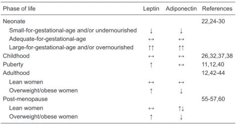
Table 1. Serum leptin and adiponectin concentrations during the female life course.
Phase of life Leptin Adiponectin References
Neonate 22,24-30
Small-for-gestational-age and/or undernourished ↓ ↓ Adequate-for-gestational-age ↔ ↔ Large-for-gestational-age and/or overnourished ↑↑ ↑↑
Childhood ↔ ↔ 26,32,37,38
Puberty ↑ ↔ 11,12,40
Adulthood 12,42-44
Lean women ↔ ↔
Overweight/obese women ↑ ↓
Post-menopause 55-57,60
Lean women ↔ ↑↓
Overweight/obese women ↑ ↓
menstrual cycle, exhibiting a monthly rhythm in ovulatory women. These variations are related to hormone changes, especially those of serum estradiol concentration along the cycle, despite the absence of variation in total body fat and weight (45). Previous studies on middle-aged, normoweight ovulatory women demonstrated that serum leptin levels were significantly lower during the follicular phase of the menstrual cycle as compared to the midcycle and luteal phases (46). Hardie et al. (47) have also demonstrated higher serum leptin concentrations during the luteal phase. However, as observed by our group, adult women show great variability in leptin levels that may mask the subtle differences observed in phases of the menstrual cycles [12.7 (6.2-16.9) vs 8.6 (3.5-22.9) ng/mL in the follicular vs
luteal phase, respectively, P = 0.436] (48). As is the case for circulating leptin, food intake also exhibits a similar monthly rhythm in ovulatory women, as documented in the premenstrual dysphoric syndrome: a nadir around the time of ovulation when estrogen is elevated, and an increase following ovulation and during the luteal phase, when pro-gesterone is dominant (49,50).
Asimakopoulos et al. (46) have not found an associa-tion between serum adiponectin levels, sex steroids, and other adipokines in middle-aged, normoweight ovulatory women, demonstrating that mean adiponectin concentra-tions remained practically stable during the menstrual cycle. In addition, no significant changes in serum adiponectin concentrations were reported by Kleiblova et al. (51) in relation to sex hormones or to the degree of insulin sensi-tivity during the menstrual cycle. In this regard, Rouen et al. (52) observed no variation in adiponectin
secretion during the menstrual cycle. These findings suggest that adiponectin is probably not related to reproductive functions in adult women. However, Galvan et al. (53) reported that serum adiponectin concentrations were lower in the postovulatory phase in healthy ovulatory women.
The way leptin and adiponectin regulate metabolic homeostasis is different in some aspects and complementary in others (54). Numerous factors, including body weight and fat composition, insulin resistance and associated metabolic disturbances, diet, and physical activity, are believed to play a role in serum leptin and adiponectin concentrations (11,12,44).
Evidence suggests that the risk for insulin resistance increases after menopause, and that it may be related to the decline in estrogen (53) and may be predominantly influenced by BMI (55,56). In fact, postmenopausal accumulation of abdominal fat and obe-sity promote abnormal adipokine secretion, particularly increasing serum leptin levels,
and contribute to insulin resistance (57). Adipose tissue accumulation in the abdominal cavity has been proposed as a mediator of obesity-related metabolic disorders (58), leading to the development of components of the metabolic syndrome and further enhancing postmenopausal cardio-vascular risk in women.
There are contradictory reports regarding serum adi-ponectin levels after menopause: some reported a decrease (55), while others found no change (57) or even detected increased serum adiponectin concentrations (59). In a cross-sectional study with amenorrheic Mexican women presenting climacteric symptoms and not on hormone replacement therapy, serum adiponectin levels showed no significant variation when compared with those of women with normal menstrual cycles (60). In addition, serum adi-ponectin levels were significantly decreased, whereas se-rum leptin levels were increased in obese postmenopausal women compared to normoweight women (57). Adiponectin concentration appears to be higher in normoweight than in overweight women in early postmenopause (52). Taken together, these data support the idea of an inhibitory role for both estrogen and body fat composition in the secretion of this adipokine.
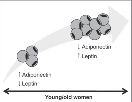
In conclusion, a progressive increase in adiposity throughout life seems to influence the relationship between leptin and adiponectin in women: serum adiponectin levels decrease, while circulating leptin levels markedly increase with body fat mass (Table 1, Figure 3). Moreover, leptin levels appear to be synchronous with hormone fluctuations and metabolic needs throughout the menstrual cycle, promoting
386 S.B. Lecke et al.
an interaction between the reproductive system, food intake and energy expenditure. Further studies are warranted to elucidate the relevance of leptin and adiponectin to fetal/ neonatal development, as well as possible interactions of these adipokines with fetal/neonatal growth and energy homeostasis. Also, while the role of leptin in puberty onset is well established, possible interactions between adiponectin and reproduction have yet to be elucidated.
Acknowledgments
Research supported by CNPq (INCT #573747/2008-3), CAPES (#1289/05) and Fundo de Apoio à Pesquisa do Hospital de Clínicas de Porto Alegre (FIPE-HCPA #340/2004), Brazil.
References
1. Chen J, Tan B, Karteris E, Zervou S, Digby J, Hillhouse EW, et al. Secretion of adiponectin by human placenta: differen-tial modulation of adiponectin and its receptors by cytokines.
Diabetologia 2006; 49: 1292-1302.
2. Henry BA, Clarke IJ. Adipose tissue hormones and the regu-lation of food intake. J Neuroendocrinol 2008; 20: 842-849. 3. Hausman GJ, Barb CR. Adipose tissue and the reproductive
axis: biological aspects. Endocr Dev 2010; 19: 31-44. 4. Kiess W, Reich A, Meyer K, Glasow A, Deutscher J, Klammt
J, et al. A role for leptin in sexual maturation and puberty?
Horm Res 1999; 51 (Suppl 3): 55-63.
5. Hassink SG, de Lancey E, Sheslow DV, Smith-Kirwin SM, O’Connor DM, Considine RV, et al. Placental leptin: an important new growth factor in intrauterine and neonatal development? Pediatrics 1997; 100: E1.
6. Zhang Y, Proenca R, Maffei M, Barone M, Leopold L, Fried-man JM. Positional cloning of the mouse obese gene and its human homologue. Nature 1994; 372: 425-432. 7. Grumbach MM. The neuroendocrinology of human puberty
revisited. Horm Res 2002; 57 (Suppl 2): 2-14.
8. Tartaglia LA, Dembski M, Weng X, Deng N, Culpepper J, Devos R, et al. Identification and expression cloning of a leptin receptor, OB-R. Cell 1995; 83: 1263-1271.
9. Schubring C, Englaro P, Siebler T, Blum WF, Demirakca T, Kratzsch J, et al. Longitudinal analysis of maternal serum leptin levels during pregnancy, at birth and up to six weeks after birth: relation to body mass index, skinfolds, sex ste-roids and umbilical cord blood leptin levels. Horm Res 1998; 50: 276-283.
10. Butte NF, Hopkinson JM, Nicolson MA. Leptin in human reproduction: serum leptin levels in pregnant and lactating women. J Clin Endocrinol Metab 1997; 82: 585-589. 11. Moschos S, Chan JL, Mantzoros CS. Leptin and
reproduc-tion: a review. Fertil Steril 2002; 77: 433-444.
12. Goumenou AG, Matalliotakis IM, Koumantakis GE, Panidis DK. The role of leptin in fertility. Eur J Obstet Gynecol Re-prod Biol 2003; 106: 118-124.
13. Scherer PE, Williams S, Fogliano M, Baldini G, Lodish HF. A novel serum protein similar to C1q, produced exclusively in adipocytes. J Biol Chem 1995; 270: 26746-26749. 14. Kadowaki T, Yamauchi T. Adiponectin and adiponectin
re-ceptors. Endocr Rev 2005; 26: 439-451.
15. Kissebah AH, Sonnenberg GE, Myklebust J, Goldstein M, Broman K, James RG, et al. Quantitative trait loci on chromo -somes 3 and 17 influence phenotypes of the metabolic syn -drome. Proc Natl Acad Sci U S A 2000; 97: 14478-14483. 16. Michalakis KG, Segars JH. The role of adiponectin in
reproduction: from polycystic ovary syndrome to assisted
reproduction. Fertil Steril 2010; 94: 1949-1957.
17. Savino F, Petrucci E, Nanni G. Adiponectin: an intriguing hormone for paediatricians. Acta Paediatr 2008; 97: 701-705.
18. Mise H, Yura S, Itoh H, Nuamah MA, Takemura M, Sagawa N, et al. The relationship between maternal plasma leptin levels and fetal growth restriction. Endocr J 2007; 54: 945-951.
19. Caprio M, Fabbrini E, Isidori AM, Aversa A, Fabbri A. Leptin in reproduction. Trends Endocrinol Metab 2001; 12: 65-72. 20. Sagawa N, Yura S, Itoh H, Kakui K, Takemura M, Nuamah
MA, et al. Possible role of placental leptin in pregnancy: a review. Endocrine 2002; 19: 65-71.
21. Caminos JE, Nogueiras R, Gallego R, Bravo S, Tovar S, Garcia-Caballero T, et al. Expression and regulation of adiponectin and receptor in human and rat placenta. J Clin Endocrinol Metab 2005; 90: 4276-4286.
22. Martos-Moreno GA, Barrios V, Saenz de Pipaon M, Pozo J, Dorronsoro I, Martinez-Biarge M, et al. Influence of prema -turity and growth restriction on the adipokine profile, IGF1, and ghrelin levels in cord blood: relationship with glucose metabolism. Eur J Endocrinol 2009; 161: 381-389. 23. Mantzoros C, Petridou E, Alexe DM, Skalkidou A, Dessypris
N, Papathoma E, et al. Serum adiponectin concentrations in relation to maternal and perinatal characteristics in new-borns. Eur J Endocrinol 2004; 151: 741-746.
24. Inoue M, Itabashi K, Nakano Y, Nakano Y, Tobe T. High-molecular-weight adiponectin and leptin levels in cord blood are associated with anthropometric measurements at birth.
Horm Res 2008; 70: 268-272.
25. Tsai PJ, Yu CH, Hsu SP, Lee YH, Chiou CH, Hsu YW, et al. Cord plasma concentrations of adiponectin and leptin in healthy term neonates: positive correlation with birthweight and neonatal adiposity. Clin Endocrinol 2004; 61: 88-93. 26. Dunger D, Ong K. Abundance of adiponectin in the newborn.
Clin Endocrinol 2004; 61: 416-417.
27. Lindsay RS, Walker JD, Havel PJ, Hamilton BA, Calder AA, Johnstone FD. Adiponectin is present in cord blood but is unrelated to birth weight. Diabetes Care 2003; 26: 2244-2249.
28. Tung WK, Lin SJ, Hwang YS, Wu CM, Wang YH, Tsai WH. Association of cord plasma leptin with birth size in term newborns. Pediatr Neonatol 2009; 50: 255-260.
29. Mellati AA, Mazloomzadeh S, Anjomshoaa A, Alipour M, Ka-rimi F, Mazloomi S, et al. Multiple correlations between cord blood leptin concentration and indices of neonatal growth.
Arch Med Res 2010; 41: 26-32.
al. Association of plasma leptin levels with maternal body weight and body mass index in premature and term new-borns. Pediatr Neonatol 2010; 51: 19-25.
31. Beltrand J, Nicolescu R, Kaguelidou F, Verkauskiene R, Si-bony O, Chevenne D, et al. Catch-up growth following fetal growth restriction promotes rapid restoration of fat mass but without metabolic consequences at one year of age. PLoS One 2009; 4: e5343.
32. Bozzola E, Meazza C, Arvigo M, Travaglino P, Pagani S, Stronati M, et al. Role of adiponectin and leptin on body de-velopment in infants during the first year of life. Ital J Pediatr
2010; 36: 26.
33. Miras M, Ochetti M, Martin S, Silvano L, Sobrero G, Castro L, et al. Serum levels of adiponectin and leptin in children born small for gestational age: relation to insulin sensitivity parameters. J Pediatr Endocrinol Metab 2010; 23: 463-471.
34. Muhlhausler B, Smith SR. Early-life origins of metabolic dysfunction: role of the adipocyte. Trends Endocrinol Metab
2009; 20: 51-57.
35. Plagemann A, Harder T. Hormonal programming in perinatal life: leptin and beyond. Br J Nutr 2009; 101: 151-152. 36. Catalano PM, Farrell K, Thomas A, Huston-Presley L,
Men-cin P, de Mouzon SH, et al. Perinatal risk factors for child-hood obesity and metabolic dysregulation. Am J Clin Nutr
2009; 90: 1303-1313.
37. Murphy MJ, Hosking J, Metcalf BS, Voss LD, Jeffery AN, Sattar N, et al. Distribution of adiponectin, leptin, and meta-bolic correlates of insulin resistance: a longitudinal study in British children; 1: Prepuberty (EarlyBird 15). Clin Chem
2008; 54: 1298-1306.
38. Darendeliler F, Poyrazoglu S, Sancakli O, Bas F, Gokcay G, Aki S, et al. Adiponectin is an indicator of insulin resistance in non-obese prepubertal children born large for gestational age (LGA) and is affected by birth weight. Clin Endocrinol
2009; 70: 710-716.
39. Bouhours-Nouet N, Dufresne S, de Casson FB, Mathieu E, Douay O, Gatelais F, et al. High birth weight and early post-natal weight gain protect obese children and adolescents from truncal adiposity and insulin resistance: metabolically healthy but obese subjects? Diabetes Care 2008; 31: 1031-1036.
40. Budak E, Fernandez SM, Bellver J, Cervero A, Simon C, Pellicer A. Interactions of the hormones leptin, ghrelin, adiponectin, resistin, and PYY3-36 with the reproductive system. Fertil Steril 2006; 85: 1563-1581.
41. Fenichel RM, Dominguez JE, Mayer L, Walsh BT, Boozer C, Warren MP. Leptin levels and luteinizing hormone pul-satility in normal cycling women and their relationship to daily changes in metabolic rate. Fertil Steril 2008; 90: 1161-1168.
42. Spritzer PM, Poy M, Wiltgen D, Mylius LS, Capp E. Leptin concentrations in hirsute women with polycystic ovary syndrome or idiopathic hirsutism: influence on LH and re -lationship with hormonal, metabolic, and anthropometric measurements. Hum Reprod 2001; 16: 1340-1346. 43. You T, Yang R, Lyles MF, Gong D, Nicklas BJ. Abdominal
adipose tissue cytokine gene expression: relationship to obesity and metabolic risk factors. Am J Physiol Endocrinol Metab 2005; 288: E741-E747.
44. Diez JJ, Iglesias P. The role of the novel adipocyte-derived
hormone adiponectin in human disease. Eur J Endocrinol
2003; 148: 293-300.
45. Cento RM, Proto C, Spada RS, Napolitano V, Ciampelli M, Cucinelli F, et al. Leptin levels in menopause: effect of es-trogen replacement therapy. Horm Res 1999; 52: 269-273. 46. Asimakopoulos B, Milousis A, Gioka T, Kabouromiti G,
Gianisslis G, Troussa A, et al. Serum pattern of circulating adipokines throughout the physiological menstrual cycle.
Endocr J 2009; 56: 425-433.
47. Hardie L, Trayhurn P, Abramovich D, Fowler P. Circulating leptin in women: a longitudinal study in the menstrual cycle and during pregnancy. Clin Endocrinol 1997; 47: 101-106. 48. Lecke SB, Toscani M, Mario FM, Wiltgen D, Morsch DM,
Spritzer PM. Influence of habitual physical activity on body composition, adipokine serum levels and gene expression on adipose tissue of PCOS women: a pilot study. Abstract book of the Androgen Excess and PCOS Society: Update Meeting.The heterogeneous nature of PCOS: Implications for treatment.Gramado: 2010.
49. Van Vugt DA. Brain imaging studies of appetite in the context of obesity and the menstrual cycle. Hum Reprod Update
2010; 16: 276-292.
50. Chung SC, Bond EF, Jarrett ME. Food intake changes across the menstrual cycle in Taiwanese women. Biol Res Nurs 2010; 12: 37-46.
51. Kleiblova P, Springer D, Haluzik M. The influence of hor -monal changes during menstrual cycle on serum adiponec-tin concentrations in healthy women. Physiol Res 2006; 55: 661-666.
52. Rouen PA, Lukacs JL, Reame NE. Adipokine concentrations in nonobese women: a study of reproductive aging, body mass index, and menstrual cycle effects. Biol Res Nurs
2010; 12: 54-61.
53. Galvan RE, Basurto L, Saucedo R, Campos S, Hernandez M, Zarate A. [Adiponectin concentrations during menstrual cycle]. Ginecol Obstet Mex 2007; 75: 435-438.
54. Xita N, Papassotiriou I, Georgiou I, Vounatsou M, Margeli A, Tsatsoulis A. The adiponectin-to-leptin ratio in women with polycystic ovary syndrome: relation to insulin resistance and proinflammatory markers. Metabolism 2007; 56: 766-771. 55. Chu MC, Cosper P, Orio F, Carmina E, Lobo RA. Insulin
resistance in postmenopausal women with metabolic syn-drome and the measurements of adiponectin, leptin, resistin, and ghrelin. Am J Obstet Gynecol 2006; 194: 100-104. 56. Donato GB, Fuchs SC, Oppermann K, Bastos C, Spritzer
PM. Association between menopause status and central adi-posity measured at different cutoffs of waist circumference and waist-to-hip ratio. Menopause 2006; 13: 280-285. 57. Hong SC, Yoo SW, Cho GJ, Kim T, Hur JY, Park YK, et al.
Correlation between estrogens and serum adipocytokines in premenopausal and postmenopausal women. Menopause
2007; 14: 835-840.
58. Antuna-Puente B, Feve B, Fellahi S, Bastard JP. Adipokines: the missing link between insulin resistance and obesity.
Diabetes Metab 2008; 34: 2-11.
59. Lee JM, Kim SR, Yoo SJ, Hong OK, Son HS, Chang SA. The relationship between adipokines, metabolic parameters and insulin resistance in patients with metabolic syndrome and type 2 diabetes. J Int Med Res 2009; 37: 1803-1812. 60. Valencia MH, Zarate A, Galvan RE. [Adiponectin levels in