Antinociception induced by stimulating
amygdaloid nuclei in rats: changes
produced by systemically administered
antagonists
Departamento de Farmacologia, Faculdade de Medicina de Ribeirão Preto, Universidade de São Paulo, Ribeirão Preto, SP, Brasil
M.A. Oliveira and W.A. Prado
Abstract
The antinociceptive effects of stimulating the medial (ME) and central (CE) nuclei of the amygdala in rats were evaluated by the changes in the latency for the tail withdrawal reflex to noxious heating of the skin. A 30-s period of sine-wave stimulation of the ME or CE produced a significant and short increase in the duration of tail flick latency. A 15-s period of 15-stimulation wa15-s ineffective. Repeated 15-stimulation of the15-se nuclei at 48-h intervals produced progressively smaller effects. The antinociception evoked from the ME was significantly reduced by the previous systemic administration of naloxone, methysergide, atro-pine, phenoxybenzamine, and propranolol, but not by mecamylamine, all given at the dose of 1.0 mg/kg. Previous systemic administration of naloxone, atropine, and propranolol, but not methysergide, phenoxy-benzamine, or mecamylamine, was effective against the effects of stimulating the CE. We conclude that the antinociceptive effects of stimulating the ME involve at least opioid, serotonergic, adrenergic, and muscarinic cholinergic descending mechanisms. The effects of stimulating the CE involve at least opioid, ß-adrenergic, and musca-rinic cholinergic descending mechanisms.
Correspondence W.A. Prado
Departamento de Farmacologia FMRP, USP
Av. Bandeirantes, 3900 14049-900 Ribeirão Preto, SP Brasil
Fax: 55 (016) 633-2301 E-mail: [email protected]
Research supported by FAPESP. M.A. Oliveira was the recipient of CAPES and CNPq fellowships.
Received July 24, 1997 Accepted February 13, 1998
Key words •Antinociception •Amygdala
•Medial nucleus of the amygdala
•Central nucleus of the amygdala
•Tail flick test
•Stimulation-produced antinociception
Introduction
Behavioral and electrophysiological stud-ies have demonstrated that at many sites in the brain electrical or chemical stimulation produces analgesia by activating centrifugal pathways that act to inhibit sensory neurons in the spinal cord (see Ref. 1). Special atten-tion has been given to the mesencephalic periaqueductal gray (PAG)/dorsal raphe nucleus (DRN) and nucleus raphe magnus (NRM) (see Ref. 2), but evidence exists for the involvement of more rostral structures,
including the amygdala, in this central pain control mechanism (see Ref. 3)
or from the observation that the microinjec-tion of some agonists into the amygdala can evoke antinociception. Lesions of the amyg-dala, mainly at its basolateral and central (CE) nuclei, attenuate several forms of envi-ronmentally induced antinociception (7-10). Bilateral lesions of the CE abolish the anti-nociceptive effects of low doses of systemi-cally administered morphine in both the rat tail flick (11) and formalin (12) tests. Micro-injection of a κ-opioid agonist (13) or neuro-tensin (14) into the amygdala evokes antino-ciception. A similar effect was demonstrated in the rat tail flick test following microinjec-tion of carbachol into various amygdaloid nuclei, including the CE and the medial (ME) nuclei (15,16). Microinjection of morphine into the corticomedial subdivision of the amygdala is effective in the flinch-jump (17,18) and hot plate (19) tests. Microinjec-tion of opioids into the CE (9,20-25) or of serotonin (26) into the basomedial part of the amygdala also induces antinociception.
Few studies, however, have been con-ducted on the effects of electrical stimula-tion of the amygdala on nociceptive re-sponses. Early studies have demonstrated that stimulation of the lateral region of the amygdala elicits antinociceptive-like effects while the medial region yields a painful re-sponse pattern (27). However, Abbott and Melzack (28) did not obtain immediate anti-nociception in rats following stimulation of the amygdala. More recently, reduction of visceral pain in cats has been reported to occur after electrical stimulation of the CE (29). Unilateral stimulation of the basolateral nucleus, CE, or ME did not affect the thresh-old for the tail withdrawal response evoked by electric shock but increased the tail flick latency (TL) to noxious heat, reduced the tonic phase of the animal response to forma-lin, and elevated the threshold for vocaliza-tion during and after the applicavocaliza-tion of an electric shock to the tail skin (30).
The present study was undertaken to ex-amine the effects of stimulating the CE or the
ME on the tail flick response evoked by noxious heating of the skin in rats. We dem-onstrate that a brief (30 s) stimulus applied to either nucleus evokes antinociception. In addition, we demonstrate that previous sys-temic administration of methysergide, nalox-one, propranolol, phenoxybenzamine or at-ropine, but not mecamylamine, is effective in inhibiting the antinociception produced by ME stimulation. Moreover, systemic naloxone, atropine and phenoxybenzamine, but not propranolol, methysergide, or meca-mylamine, are effective against CE stimula-tion-produced antinociception.
Material and Methods
Subjects and surgery
The experiments were conducted on male Wistar rats (140-160 g) housed two to a cage with free access to food and water and main-tained at an average ambient temperature of 24oC with a 12-h light-dark cycle before and after surgery. The proposals of the Commit-tee for Research and Ethical Issues of IASP (31) were followed throughout the experi-ments. Each animal was anesthetized with sodium thiopentone (50 mg/kg, ip) and a Teflon-insulated monopolar electrode (OD = 0.007) was stereotaxically implanted into the skull to lie in the CE or ME nuclei. The coordinates used were: AP = +5.8, L = 3.5, and H = -3.2 mm, for the ME, and AP = +5.8, L = 4.4, and H = -5.5 mm, for the CE, as proposed elsewhere (32). The electrode was then fixed to the skull with two steel screws and dental cement. One of these screws was used as the indifferent electrode. After re-ceiving penicillin (50 mg/kg, im) the animal was allowed to recover for at least one week before the experiments.
Tail flick test
with the tail laid across a small wire that was at room temperature (23 ± 2oC). The coil temperature was then raised by the passage of electric current, which was previously adjusted to ensure a tail withdrawal reflex within 2.5-3.5 s. A cut-off time of 6 s was established to minimize the probability of skin damage. Tail flick latencies were meas-ured at 10-min intervals until a stable baseline (BL) was obtained over three consecutive trials. Only rats showing a stable BL after six trials were used in each experiment. Each TL was normalized by an index of antinocicep-tion (IA) using the formula IA = (TL - aver-age BL)/(6 - averaver-age BL).
Stimulation procedures
Immediately after BL determination the animal was placed inside a glass-walled box (20 x 30 x 35 cm), a 60-Hz sine-wave alter-nating current was applied to the electrode for 15 or 30 s and the TL determined within 10 s and the procedure was repeated at 10-min intervals over a period of 30 10-min. Dur-ing the stimulation period the drop in voltage across a 1-kΩ resistor in series with the electrode was continuously monitored on an oscilloscope. No attempt was made to test for the presence of antinociception during the stimulation. Two groups of 5 animals each with electrodes implanted in the ME or CE were used as sham-stimulated rats.
A group of 18 rats with electrodes im-planted in the ME or CE was preliminarily used for the determination of the CI50, i.e., the current intensity producing an antinoci-ceptive effect in 50% of the animals in the experimental group. For calculation, antino-ciception was arbitrarily considered to occur whenever IA ≥0.5 was obtained. Immedi-ately after BL determination each animal received30 s of brain stimulation, and the TL was determined up to 10 s later. During this first stimulation period the lowest cur-rent of 1.4 µA root mean square (rms) was used and then increased to 3.5 µA in a
sec-ond test 5 min later and to a maximum of 35 µA in a stepped sequence of 1.4, 3.5, 7.0, 10.0, 14.0, 21.0, 35.0 µA rms. The animals were spared further stimulation whenever IA = 1.0 was obtained.
Histology
At the end of the experiment the animal was killed with an overdose of sodium thio-pentone and perfused through the heart with formalin. Electrode tracks were localized on 50-µm serial coronal sections stained with neutral red, and identified on diagrams from the atlas of König and Klippel (32).
Statistical analysis
The CI50 was calculated by the method of Litchfield and Wilcoxon (33). The results of the remaining studies are reported as graphs of averaged IA (± SEM) values against time of reading for a group of rats. The effects of different treatments on IA were analyzed statistically by multivariate analysis of vari-ance (MANOVA) with repeated measures to compare the groups over all times. The factors analyzed in the experiments of ME or CE stimulation were treatments, time and treatment x time interaction. In the case of significant treatment x time interactions a one-way ANOVA followed by the Duncan test was performed for each time. The anal-ysis was performed using the statistical soft-ware package SPSS/PC+, version 3.0, and the level of significance was set at P<0.05.
Drugs
bimaleate was from Sandoz (Basel, Switzer-land). The antagonists were all dissolved in saline and given at the dose of 1.0 mg/kg, 10 min (naloxone, atropine, and propranolol), 15 min (methysergide and mecamylamine), or 3 h (phenoxybenzamine) before intracere-bral stimulation. A longer interval had to be
used between phenoxybenzamine adminis-tration and brain stimulation because the central effects of this drug develop slowly (34).
Results
Determination of the current intensity applied to the ME and CE for the production of antinociception
The latency for the tail flick reflex was increased by electrical stimulation of ME or CE at the sites depicted in Figure 1A. The current intensity for the maximal possible effect in the test (IA = 1.0), however, varied widely. One of eleven animals stimulated in the ME with a current intensity of 1.4 µA rms yielded IA = 1.0, whereas other rats showed full antinociception after stimula-tion with current intensities of 3.5 (2 ani-mals), and 7.0, 10.0, 14.0, and 21.0 µA rms (1 animal at each intensity). Four animals did not show a full effect even after stimula-tion at 35 µA rms. Similar results were ob-tained for 7 rats stimulated in the CE. Full antinociception was obtained at current in-tensities of 1.4 and 10.0 µA rms (2 animals each) and at 3.5 and 7.0 µA rms (1 animal each). The remaining animal did not show a full effect even at the current intensity of 35.0 µA. The CI50 were 10.11 (confidence limits = 7.99 and 11.87) and 5.16 (3.74 and 6.29) µA rms for the ME and CE, respec-tively. We therefore decided to stimulate systematically these nuclei with 21.0 µA rms.
Time-course of the effects of stimulating the ME and CE. Influence of duration of the stimulation and repeated stimulation
Stimulation of the ME (Figure 1B) or CE (Figure 1C) with 21.0 µA over a period of 15 s produced a slight increase in the index of antinociception (26% and 35% for ME and CE stimulation, respectively) and the effects
Index of antinociception
1.0
0.8
0.6
0.4
0.2
0.0
-0.2
1.0
0.8
0.6
0.4
0.2
0.0
-0.2
0 h 48 h 96 h Sham
-20 -10 0 10 20 30 -20 -10 0 10 20 30 Time (min) Time (min)
B D
C E
A
A 5150 A 4890 A 4380 A 4230
Figure 1 - Antinociceptive effects of stimulating the medial or central nuclei of the amygdala. (A) Cross sections taken from the atlas of König and Klippel (32), at the indicated AP levels, showing the location of the sites stimulated during experiments for the determination of current thresholds. The remaining graphs show the time-course of the effects of stimulating the medial (B and D) or the central (C and E) nuclei of the amygdala for 15 or 30 s, respectively, on three different occasions at 48-h intervals. N = 4 for the curves of graph C and 5 for the remaining graphs. *P<0.05 compared to sham-stimulated animals (Duncan test).
*
did not change significantly for experiments repeated 48 or 96 h later. The effects were stronger and of short duration following stim-ulation of the ME (Figure 1D) or CE (Figure 1E) with the same current intensity applied over a period of 30 s (58% and 54% for ME and CE stimulation, respectively), but the effects were smaller when the stimulation was repeated 48 and 96 h later. The curves in Figure 1B and C did not differ significantly regarding the different occasions of stimula-tion (F3,16 = 0.97, P = 0.43, and F3,16 = 1.15, P = 0.36, respectively) nor did they show significant effect x time interactions (F21,112 = 0.96, P = 0.52, and F21,112 = 1.01, P = 0.46, respectively). The curves in Figure 1D did not differ when the different occasions of stimulation were compared (F3,13 = 1.73, P = 0.21) and showed no significant effect x time interaction (F21,91 = 1.10, P = 0.35). The curves in Figure 1E were significantly dif-ferent (F3,16 = 3.89, P = 0.029) and showed a significant effect x time interaction (F21,112 = 2.42, P = 0.002). The effects obtained for animals stimulated in the CE on the first occasion were significantly different from those obtained for sham-stimulated rats at times 0 and 2 min (ANOVA followed by the Duncan test). The subsequent experiments were then conducted on animals stimulated only once with a current intensity of 21.0 µA rms applied to each nucleus over a period of 30 s.
Some rats stimulated in the CE presented aversive-like behaviors during stimulation, including vocalization, masticatory move-ments and attempts to escape from the box. Apparently, the frequency of behaviors evoked by stimulating these nuclei was not changed by increasing the duration of the stimulation. Escape was also observed in some rats stimulated in the ME. These be-haviors were more frequent during longer periods of stimulation. After the end of stim-ulation, no gross motor disturbance was de-tected. The animals walked and responded normally to innocuous stimuli. No attempt
was made to quantify these behaviors in the present study.
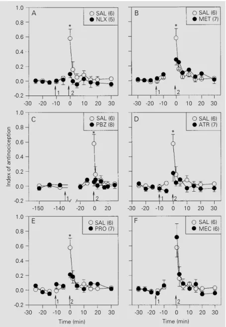
Effects of ip administration of antagonists on the antinociception induced by stimulation of the ME
Six groups of rats were treated by ip administration of antagonists, 10 min (nalox-one, atropine, and propranolol), 15 min (methysergide and mecamylamine) or 3 h (phenoxybenzamine) before intracerebral stimulation. All drugs were given at the dose of 1 mg/kg. A group of rats treated with saline (0.1 ml/kg, ip) was used as control.
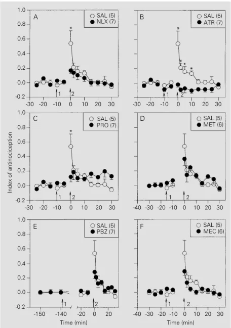
Naloxone (Figure 2A), methysergide (Fig-ure 2B), phenoxybenzamine (Fig(Fig-ure 2C), atropine (Figure 2D) and propranolol (Fig-ure 2E), but not mecamylamine (Fig(Fig-ure 2F), significantly inhibited the antinociceptive effects of stimulating the ME. The curves in Figure 2 did not differ significantly regard-ing treatments (F6,39 = 2.33, P = 0.05) but showed a significant treatment x time inter-action (F42,273 = 2.44, P<0.001). On the other hand, naloxone (Figure 3A), atropine (Fig-ure 3B), and propranolol (Fig(Fig-ure 3C), but not methysergide (Figure 3D), mecamyla-mine (Figure 3E), or phenoxybenzamecamyla-mine (Fig-ure 3F), were effective against the antinoci-ception induced by stimulation of the CE. The curves in Figure 3 differed significantly regarding treatments (F6,38 = 5.10, P = 0.001) and showed a significant treatment x time interaction (F42,266 = 2.31, P<0.001). The antagonists alone had no significant effect on tail flick latency.
Discussion
Masti-catory movements during stimulation of the CE have also been reported elsewhere (35). The antinociceptive effects of stimulat-ing the ME or the CE were dependent on the pattern of electrical stimulation. The current intensity required for a full antinociceptive effect was variable, the CE being more
sen-sitive than the ME. Electrical stimulation of these nuclei was more effective when ap-plied for 30 s than when apap-plied for 15 s. The small monopolar electrodes used in these experiments reduce the risk of tissue lesion and the occurrence of edema at the site reached by the electrode tip. Moreover, the biphasic alternating current applied to mon-opolar electrodes allows a more focal stimu-lation of the target structure (36).
We have also shown that the antinoci-ception evoked from the ME or CE was progressively weaker when the stimulation was repeated at 48-h intervals. The repeated stimulation of the amygdala may somehow cause irreversible or long-lasting functional changes at the site of stimulation. Repeated stimulation of the amygdala may produce kindling, a phenomenon that may change the animals responsiveness to pain (37). An alternative explanation for the phenomenon could be the development of tolerance to the stimulation. Similar changes induced by re-peated stimulation of the PAG have been previously demonstrated, and probably in-volve the participation of endogenous opioid modulation (38). In fact, opioid mechanisms may participate in the antinociception evoked by amygdaloid stimulation. The ME and CE express mRNA for µ- and κ-opioid receptors (39,40). Fibers and terminals immunoreac-tive to ß-endorphins (41) or enkephalins (42,43) were demonstrated in the ME and CE, respectively. Our data, however, do not allow us to conclude about the mechanism involved in the reduced effectiveness of re-peated stimulation of the ME or CE.
The antinociceptive effects of stimulat-ing the ME were significantly inhibited by the previous systemic administration of naloxone (an opioid receptor antagonist), methysergide (a 5-HT receptor antagonist), atropine (a muscarinic cholinergic receptor
antagonist), phenoxybenzamine (an α
-adrenoceptor antagonist), and propranolol (a ß-adrenoceptor antagonist), but not by mecamylamine (a nicotinic cholinergic
re-Index of antinociception
1.0
0.8
0.6
0.4
0.2
0.0
-0.2
1.0
0.8
0.6
0.4
0.2
0.0
-0.2
-30 -20 -10 10 20 30
A B
0 -30 -20 -10 0 10 20 30
-150 -140 -20 0 20 -30 -20 -10 0 10 20 30
-30 -20 -10 0 10 20 30 -30 -20 -10 0 10 20 30 1.0
0.8
0.6
0.4
0.2
0.0
-0.2
SAL (6)
NLX (5) SAL (6)MET (7)
1 2
SAL (6) PBZ (8)
1 2
SAL (6) PRO (7)
1 2
SAL (6) ATR (7)
1 2
SAL (6) MEC (6)
1 2
Time (min) Time (min)
* *
* *
*
C D
E F
Figure 2 - Effects of intraperitoneal administration (arrow 1) of saline (SAL; 0.1 ml/kg) or antagonists (1 mg/kg) on the antinociception induced by electrical stimulation (arrow 2) of the medial nucleus of the amygdala of rats. A, Effect of naloxone (NLX); B, effect of methysergide (MET); C, effect of phenoxybenzamine (PBZ); D, effect of atropine (ATR); E, effect of propranolol (PRO); F, effect of mecamylamine (MEC). The number of rats per curve is given in parentheses. Data are reported as mean ± SEM for each group of rats. *P<0.05 compared to drug-treated animals (Duncan test).
ceptor antagonist). Naloxone, atropine, and propranolol, but not methysergide, phenoxy-benzamine, or mecamylamine, were signifi-cantly effective against the antinociception induced by stimulating the CE. These an-tagonists were all used at doses already known to be effective against similar effects in-duced by the stimulation of other brain struc-tures known to participate in the descending control of pain (44-48). The effectiveness of propranolol against the stimulation-produced antinociception from the ME or CE is in-dicative that ß-adrenergic mechanisms may be involved in the phenomenon. Propranolol exhibits local anesthetic properties and has affinity also for a range of serotonergic re-ceptor subtypes (49). A local anesthetic ef-fect of propranolol seems to depend on higher drug concentrations (see 50) and is, there-fore, unlikely to be the reason for its inhibi-tory effect found in this study. The present results do not allow us to exclude that the effectiveness of propranolol against the stim-ulation-produced antinociception from the ME derives from its 5-HT antagonist prop-erty. However, the nonspecific 5-HT an-tagonist methysergide was effective against the effect of stimulating the ME, but not the CE. Thus, ß-adrenergic mechanisms may also be involved in the descending mech-anism activated from the CE. The different profiles of effectiveness of the antagonists used in this study provide evidence that the ME and CE function separately to produce inhibition of the tail flick reflex.
The tail flick escape from noxious heat is a spinal reflex (51) and its inhibition by stimulating supraspinal structures indicates that this action may somehow inhibit spinal mechanisms. Motor impairment produced by intracerebral stimulation could be one reason for the inhibition of the tail flick reflex. Objective tests for motor changes were not conducted in the present study. However, no gross motor disturbance was detected throughout the experiments. The animals walked normally after the
stimula-tion period and responded to innocuous stimuli.
Few reports are available regarding di-rect projections from the amygdala to the spinal cord. A sparse population of CE neu-rons in monkeys (52) and cats (53) projects
Index of antinociception
1.0
0.8
0.6
0.4
0.2
0.0
-0.2
1.0
0.8
0.6
0.4
0.2
0.0
-0.2
-30 -20 -10 10 20 30
A B
0 -30 -20 -10 0 10 20 30
-30 -20 -10 0 10 20 30 -30 -20 -10 0 10 20 30
-30 -20 -10 0 10 20 30 1.0
0.8
0.6
0.4
0.2
0.0
-0.2
SAL (5)
NLX (7) SAL (5)ATR (7)
1 2
SAL (5) PRO (7)
1 2
SAL (5) PBZ (7)
1 2
SAL (5) MET (6)
1 2
SAL (5) MEC (6)
1 2
Time (min) Time (min)
* *
*
C D
E F
-150 -140 -20 0 20
** *
-40
-40
Figure 3 - Effects of intraperitoneal administration (arrow 1) of saline (SAL; 0.1 ml/kg) or antagonists (1 mg/kg) on the antinociception induced by electrical stimulation (arrow 2) of the central nucleus of the amygdala of rats. A, Effect of naloxone (NLX); B, effect of atropine (ATR); C, effect of propranolol (PRO); D, effect of methysergide (MET); E, effect of phenoxybenzamine (PBZ); F, effect of mecamylamine (MEC). The number of rats per curve is given in parentheses. Data are reported as means ± SEM for each group of rats. *P<0.05 compared to drug-treated animals (Duncan test).
to the cervical spinal cord. Alternatively, anatomical studies have demonstrated direct reciprocal projections between the amyg-dala and the PAG (54-59). The CE (56,60) and ME (55,56,61) have direct and indirect (via the hypothalamus) connections with the PAG. The CE also sends projections to the parabrachial nucleus (62,63) and locus coe-ruleus (60,64), which are structures also known to exert antinociceptive effects when electrically stimulated (65,66). Thus, the depression of the tail flick reflex by stimulat-ing the ME or CE probably involves activa-tion of descending pathways that utilize re-lay stations before reaching the spinal cord.
In summary, this study demonstrates that brief electrical stimulation of the ME and CE amygdaloid nuclei increases the tail flick response latency. The effect obtained from the ME involves at least opioid, serotoner-gic, adrenerserotoner-gic, and muscarinic cholinergic mechanisms. The effect from the CE seems to depend on at least opioid, ß-adrenergic, and muscarinic cholinergic mechanisms.
Acknowledgments
We thank P.R. Castania for technical sup-port.
References
1. Basbaum AI (1982). Anatomical sub-strates for the descending control of noci-ception. In: Sjölund B & Björklund A (Edi-tors), Brain Stem Control of Spinal Mecha-nisms. Elsevier, Amsterdam, 119-133. 2. Besson JM & Chaouch A (1987).
Periph-eral and spinal mechanisms of nocicep-tion. Pharmacological Reviews, 67: 67-186.
3. Kuypers HGJM (1982). A new look at the organization of the motor system. In: Kuypers HGJM & Martin GF (Editors), Anatomy of Descending Pathways to the Spinal Cord. Progress in Brain Research. Vol. 57. Elsevier, Amsterdam, 381-403. 4. Zhou ZF, Du MY, Jiang Y & Han JS (1981).
Effect of intracerebral microinjection of naloxone on acupuncture and morphine analgesia in the rabbit. Scientia Sinica, 24: 1166-1178.
5. LeDoux JE (1987). Emotion. In: Plum F (Editor), Handbook of Physiology: Nervous System V. American Physiological Soci-ety, Washington, DC, 419-459.
6. Good AJ & Westbrook RF (1995). Effects of a microinjection of morphine into the amygdala on the acquisition and expres-sion of conditioned fear and hypoalgesia in rats. Behavioral Neuroscience, 109: 631-641.
7. Helmstetter F (1992). The amygdala is essential for the expression of condi-tioned hypoalgesia. Behavioral Neurosci-ence, 106: 518-528.
8. Helmstetter F & Bellgowan PS (1993).
Lesions of the amygdala block conditional hypoalgesia in the tail flick test. Brain Re-search, 612: 253-257.
9. Watkins LR, Wiertelak EP & Maier SF (1993). The amygdala is necessary for the expression of conditioned but not uncon-ditioned analgesia. Behavioral Neurosci-ence, 107: 402-405.
10. Fox RJ & Sorenson CA (1994). Bilateral lesions of the amygdala attenuate analge-sia induced by diverse environmental chal-lenges. Brain Research, 648: 215-221. 11. Manning BH & Mayer DJ (1995). The
cen-tral nucleus of the amygdala contributes to the production of morphine antinoci-ception in the rat tail-flick test. Journal of Neuroscience, 15: 8199-8213.
12. Manning BH & Mayer DJ (1995). The cen-tral nucleus of the amygdala contributes to the production of morphine antinoci-ception in the formalin test. Pain, 63: 141-152.
13. Helmstetter FA, Brozoski EL & Frost JÁ (1991). Kappa opioid agonists produce hypoalgesia in the tail flick test after appli-cation to the amygdala. Society for Neuro-science Abstracts, 17: 296.
14. Kalivas PW, Gau BA, Nemeroff CB & Prange AJ (1982). Antinociception after microinjection of neurotensin into the central amygdaloid nucleus of the rat. Brain Research, 243: 279-286.
15. Klamt JG & Prado WA (1991). Antinoci-ception and behavioral changes induced by carbachol microinjected into identified
sites of the rat brain. Brain Research, 549: 9-18.
16. Oliveira MA & Prado WA (1994). Antinoci-ception and behavioral manifestations in-duced by intracerebroventricular or intra-amygdaloid administration of cholinergic agonists in the rat. Pain, 57: 383-391. 17. Rodgers RJ (1977). Elevation of aversive
threshold in rats by intra-amygdaloid in-jection of morphine sulfate. Pharmacolo-gy, Biochemistry and Behavior, 6: 385-390.
18. Rodgers RJ (1978). Influence of intra-amygdaloid opiate injections on shock thresholds, tail flick latencies and open field behavior in rats. Brain Research, 153: 211-216.
19. Yaksh TL, Al-Rodhan NRF & Jensen TS (1988). Sites of action of opiates in pro-duction of analgesia. In: Fields HL & Besson JM (Editors), Progress in Brain Research. Vol. 77. Elsevier, New York, 371-394.
20. File SE & Rodgers RJ (1979). Partial anxio-lytic action of morphine sulphate follow-ing microinjection into the central nucleus of the amygdala in rats. Pharmacology, Biochemistry and Behavior, 11: 313-318. 21. Rodgers RJ & File SE (1979). Exploratory
behavior and aversive thresholds follow-ing intra-amygdaloid application of opiates in rats. Pharmacology, Biochemistry and Behavior, 11: 505-511.
ha-benula, nucleus accumbens or amygdala of rabbits. Acta Pharmacologica Sinica, 5: 150-153.
23. Davis M (1986). The role of the amygdala in conditioned fear. In: Aggleton JP (Edi-tor), The Amygdala: Neurobiological As-pects of Emotion, Memory, and Mental Dysfunction. Wiley-Liss, New York, 339-351.
24. al-Rodhan N, Chipkin R & Yaksh TL (1990). The antinociceptive effects of SCH-32615, a neutral endopeptidase (enkeph-alinase) inhibitor, microinjected into the periaqueductal, ventral medulla and amyg-dala. Brain Research, 520: 123-130. 25. Ma QP, Yin GF, Ai MK & Han JS (1991).
Serotonergic projections from the nucleus raphe dorsalis to the amygdala in the rat. Neuroscience Letters, 134: 21-24. 26. Plasnik A, Danysz W & Kostowski W
(1985). Some behavioral effects of micro-injections of noradrenaline and serotonin into the amygdaloid body of the rat brain. Physiology and Behavior, 34: 481-487. 27. Lico MC, Hoffmann A & Covian MR
(1974). Influence of some limbic struc-tures upon somatic and autonomic mani-festations of pain. Physiology and Behav-ior, 12: 805-811.
28. Abbott FV & Melzack R (1978). Analgesia produced by stimulation of limbic struc-tures and its relation to epileptiform after-discharges. Experimental Neurology, 62: 720-734.
29. Sha L, Huang P, Ding W & Teng G (1993). The inhibitory effects of stimulating AC, AL, BNST and AHL on visceral pain. Chen Tzu Yen Chiu Acupuncture Research, 18: 37-43.
30. Mena NB, Mathur R & Nayar U (1995). Amygdalar involvement in pain. Indian Journal of Physiology and Pharmacology, 39: 339-346.
31. Zimmermann M (1983). Ethical guidelines for investigation of pain in conscious ani-mals. Pain, 16: 109-110.
32. König JFR & Klippel RA (1974). The Rat Brain, a Stereotaxic Atlas. Krieger, New York.
33. Litchfield JT & Wilcoxon F (1949). A sim-plified method of evaluating dose-effect experiments. Journal of Pharmacology and Experimental Therapeutics, 96: 99-113.
34. Azami J, Llewelyn MB & Roberts MHT (1982). The contribution of nucleus reticularis paragigantocellularis and nucleus raphe magnus to the analgesia produced by systemically administered morphine, investigated with the microin-jection technique. Pain, 12: 229-246.
35. Applegate CD, Kapp BS, Underwood MD & McNalli CL (1983). Autonomic and somatomotor effects of amygdala central nucleus. Stimulation in awake rabbits. Physiology and Behavior, 31: 353-360. 36. Thorn BE, Applegate L & Jones K (1990).
The relative efficacy of monopolar vs. bi-polar electrodes in stimulation-produced analgesia. Experimental Brain Research, 79: 266-270.
37. Frenk H & Yitzhaky J (1981). Effects of amygdaloid binding on the pain threshold of the rat. Experimental Neurology, 71: 487-496.
38. Mayer DJ & Hayes RL (1975). Stimula-tion-produced analgesia: development of tolerance and cross-tolerance to mor-phine. Science, 188: 941-943.
39. Mansour A, Fox CA, Burke S, Meng F, Thompson RC, Akil H & Watson SJ (1994). Mu, delta, and kappa opioid receptor ex-pression in the rat CNS: an in situ hybrid-ization study. Journal of Comparative Neu-rology, 350: 412-438.
40. Arvidsson U, Riedl M, Chakrabarti S, Lee JH, Nakano AH, Dado RJ, Loh HH, Law PY, Wessendorff W & Elde R (1995). Dis-tribution and targeting of a µ-opioid recep-tor (MOR 1) in brain and spinal cord. Jour-nal of Neuroscience, 15: 3328-3341. 41. Gray TS, Cassell MD & Kiss JZ (1984).
Distribution of pro-opiomelanocortin de-rived peptides and enkephalins in the rat central nuclei of the amygdala. Brain Re-search, 306: 354-358.
42. Roberts GW, Woodhams PL, Polak JM & Crow TJ (1982). Distribution of neuropep-tides in the limbic system of the rat: the amygdaloid complex. Neuroscience, 7: 99-131.
43. Cassell MD & Gray TS (1989). Morpholo-gy of peptide-immunoreactive neurons in the rat central nucleus of the amygdala. Journal of Comparative Neurology, 281: 320-333.
44. Akil H, Mayer DJ & Liebeskind JC (1976). Antagonism of stimulation-produced anal-gesia by naloxone, a narcotic antagonist. Science, 191: 961-962.
45. Carstens E, Fraunhoffer M & Zimmer-mann M (1981). Serotonergic mediation of descending inhibition from midbrain periaqueductal gray, but not reticular for-mation, of spinal nociceptive transmission in the cat. Pain, 10: 149-167.
46. Carstens E, Culhane ES & Banisadr R (1990). Partial involvement of mono-amines and opiates in the inhibition of rat spinal nociceptive neurons evoked by stimulation in midbrain periaqueductal gray or lateral reticular formation. Brain
Research, 522: 7-13.
47. Fang F & Proudfit HK (1996). Spinal cho-linergic and monoamine receptors medi-ate the antinociceptive effect of morphine microinjected in the periaqueductal gray on the rat tail, but not the feet. Brain Research, 722: 95-108.
48. Guimarães APC & Prado WA (1994). Anti-nociceptive effects of carbachol microin-jected into different portions of the mes-encephalic periaqueductal gray matter of the rat. Brain Research, 647: 220-230. 49. Saxena PR (1995). Serotonin receptors:
subtypes, functional responses and thera-peutic relevance. Pharmacological Thera-peutics, 66: 339-368.
50. Phillips WJ, Enyeart JJ & Hinkle PM (1989). Pituitary thyrotropin-releasing hor-mone receptors: local anesthetic effects on binding and responses. Molecular En-docrinology, 3: 1345-1351.
51. Mayer DJ & Liebeskind JC (1974). Pain reduction by focal electrical stimulation of the brain: an anatomical and behavioral analysis. Brain Research, 68: 73-93. 52. Mizuno N, Takahashi O, Satoda T &
Matsushima R (1985). Amygdalospinal projections in the macaque monkey. Neu-roscience Letters, 53: 327-330.
53. Sandrew BB, Edwards DL, Poletti CE & Foote WE (1986). Amygdalospinal projec-tions in the cat. Brain Research, 373: 235-239.
54. Beitz AJ (1982). The organization of affer-ent projections to the midbrain periaque-ductal gray of the rat. Neuroscience, 7: 133-159.
55. Eberhart JÁ, Morrell JI, Krieger MS & Pfaff DW (1985). An autoradiographic study of projections ascending from the midbrain central gray, and from the region lateral to it, in the rat. Journal of Comparative Neu-rology, 241: 285-310.
56. Rizvi TA, Ennis M, Behbehani MM & Shipley MT (1991). Connections between the central nucleus of the amygdala and the midbrain periaqueductal gray: topog-raphy and reciprocity. Journal of Compara-tive Neurology, 303: 121-131.
57. Li YQ, Takada M, Matsuzaki S, Shinonasa Y & Mizuno N (1993). Identification of periaqueductal gray and dorsal raphe nucleus projecting to both the trigeminal sensory complex and forebrain structures: a fluorescent retrograde double-labeling study in the rat. Brain Research, 623: 267-277.
through NMDA receptors. Brain Re-search, 635: 187-195.
59. Cameron AA, Khan IA, Westlund KN, Cliffer KD & Willis WD (1995). The effer-ent projections of the periaqueductal gray in the rat: a Phaseolus vulgaris -leucoag-glutinin study. I. Ascending projections. Journal of Comparative Neurology, 351: 568-584.
60. Hopkins DA & Holstege G (1978). Amyg-daloid projections to the mesencephalon, pons and medulla oblongata in the cat. Experimental Brain Research, 32: 529-547.
61. Canteras NS, Simerly RB & Swanson LW (1995). Organization of projections from
the medial nucleus of the amygdala: a PHAL study in the rat. Journal of Com-parative Neurology, 360: 213-245. 62. Krettek JE & Price JL (1978). Amygdaloid
projections to subcortical structures within the basal forebrain and brainstem in the rat and cat. Journal of Comparative Neurology, 178: 225-253.
63. Veening JG, Swanson LW & Sawchenko PE (1984). The organization of projections from the central nucleus of the amygdala to brainstem sites involved in central au-tonomic regulation: a combined retro-grade transport-immunohistochemical study. Brain Research, 303: 337-357. 64. Wallace DM, Magnuson DJ & Gray TS
(1989). The amygdalo-brainstem pathway: selective innervation of dopaminergic, noradrenergic and adrenergic cells in the rat. Neuroscience Letters, 97: 252-258. 65. DeSalles AF, Katayama Y, Becher MP &
Hayes RL (1985). Pain suppression in-duced by electrical stimulation of the pon-tine parabrachial region. Journal of Neuro-surgery, 62: 397-407.