Monounsaturated fats and
immune function
Division of Human Nutrition, School of Biological Sciences, University of Southampton, Southampton, UK
P. Yaqoob
Abstract
Animal studies suggest that olive oil is capable of modulating func-tions of cells of the immune system in a manner similar to, albeit weaker than, fish oils. There is some evidence that the effects of olive oil on immune function in animal studies are due to oleic acid rather than to trace elements or antioxidants. Importantly, several studies have demonstrated effects of oleic acid-containing diets on in vivo immune responses. In contrast, consumption of a monounsaturated fatty acid (MUFA)-rich diet by humans does not appear to bring about a general suppression of immune cell functions. The effects of this diet in humans are limited to decreasing aspects of adhesion of peripheral blood mononuclear cells, although there are trends towards decreases in natural killer cell activity and proliferation. The lack of a clear effect of MUFA in humans may be attributable to the higher level of monounsaturated fat used in the animal studies, although it is ulti-mately of importance to examine the effects of intakes which are in no way extreme. The effects of MUFA on adhesion molecules are poten-tially important, since these molecules appear to have a role in the pathology of a number of diseases involving the immune system. This area clearly deserves further exploration.
Correspondence P. Yaqoob
Division of Human Nutrition School of Biological Sciences Biomedical Sciences Building University of Southampton Bassett Crescent East Southampton SO16 7PX UK
Fax: 44 1703 594383 E-mail: [email protected]
Presented at the XII Annual Meeting of the Federação de Sociedades de Biologia Experimental, Caxambu, MG, Brasil, August 27-30, 1997.
Received August 21, 1997 Accepted September 8, 1997
Key words
•Fatty acids •Lipids •Immunity •Olive oil
•Adhesion molecules
Introduction
The Mediterranean diet has become a cultural model for dietary improvement. Since the 1950s there has been growing evi-dence that Mediterranean countries display rates of chronic diseases that are amongst the lowest in the world and life expectancies that are amongst the highest (1). The Mediterra-nean diet is characterised by the use of olive oil as the major culinary fat and, although the total intake of fat may be relatively high, this is strongly correlated with a low intake of saturated fat (2). As the Seven Countries Study (2) clearly showed, it is the type of fat rather than the level of fat consumed in the diet that is most closely related to the
inci-dence of coronary heart disease and subse-quent studies have shown that the replace-ment of saturated fatty acids (SFA) with either monounsaturated fatty acids (MUFA) or polyunsaturated fatty acids (PUFA) may be beneficial (3,4).
Since the consumption of diets rich in MUFA has been linked with a low preva-lence of atherosclerosis, there has been great interest in the effects of MUFA on lipopro-tein metabolism (4). Less attention has been paid to the effects of MUFA on the immune system, although cells of the immune system are an inherent part of the inflammatory events which are involved in the develop-ment and progression of atherosclerosis.
placebo treatment in studies investigating the effects of fish oils on immune function and in various human disease conditions, since MUFA have typically been regarded as being neutral (5,6). However, a number of clinical trials have reported effects of the olive oil treatment which are equal or similar to the effects of the treatment under test (usually fish oil). One such study reported no significant difference between fish oil supplements and an olive oil placebo in pre-venting restenosis after coronary angioplasty (7); a subsequent letter to The Lancet sug-gests that “...future studies of oil supple-ments should not consider olive oil as a placebo until there are more data evaluating
the role of MUFA ...” (8).
A smaller number of studies have sug-gested that there may be beneficial effects of olive oil consumption on rheumatoid arthri-tis (9), an autoimmune disease characterised by infiltration of synovial tissues and fluid by cells of the immune system and vigorous overactivity and inflammation therein. In particular, a much-cited study by Linos et al. (10) shows that frequent consumption of olive oil decreases the relative risk for devel-oping rheumatoid arthritis. It is proposed that the suppressive effect of olive oil on the development of rheumatoid arthritis may be exerted via an effect on the immune system. Studies investigating the effects of MUFA-rich diets on immune functions have often been overshadowed by those which investigate the feeding of diets rich in n-6 (such as maize, soybean, safflower or
sun-flower oils) or n-3 PUFA (such as linseed or fish oils). In general, the n-6 PUFA are be-lieved to enhance immune function and the n-3 PUFA to suppress it (11). However, there is now growing evidence that MUFA-rich oils, which were previously thought to be neutral with respect to immune function, have effects which are similar to fish oils. The purpose of this review is to discuss and evaluate the evidence that monounsaturated fats can influence the composition and func-tions of cells of the immune system.
Effects of olive oil on ex vivo lymphocyte proliferation
The in vitro effects of fatty acids on
lymphocyte proliferation have been studied since the early 1970s and have been re-viewed in detail elsewhere (11,12). These studies have investigated the effects of a large range of fatty acids, including oleic acid, the major fatty acid contained in olive oil, but the results are disparate and compari-sons between studies are made difficult by the differences in the concentrations of fatty acids used, the cell type studied, the means by which they were presented to cells and the conditions of incubation. The in vitro studies therefore remain contradictory, some showing no effect of oleic acid and some showing a suppression of lymphocyte prolif-eration (11,12).
In order to obtain information about the effects of dietary lipids, we have investi-gated the effects of feeding rats a range of high-fat (200 g/kg) diets, each with a charac-teristic fatty acid composition, or a low-fat (25 g/kg corn oil) diet on lymphocyte fatty acid composition and on a number of lym-phocyte functions. In these studies, the ani-mals were fed on hydrogenated coconut oil, olive oil, safflower oil, evening primrose oil or fish (menhaden) oil for a period of 10 weeks. The first of these studies reported a significant suppressive effect of olive oil on
the ex vivo proliferation of mesenteric lymph
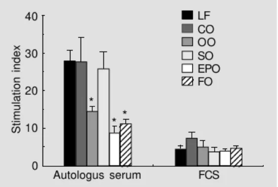
Figure 1 - Effect of dietary lipid manipulation on proliferation of rat mesenteric lymph node lym-phocytes cultured in autologous serum or foetal calf serum (FCS). Rats were fed for 10 weeks on either a low-fat diet (LF) or diets containing 200 g/kg coconut oil (CO), olive oil (OO), safflower oil (SO), evening primrose oil (EPO) or fish oil (FO). Proliferation in response to 5 µg/ml Con A was measured by incorporation of [3H]thymidine. *P<0.05
com-pared to the low-fat diet (one-way analysis of variance). Data taken from Yaqoob et al. (13), with permission of Blackwell Science Ltd.
Stimulation index
40 30 20 10 0
Autologus serum FCS LF CO OO SO EPO FO
* **
123 123
123 123 123 123 123 123 123 123 123
node lymphocytes in response to the T-cell mitogen, concanavalin A (Con A), when compared with feeding a low-fat diet or diets rich in hydrogenated coconut oil or safflower oil (13; Figure 1). The effect of the olive oil diet was similar in magnitude to those of the fish oil or evening primrose oil diets (13; Figure 1) and was also demonstrated in whole blood cultures stimulated with sub-optimal concentrations of Con A (14; Figure 2). All of the high-fat diets were shown to modulate the fatty acid composition of lymphocytes, resulting in characteristic profiles for each dietary group (15). Only one other study has examined the effects of feeding an olive oil-rich diet on lymphocyte proliferation in ro-dents. The study of Berger et al.(16) com-pared the effects of feeding a low-fat (30 g/ kg), olive oil (100 g/kg), safflower oil (100 g/ kg), linseed oil (100 g/kg) or fish plus saf-flower oil (90 + 10 g/kg) diet to dams for 5 months on the proliferation of Con A-stimu-lated murine spleen lymphocytes of their pups prior to weaning. In contrast to the studies described above, they found no ef-fect of dietary manipulation (16). There are several possible reasons for this. First, the high-fat diets used by Berger et al. (16) contained half the total amount of fat used in our studies. Second, Berger et al. fed murine dams on each of the diets and then tested lymphocyte proliferation using cells from the pups before weaning, whereas our stud-ies used rats fed for 10 weeks immediately after weaning. It is possible that in the study of Berger et al. the suckling pups may not have been exposed to milk of sufficiently differing fatty acid composition to allow dietary lipid manipulation to occur through this transition. Third, in the study by Berger et al., lymphocytes were cultured in foetal calf serum, whereas in our studies cells were cultured in autologous serum or as whole blood cultures; we have shown that culturing cells for 48 h in foetal calf serum, but not autologous serum, reverses the changes in fatty acid composition brought about by
di-etary lipid manipulation (15) and masks any effects of dietary lipid manipulation on cell function (13; Figure 1).
Since olive oil contains a number of anti-oxidants, sterols, hydrocarbons and alcohols (17), it seemed important to determine whether the effects observed following feed-ing of this diet to rats were due to oleic acid or to some other component of the oil. This was investigated using a diet rich in high-oleic acid sunflower oil (18); in earlier stud-ies, feeding the safflower oil diet had no effect on proliferation of lymph node lym-phocytes (13; Figure 1) or of whole blood cultures (14; Figure 2). The effects of feed-ing a diet containfeed-ing high-oleic acid sun-flower oil were therefore compared with the effects of feeding the low-fat, olive oil or safflower oil diet (18). The fatty acid compo-sition of spleen lymphocyte lipids was strongly affected by that of the diets fed; importantly, the high-oleic acid sunflower oil and olive oil diets resulted in a signifi-cantly higher proportion of oleic acid in
Stimulation index
800
600
400
200
0 * * *
* * *
* *
* *
1 5 10 25 50 Con A (µg/ml)
Figure 2 - The effect of dietary lipid manipulation on the proliferation of Con A-stimulated whole blood cultures. Rats were fed for 10 weeks on either a low-fat diet (LF) or diets containing 200 g/kg coconut oil (CO), olive oil (OO), safflower oil (SO), evening primrose oil (EPO) or fish oil (FO). Proliferation of whole blood (heparinised blood diluted 1:10 with culture medium containing RPMI, 2 mM glutamine and antibiotics) in response to Con A was measured by incorporation of [3H]thymidine. *P<0.05
com-pared to the low-fat diet (one-way analysis of variance). Data taken from Yaqoob et al. (14), with permission of Elsevier Science Inc.
123 123
LF CO OO SO EPO FO
123 123 123
12 12 12 12
12 12 12 12 12 12 12 12
123 123 123 123 123 123 123 123 123 123 123 123 123 123 123 123 123 123 123 123 123 123
lymphocyte lipids than the low-fat or saf-flower oil diet (18). Feeding either the olive oil or the high-oleic acid sunflower oil diet significantly decreased the proliferation of spleen lymphocytes compared with feeding the low-fat or safflower oil diet; the effects of the olive oil and high-oleic acid sunflower oil diets were not significantly different from one another (18; Figure 3). This suggests that the effects of the olive oil diet are likely to be due to oleic acid rather than to other components of olive oil.
The studies outlined above have used relatively large amounts of a single oil and as such they represent very extreme diets, which are unlikely to be encountered by free-living human beings. Furthermore, the use of such oils inevitably results in variation in the lev-els of several fatty acids together and not only of the one under investigation. A fur-ther study fur-therefore investigated the effects of relatively small changes in the levels of commonly consumed fatty acids in a con-trolled manner in which one fatty acid was exchanged for another, without altering the
levels of other fatty acids in the diet (19). The nine diets used in this study contained 178 g/kg fat, and differed in their propor-tions of palmitic, oleic, linoleic and α -lino-lenic acids whilst maintaining a constant n-6 PUFA:n-3 PUFA ratio of 7 (Table 1). The effect on lymphocyte proliferation of replac-ing one fatty acid with another appeared to be influenced by the level of other fatty acids in the diet. On further examination, there was a significant inverse linear relationship between proliferation (reported as stimula-tion index) and the oleic acid:linoleic acid ratio of the diet (r = 0.331; P = 0.028). However, the best-fit relationship was of the form:
stimulation index =
a0 + b0 log (oleic acid:linoleic acid)
where a0 = 75.8 and b0 = -18.56 (P<0.05 for both values). This relationship is illustrated in Figure 4; the authors showed that lympho-cyte proliferation is decreased with increas-ing dietary oleic acid levels up to an oleic acid level of 35.6 g/100 g fatty acids (diet E), but increasing the oleic acid level above this value does not result in any further increase (19).
Studies carried out in our laboratory have recently been extended to human work; we have performed a study to investigate the effects of consumption of a MUFA-rich diet for 2 months on immune function in healthy
Stimulation index
400 300 200 100 0
LF OO SO HOSO Diet
Figure 3 - The effect of feeding a high-oleic acid sunflower oil diet on proliferation of rat spleen lym-phocytes. Lymphocytes were cultured in autologous serum in the absence or presence of Con A; lymphocyte proliferation is expressed as stimulation index. Statistical significance is indi-cated as: avs low-fat (LF); bvs
ol-ive oil (OO); cvs safflower oil
(SO); dvs high-oleic acid
sun-flower oil (HOSO). Data taken from Jeffery et al. (18), with per-mission of S. Karger AG, Basel.
Table 1 - Fatty acid composition of diets used in the study of Jeffery et al. (19).
Fatty acid (g/100 g total fatty acids) Diet 16:0 18:1n-9 18:2n-6 18:3n-3 A 56.6 18.5 15.4 2.1 B 37.9 19.8 30.9 4.4 C 21.9 19.7 45.8 6.3 D 10.3 25.2 51.7 6.8 E 6.2 35.6 46.2 6.6 F 22.4 36.8 30.3 4.2 G 22.4 53.1 15.6 2.2 H 5.2 53.7 30.9 4.4 I 4.5 71.6 15.4 2.2
Stimulation index
120 100 80 60 40 0
OA:LA ratio of diet
1 2 3 4 5 Figure 4 - Relationship between
the oleic acid:linoleic acid (OA:LA) ratio in the diet and spleen lymphocyte proliferation in rats. Data taken from Jeffery et al. (19), with permission of the Nutrition Society.
bd
ac
bd
ac
123456789 123456789 123456789 123456789 123456789 123456789 123456789 123456789 123456789 123456789 123456789 123456789 123456789 123456789 123456789 123456789 123456789 123456789 123456789 123456789 123456789 123456789 123456789
123456789 123456789 123456789 123456789 123456789 123456789 123456789 123456789 123456789 123456789 123456789
123456789 123456789 123456789 123456789 123456789 123456789 123456789 123456789 123456789 123456789 123456789 123456789 123456789 123456789 123456789 123456789 123456789 123456789 123456789
12345678 12345678 12345678 12345678 12345678 12345678 12345678 12345678 12345678 12345678 12345678
• •
• •• • • •
middle-aged men. Middle-aged men (mean age 48 years; range 41-56 years; BMI range 21.9-30.7 kg/m2) were randomly assigned to consume either a control diet (designed to reproduce the current UK diet fatty acid composition) or a diet containing foods en-riched with highly refined olive oil for 8 weeks. Foods provided for subjects included the main meal of the day (as a frozen recipe meal), cooking oils and spreads, biscuits and puddings. Subjects on the MUFA diet con-sumed significantly less saturated fat (% energy) compared with those on the control diet and significantly more MUFA; MUFA contributed 18.4% energy in this group com-pared with 11.3% in the control group. Con-sumption of a MUFA-rich diet did not affect the proliferative response of either whole blood cultures (Figure 5) or peripheral blood mononuclear cells (PBMNC; Figure 6) to the T-cell mitogen, concanavalin A (20). This observation contrasts with results ob-tained using laboratory animals (20). The lack of a clear effect of MUFA may be attributable to the higher level of monoun-saturated fat used in the animal studies; in these studies rats were fed for 10 weeks on diets containing 200 g/kg olive oil (MUFA therefore contributed approximately 30% of total energy intake), whereas in the human study, MUFA contributed approximately 18% of the total energy intake. While it is possible that a higher level of dietary MUFA may have resulted in suppression of prolif-eration, the purpose of the study was to examine the effects of intakes which are in no way extreme; the intakes employed in the human study closely correspond to current Mediterranean intakes (21) and can readily be achieved through consumption of meals which use olive oil as the primary cooking fat.
Effects of olive oil on ex vivo natural killer cell activity
One of the most important mechanisms
by which the immune system deals with foreign cells is to damage or destroy them. Typical target cells include malignant cells, normal cells of the host that are infected with viruses or other microorganisms and normal cells from individuals unrelated to the re-sponding host. Natural killer (NK) cells are a subset of lymphocytes found mainly in blood and the spleen (22). They are derived from the bone marrow, but are neither T cells nor B cells and they do not undergo thymic maturation. Killing by NK cells is part of natural rather than specific immunity, since it is not induced by a specific antigen and is
Stimulation index
200 160 120 80 40 0
Con A (µg/ml)
20 40 60 80 100
B
Stimulation index
200
160
120
80 40 0
Con A (µg/ml)
20 40 60 80 100
A Figure 5 - Effect of MUFA con-sumption on proliferation of leukocytes in whole blood in healthy middle-aged men. A, Control group; B, MUFA group. (squares, baseline; circles, 1 month; triangles, 2 months). Pro-liferation, measured as thymi-dine incorporation, is expressed as stimulation index. Data are reported as mean ± SEM of 22-29 subjects in each group. There were no significant effects of the MUFA diet as analysed by two-way repeat-measures ANOVA. Data taken from Yaqoob et al. (20), with permis-sion.
Stimulation index
120 100 80 60 40
Baseline 20
0
1 month 2 months Control MUFA
Figure 6 - Effect of MUFA con-sumption on proliferation of PBMNC in healthy middle-aged men. Proliferation, in response to 15 µg/ml concanavalin A, was assessed as [3H]thymidine
incor-poration and is expressed as stimulation index. Data are re-ported as mean ± SEM of 21-29 subjects in each group. Signifi-cant effects of time and diet and their interactions were analysed by two-way repeat-measures ANOVA (no significant differ-ences found). Data taken from Yaqoob et al. (20), with permis-sion.
• •
• •
not restricted by MHC molecules.
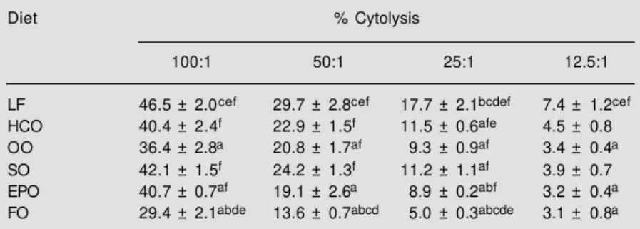
Feeding rats for 10 weeks on a diet con-taining 200 g/kg olive oil results in signifi-cant suppression of NK cell activity com-pared with feeding a low-fat diet or diets containing 200 g/kg hydrogenated coconut oil or safflower oil (23; Table 2). The inhibi-tion of NK cell activity is greater than that produced by feeding a diet rich in evening primrose oil (23; Table 2), but not as great as that resulting from feeding a diet containing 200 g/kg fish oil (23; Table 2). As with the experiments on lymphocyte proliferation de-scribed above, when the effects of high-oleic acid sunflower oil were compared with those of olive oil, NK cell activity was significant-ly lower for spleen significant-lymphocytes from rats fed the olive oil or the high-oleic acid sun-flower oil than for those from rats fed the low-fat or the safflower oil diets (18; Figure 7); the effects of the olive and high-oleic acid sunflower oils were not significantly different from one another (18; Figure 7). Once again, this suggests that the effects of the olive oil diet are likely to be due to oleic acid rather than to other components of olive oil. In a study comparing the effects of diets A-I (Table 1) on NK cell activity, there was a significant negative linear relationship be-tween the oleic acid content of the diet and NK cell activity, suggesting that dietary oleic acid causes diminished NK cell activity (19; Figure 8). Furthermore, there was a negative relationship between the oleic acid:linoleic acid ratio in the diet and NK cell activity and a weak negative relationship (r = -0.289; P = 0.092) between NK cell activity and the level of oleic acid in spleen lymphocytes (19).
The study by Berger et al. (16) also exam-ined the effects of a range of high-fat diets on NK cell activity. This study showed no ef-fect of an olive oil-rich diet on NK cell activity in mice when compared with a low-fat, safflower oil or linseed oil diet (16); once again, the reasons for the lack of effect may be attributable to the amount of fat in
Table 2 - The effect of dietary lipid manipulation on natural killer cell activity in freshly prepared rat spleen lymphocytes.
Cytolysis of YAC-1 (target) cells by rat spleen lymphocytes (effector cells) was measured by release of 51Cr by pre-loaded YAC-1 cells at various ratios of effector:target cells
(100:1, 50:1, 25:1, 12.5:1). Results are reported as % cytolysis. Statistical significance for P<0.05 is indicated as follows: avs low-fat (LF); bvs hydrogenated coconut oil (HCO); cvs olive oil (OO); dvs safflower oil (SO); evs evening primrose oil (EPO); fvs fish oil (FO).
Data taken from Yaqoob et al. (23), with permission of Elsevier Science. Diet % Cytolysis
100:1 50:1 25:1 12.5:1 LF 46.5 ± 2.0cef 29.7 ± 2.8cef 17.7 ± 2.1bcdef 7.4 ± 1.2cef
HCO 40.4 ± 2.4f 22.9 ± 1.5f 11.5 ± 0.6afe 4.5 ± 0.8
OO 36.4 ± 2.8a 20.8 ± 1.7af 9.3 ± 0.9af 3.4 ± 0.4a
SO 42.1 ± 1.5f 24.2 ± 1.3f 11.2 ± 1.1af 3.9 ± 0.7
EPO 40.7 ± 0.7af 19.1 ± 2.6a 8.9 ± 0.2abf 3.2 ± 0.4a
FO 29.4 ± 2.1abde 13.6 ± 0.7abcd 5.0 ± 0.3abcde 3.1 ± 0.8a
% Cytolysis 40 30 20 10 0
LF OO SO HOSO Diet
Figure 7 - The effect of feeding a high-oleic acid sunflower oil diet on rat spleen lymphocyte natural killer cell activity. Cytolysis of YAC-1 (target) cells by rat spleen lymphocytes (effector cells) was measured by release of 51Cr by
pre-loaded YAC-1 cells at a ratio of 100:1 effector:target cells. Results are expressed as % cy-tolysis. Statistical significance is indicated as: avs low-fat (LF); bvs
olive oil (OO); cvs safflower oil
(SO); dvs high-oleic acid
sun-flower oil (HOSO). Data taken from Jeffery et al. (18), with per-mission of S. Karger AG, Basel.
NK cell activity (% cytolysis)
45 40 35 30 25
10 20 30 40 50 60 20
70 80
Oleic acid content of diet (g/100 g fatty acids)
the diet and/or the protocol used (dams fed for 5 months and pups subsequently used prior to weaning).
In a study investigating the effects of MUFA on immune function in healthy, middle-aged men (20), consumption of the MUFA diet produced a small decrease in NK cell activity at 2 months, but not at 1 month (20; Table 3). However, this was not statistically significant when compared ei-ther with the baseline or with the control group, largely due to the small sample size for the control group at 2 months (20; Table 3). Natural killer cell activity was unaffected by consumption of the control diet (20; Table 3). As was seen with the effects of olive oil on lymphocyte proliferation, this observa-tion contrasts with animal studies, which have shown a strong suppression of NK cell activity by an olive oil-rich diet (23) and may be attributable to the higher level of mono-unsaturated fat used in the animal studies. It is interesting to note, however, that the small changes in NK cell activity and proliferation observed after 2 months of consumption of the MUFA diet were accompanied by a small, but significant increase in the proportion of oleic acid in plasma phospholipids (20) and in PBMNC (20).
Effects of olive oil on expression of adhesion molecules
Recent advances in our understanding of basic mechanisms of inflammation, of cell-cell interactions and of leukocyte trafficking have highlighted the importance of adhesive interactions between leukocytes and cellular or extracellular components of tissues. There has been a significant expansion of knowl-edge regarding the role of cell surface adhe-sion molecules in these processes over the last 10 years and a number of adhesion mol-ecules have been classified into families ac-cording to sequence homology and func-tions. It has been suggested that some adhe-sion molecules may have pathophysiologi-cal as well as physiologipathophysiologi-cal roles; some ad-hesion molecules have been implicated in the transendothelial migration of leukocytes into synovial tissue and fluid in rheumatoid arthritis (24) and in leukocyte-endothelium interactions which lead to the formation of atherosclerotic plaques (24). There has con-sequently been a great deal of interest in recent years in the potential modulation of the expression and/or functions of adhesion molecules by fatty acids, particularly those of the n-3 PUFA family. Once again, the
Table 3 - Effect of MUFA consumption on natural killer cell activity of PBMNC in healthy middle-aged men. NK cell activity was determined by measuring release of lactate dehydrogenase from target cells (K562) as a result of lysis by effector cells (PBMNC). Data are reported as mean ± SEM of the indicated number of analyses. Two-way repeat-measures ANOVA showed no significant effect of diet or time. Data taken from Yaqoob et al. (20), with permission.
% Cytolysis
Baseline 1 month 2 months E/T ratio Control MUFA Control MUFA Control MUFA
intense interest in fish oils and n-3 PUFA has overshadowed some potentially exciting ef-fects of MUFA on adhesion molecule ex-pression.
In a study by De Caterina et al. (25), human saphenous vein endothelial cells (HSVEC) were preincubated for 24 h with 10 µM arachidonic acid (AA), docosahexa-enoic acid (DHA), eicosapentadocosahexa-enoic acid (EPA) or oleic acid prior to 6 h of stimulation with tumour necrosis factor-α (TNFα) and subsequent measurement of the surface ex-pression of vascular cell adhesion molecule-1 (VCAM-molecule-1). It was reported that DHA (but not EPA) and oleic acid significantly de-creased the expression of VCAM-1 by HSVEC (25; Figure 9). The authors con-cluded that since DHA produced the greatest inhibition, further experiments in the study would focus exclusively on this fatty acid. They went on to report time- and
concentra-tion-dependent inhibition of the expression of E-selectin and intercellular adhesion mol-ecule-1 (ICAM-1) by DHA, a reduction in the accumulation of VCAM-1 mRNA and an inhibition of the adhesion of human mono-cytic cells (U937) to DHA-treated HSVEC (25). Since oleic acid had the same effect on VCAM-1 expression as DHA in the initial experiment (25; Figure 9), it is unfortunate that the remainder of the study focussed solely on DHA.
In a dietary study comparing the effects of low-fat (25 g/kg) and high-fat diets con-taining 200 g/kg hydrogenated coconut oil, olive oil, safflower oil, evening primrose oil or fish oil, the level of expression of the adhesion molecules CD2, ICAM-1 and LFA-1 on rat spleen lymphocytes was decreased by both olive oil and fish oil (26). In the human study carried out by our group (20), a MUFA-rich diet resulted in a significant de-crease in the expression of the leukocyte adhesion molecule, ICAM-1, after 2 months of consuming the diet compared both with baseline values and with those from the con-trol group (20; Table 4). The expression of ICAM-1 did not change during the consump-tion of the control diet (20; Table 4). The MUFA diet also decreased the expression of a monocyte/macrophage-associated adhesion molecule, Mac-1, by approximately 25%
500 400 300 200 100 0
VCAM-1 expression (mU of absorbance)
Control OA AA EPA DHA Figure 9 - Effect of preincubation
(24 h) with fatty acids on surface expression of VCAM-1 by HSVEC in response to stimula-tion (6 h) with TNFα. VCAM-1 expression is measured in mU of absorbance by a cell surface enzyme immunoassay. Expres-sion of VCAM-1 by cells preincu-bated with oleic acid was signifi-cantly lower than in control or AA-treated cells and expression of VCAM-1 by cells preincubated with DHA was significantly lower than that of all other groups, apart from OA-treated cells. OA, Oleic acid; AA, arachidonic acid; EPA, eicosapentaenoic acid; DHA, do-cosahexaenoic acid. Data taken from De Caterina et al. (25), with permission of the American Heart Association.
Table 4 - Effect of MUFA consumption on expression of surface molecules on PBMNC in healthy middle-aged men.
Data are reported as mean ± SEM of the indicated number of analyses. ‘ns’ indicates no significant effect of time or diet or interaction between time and diet as analysed by two-way repeat-measures ANOVA. ‘a’ denotes a significant effect of diet (P = 0.035) and time (P = 0.049), with a significant interaction (P = 0.046). Significant differences between groups (Student t-test) are indicated as follows: *P<0.05 vs control group;
+P<0.01 vs baseline. Data taken from Yaqoob et al. (20), with permission.
% Marker-positive cells
Baseline 1 month 2 months
Control MUFA Control MUFA Control MUFA ANOVA N = 18-20 N = 19-20 N = 18-23 N = 20-23 N = 20-23 N = 19-21
CD54 19.0 ± 1.3 20.8 ± 1.4 19.1 ± 1.2 16.4 ± 1.4 20.0 ± 1.5 15.9 ± 1.1*+ a
CD11b 15.5 ± 1.2 17.0 ± 1.8 15.4 ± 1.4 15.4 ± 1.3 13.7 ± 1.3 12.6 ± 1.5 ns
compared with baseline values. However, this decrease was not statistically signifi-cant, either compared with the baseline or with the control group (20; Table 4). The decrease in the expression of ICAM-1 by the MUFA diet is an important finding. ICAM-1 is a member of the Ig superfamily of adhe-sion molecules and is involved in leukocyte-leukocyte adhesion (27) as well as adhesion of leukocytes to endothelial cells (24) and to fibrinogen, a plasma adhesive protein (28). ICAM-1 is expressed on mononuclear cells that infiltrate inflamed synovium in patients suffering from rheumatoid arthritis (29) and in some cases, such patients may also have high levels of serum soluble ICAM-1 (29). The formation of plaques in atherosclerosis shows many features which are common to the inflammation seen in rheumatoid arthri-tis, such as adhesive interactions between endothelial cells and leukocytes and extravas-cular leukocyte accumulation and ICAM-1 is thought to play a pivotal role in the recruit-ment of mononuclear cells to, and therefore the growth of, the atherosclerotic plaque (30). Recent evidence suggests that a MUFA-rich diet may indeed affect the process of cellular adhesion. In an interesting study by Mata et al. (31), healthy men and women living in a religious community were sub-jected to four consecutive dietary periods (isocaloric) differing in the fat content of SFA, MUFA and n-3 and n-6 PUFA. It was reported that LDL-induced monocyte adhe-sion to endothelial cells was lower during the MUFA period than each of the others and that resistance of LDL to oxidation was great-est during the MUFA period (31). The au-thors suggest that the modulation of LDL fatty acid composition was responsible for the differences in adhesion and showed a significant negative correlation between monocyte adhesion to endothelial cells and the oleic acid content of LDL (31). Expres-sion of adheExpres-sion molecules by either cell type was not measured, but it is possible that this too may have played some role in the
decreased adhesion during the MUFA pe-riod.
The fact that a high MUFA diet appears to decrease the expression of ICAM-1 on circulating PBMNC by up to 20% opens up the exciting possibility that the low preva-lence of atherosclerosis, and perhaps other inflammatory conditions, in Mediterranean populations may at least partly involve the effects of dietary MUFA on adhesion mole-cule expression; this is a potentially impor-tant area which clearly deserves further ex-ploration.
Effects of olive oil on in vivo immune responses
The ex vivo studies described so far, whilst
other diets (32). There was also no increase in the rate of protein synthesis in response to TNFα in the livers of animals fed the butter diet, whereas animals fed on the corn oil and coconut oil diets demonstrated the normal increase in protein synthesis associated with the acute-phase response (32). In a subse-quent study, it was demonstrated that diets containing 50 or 100 g/kg butter or olive oil completely suppressed the increases in tis-sue zinc content, liver protein synthesis and serum caeruloplasmin levels in response to subcutaneous Escherichia coli endotoxin, when compared with a maize oil diet or standard laboratory chow (33). In both stud-ies, the butter and olive oil diets decreased the intensity of anorexia induced by TNFα or endotoxin (33), demonstrating clearly the diminished susceptibility to the lethal effects of both agents in experimental animals.
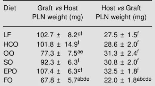
An alternative experimental model for in vivo immune responses is the ‘graft versus host’ (Gv.H) response, which can be elicited in rodents by injection of allogenic cells into the footpad of a host. The response primarily involves the polyclonal activation and pro-liferation of host B cells. The ‘host versus
graft’ (Hv.G) response, on the other hand, is a T cell-mediated response, in which cyto-toxic T lymphocytes of the host recognise MHC antigens on the injected cells. In both cases the enlargement of the popliteal lymph nodes (more than 15-fold in the Gv.H re-sponse and 4-fold in the Hv.G rere-sponse) is due largely to proliferation of activated host cells within the lymph node, although there is also recruitment of cells from the blood-stream. Using this assay, Sanderson et al. (34) demonstrated that feeding rats a diet containing 200 g/kg fish oil suppressed the Gv.H response compared with feeding a low-fat diet or diets containing 200 g/kg coconut oil, safflower oil or evening primrose oil; feeding a diet containing 200 g/kg olive oil had a similar effect, although the response was depressed only compared with the low-fat and evening primrose oil diets (34; Table 5). The expression of the adhesion mol-ecules LFA-1 and ICAM-1 on lymphocytes from popliteal lymph nodes following a Gv.H response was significantly lower in animals fed the olive oil or fish oil diet compared with those fed the low-fat or coconut oil diet (34). It was speculated that the smaller popliteal lymph node size in animals fed the fish oil or olive oil diet may result from a suppression of both the activation of cells within the node and of the movement of cells from the bloodstream into the nodes (34). Interestingly, while the fish oil diet had a similar suppressive effect on the Hv.G re-sponse as that on the Gv.H rere-sponse, the olive oil diet had no effect on the Hv.G response (33; Table 5); it appears, therefore, that in this model, olive oil is able to modu-late in vivo responses involving B cells, but not those involving cytotoxic T lymphocytes. Although there are no published human studies which have set out to examine the effects of olive oil on in vivo immune re-sponses, at least one study investigating the effects of fish oil supplements on immuno-logical parameters (including the systemic humoral response to tetanus toxoid) in healthy
Table 5 - Effect of dietary lipids on popliteal lymph node (PLN) weight following the graft versus host and host versus graft response in the rat. This in vivo immune response was measured as described in the text. Statistical significance (one-way ANOVA) for P<0.05 at least is indicated as follows: avs LF, bvs HCO, cvs OO, dvs SO, evs
EPO, fvs FO. LF, Low-fat; HCO, hydrogenated
coconut oil; OO, olive oil; SO, safflower oil; EPO, evening primrose oil; FO, fish oil. Data taken from Sanderson et al. (34), with permission of Academ-ic Press, Inc.
Diet Graft vs Host Host vs Graft PLN weight (mg) PLN weight (mg) LF 102.7 ± 8.2cf 27.5 ± 1.5f
HCO 101.8 ± 14.9f 28.6 ± 2.0f
OO 77.3 ± 7.5ae 31.3 ± 2.4f
SO 92.3 ± 6.3f 30.8 ± 2.0f
EPO 107.4 ± 6.3cf 32.5 ± 1.8f
volunteers has used olive oil as a placebo treatment (5). The authors claim to demon-strate an immunosuppressive effect of fish oil compared with olive oil, but the protocol is far from satisfactory. Six volunteers were involved in the study, only two of whom received the olive oil treatment and if the data are scrutinised, it is clear that a larger number of subjects may have produced dif-ferent results (5), particularly since meas-urements of human immune responses are prone to substantial inter-individual variation.
Olive oil and autoimmune disorders
A much-cited study by Linos et al. (10) has suggested that there may be beneficial effects of olive oil consumption on rheuma-toid arthritis (RA). This study compared the relative risk of development of RA in rela-tion to lifelong consumprela-tion of olive oil in a Greek population and demonstrated that high consumers of olive oil (almost every day throughout life) were 4 times less likely to develop RA than those who consumed olive oil less than 6 times per month on average throughout their lives (10). Interestingly, the effect of fish consumption on relative risk for RA was also tested, but the effect was not statistically significant (10). The study, al-though of great interest, has the drawback that the population studied consisted of a very large proportion of high consumers of olive oil. However, there is further evidence that olive oil may have beneficial effects relating to RA. In a study by Kremer et al. (9) examining the effects of fish oil supplemen-tation on the severity and progression of RA, olive oil was used as a placebo treatment, but clinical evaluations and immunologic tests showed it to have effects which were similar to those of fish oil. A total of 5 out of 45 clinical measures were significantly changed from baseline in the olive oil group, 8 out of 45 in a low-dose fish oil group and 21 out of 45 in a high-dose fish oil group (9). Produc-tion of interleukin-1 by macrophages was
decreased in the olive oil group, although not to the same extent as either of the fish oil groups (9). The authors concluded that “ di-etary supplementation with olive oil is also associated with certain changes in immune function, which require further
investiga-tion.”
Conclusion
Animal studies, depending on the proto-col, species and type of measurement, gener-ally support the idea that olive oil is capable of modulating functions of cells of the im-mune system. The effects appear to be simi-lar to, albeit weaker than, those seen follow-ing feedfollow-ing of diets containfollow-ing fish oils. There is some evidence that the effects of olive oil on immune function in animal studies are due to oleic acid rather than to trace elements or antioxidants. Importantly, several studies have demonstrated effects of oleic acid-con-taining diets on in vivo immune responses.
In contrast, consumption of a MUFA-rich diet by humans does not appear to bring about a general suppression of immune cell functions. The effects of this type of diet in humans are limited to decreasing the expres-sion of adheexpres-sion molecules on PBMNC (20) and decreasing LDL-induced adhesion of monocytes to endothelial cells (31), although there are trends towards decreases in NK cell activity and proliferation (20). The lack of a clear effect of MUFA in humans may be attributable to the higher level of monoun-saturated fat used in the animal studies, as discussed previously; however, it is ultimately of importance to examine the effects of in-takes which are in no way extreme. The intakes employed in the two human studies discussed closely correspond to current Medi-terranean intakes and can readily be achieved through consumption of meals which use olive oil as the primary cooking fat.
antioxi-dants varied between the diets and/or sub-jects cannot be excluded. Therefore, the sug-gestion that the effects observed in these studies are due to specific modulation of dietary oleic acid is favourable (given the changes in fatty acid composition in both), but not conclusive. Similarly, it is extremely difficult to determine conclusively whether
the effects observed are indeed due to an increased level of MUFA or to a decreased level of SFA. The effects of MUFA on adhe-sion molecules are potentially important, since they appear to have a role in the pathol-ogy of a number of diseases involving the immune system. This area clearly deserves further exploration.
References
1. Nestle M (1995). Mediterranean diets: his-torical and research overview. American Journal of Clinical Nutrition, 61 (Suppl): 1313S-1320S.
2. Keys A (1970). Coronary heart disease in seven countries. Circulation, 41: 1-211. 3. Mensink RP & Katan MB (1987). Effect of
monounsaturated fatty acids versus com-plex carbohydrates on high-density lipo-proteins in healthy men and women. Lan-cet, i: 122-124.
4. Mata P, Alvarez-Silva LA, Rubio MJ, Nuno J & De Oya M (1992). Effects of long-term monounsaturated vs polyunsaturated-en-riched diets on lipoproteins in healthy men and women. American Journal of Clinical Nutrition, 55: 846-850.
5. Virella G, Fourspring K, Hyman B, Haskill-Stroud R, Long L, Virella I, La Via M, Gross AJ & Lopes-Virella M (1991). Immunosup-pressive effects of fish oil in normal hu-man volunteers: correlation with the in vitro effects of eicosapentaenoic acid on human lymphocytes. Clinical Immunology and Immunopathology, 61: 161-176. 6. Cleland LG, French JK, Betts WH, Murphy
GA & Elliot MJ (1988). Clinical and bio-chemical effects of dietary fish oil supple-ments in rheumatoid arthritis. Journal of Rheumatology, 15: 1471-1475.
7. Dehmer GJ, Popma JJ, van den Berg EK, Eichhorn EJ, Prewitt JB, Campbell WB, Jennings L, Willerson JT & Schmitz JM (1988). Reduction in the rate of early re-stenosis after coronary angioplasty by a diet supplemented with n-3 fatty acids. New England Journal of Medicine, 319: 733-740.
8. Milner MR (1989). Fish oil for preventing coronary restenosis. Lancet, II: 693. 9. Kremer JM, Lawrence DA, Jubiz W,
DiGiacomo R, Rynes R, Bartholomew LE & Sherman M (1990). Dietary fish oil and olive oil supplementation in patients with rheumatoid arthritis. Arthritis and Rheu-matism, 33: 810-820.
10. Linos A, Kaklamanis E, Kontomerkos A, Koumantaki Y, Gazi S, Vaiopoulos G,
Tsokos GC & Kaklamanis PH (1991). The effect of olive oil and fish consumption on rheumatoid arthritis - a case control study. Scandinavian Journal of Rheumatology, 20: 419-426.
11. Yaqoob P & Calder PC (1993). The effects of fatty acids on lymphocyte functions. International Journal of Biochemistry, 25: 1705-1714.
12. Gurr MI (1983). The role of lipids in the regulation of the immune system. Prog-ress in Lipid Research, 22: 257-287. 13. Yaqoob P, Newsholme EA & Calder PC
(1994). The effect of dietary lipid manipu-lation on rat lymphocyte subsets and pro-liferation. Immunology, 82: 603-610. 14. Yaqoob P, Newsholme EA & Calder PC
(1995). The effect of fatty acids on leuko-cytes subsets and proliferation in whole blood. Nutrition Research, 15: 279-287. 15. Yaqoob P, Newsholme EA & Calder PC
(1995). Influence of cell culture conditions on diet-induced changes in lymphocyte fatty acid composition. Biochimica et Bio-physica Acta, 1255: 333-340.
16. Berger A, German JB, Chiang BL, Ansari AA, Keen CL, Fletcher MP & Gershwin MR (1993). Influence of feeding unsatur-ated fats on growth and immune status of mice. Journal of Nutrition, 123: 225-233. 17. Gunstone FD, Harwood JL & Padley FB
(1994). The Lipid Handbook. 2nd edn. Chapman & Hall, London, 79-82. 18. Jeffery NM, Yaqoob P, Newsholme EA &
Calder PC (1996). The effects of olive oil upon rat serum lipid levels and lympho-cyte functions are due to oleic acid. An-nals of Nutrition and Metabolism, 40: 71-80.
19. Jeffery NM, Cortina M, Newsholme EA & Calder PC (1997). Effects of variations in the proportions of saturated, monounsat-urated and polyunsatmonounsat-urated fatty acids in the rat diet on spleen lymphocyte func-tions. British Journal of Nutrition, 77: 805-823.
20. Yaqoob P, Knapper JA, Webb DH, Wil-liams CM, Newsholme EA & Calder PC
(1998). The effect of olive oil consump-tion on immune funcconsump-tions in middle-aged men. American Journal of Clinical Nutri-tion (in press).
21. Ferro-Luzzi A & Branca F (1995). The Mediterranean diet, Italian style: proto-type of a healthy diet. American Journal of Clinical Nutrition, 61 (Suppl): 1338S-1345S.
22. Herberman RB (1988). Lymphocytes: Cy-totoxic activities. In: Gallin JI, Goldstein IM & Snyderman R (Editors), Inflamma-tion: Basic Principles and Clinical Corre-lates. Raven Press, New York, 613-630. 23. Yaqoob P, Newsholme EA & Calder PC
(1994). Inhibition of natural killer cell activ-ity by dietary lipids. Immunology Letters, 41: 241-247.
24. Munro JM (1993). Endothelial-leukocyte adhesive interactions in inflammatory dis-eases. European Heart Journal, 14 (Suppl K): 72-77.
25. De Caterina R, Cybulsky MI, Clinton SK, Gimbrone Jr MA & Libby P (1994). The omega-3 fatty acid docosahexaenoate re-duces cytokine-induced expression of proatherogenic and proinflammatory pro-teins in human endothelial cells. Arterio-sclerosis and Thrombosis, 14: 1829-1836. 26. Sanderson P, Yaqoob P & Calder PC (1995). Effects of dietary lipid manipula-tion upon rat spleen lymphocyte funcmanipula-tions and the expression of lymphocyte surface molecules. Journal of Nutritional and En-vironmental Medicine, 5: 119-132. 27. Chapman PT & Haskard DO (1995).
Leu-kocyte adhesion molecules. British Medi-cal Bulletin, 51: 296-311.
28. Languino LR, Duperray A, Joganic KJ, Fornaro M, Thornton GB & Altieri DC (1995). Regulation of leukocyte-endothe-lium interaction and leukocyte transendo-thelial migration by intercellular adhesion molecule 1-fibrinogen recognition. Pro-ceedings of the National Academy of Sci-ences, USA, 92: 1505-1509.
arthri-tis. Current Opinion in Rheumatology, 6: 300-304.
30. Poston RN, Haskard DO, Coucher JR, Gall NP & Johnson-Tidy RR (1992). Expression of intercellular adhesion molecule-1 in ath-erosclerotic plaques. American Journal of Pathology, 140: 665-673.
31. Mata P, Alonso R, Lopez-Farre A, Ordovas JM, Lahoz C, Garces C, Caramelo C, Codoceo R, Blazquez E & de Oya M
(1996). Effect of dietary fat saturation on LDL oxidation and monocyte adhesion to human endothelial cells in vitro. Arterio-sclerosis, Thrombosis, and Vascular Biol-ogy, 16: 1347-1355.
32. Mulrooney HM & Grimble RF (1993). In-fluence of butter and of corn, coconut and fish oils on the effects of recombinant human tumour necrosis factor-α in rats. Clinical Science, 84: 105-112.
33. Besler HT & Grimble RF (1995). Compari-son of the modulatory influence of maize and olive oils and butter on metabolic re-sponses to endotoxin in rats. Clinical Sci-ence, 88: 59-66.