Effect of bullfrog (
Rana catesbeiana
)
oil administered by gavage on the fatty
acid composition and oxidative stress
of mouse liver
Departamentos de 1Genética e Morfologia, and 2Biologia Celular,
Instituto de Ciências Biológicas, Universidade de Brasília, Brasília, DF, Brasil 3Departamento de Alimento e Nutrição, Faculdade de Engenharia de Alimentos, Universidade Estadual de Campinas, Campinas, SP, Brasil
4Centro de Ciências Biológicas e Saúde, Universidade Cruzeiro do Sul, São Paulo, SP, Brasil
5Departamento de Fisiologia e Biofísica, Instituto de Ciências Biomédicas, Universidade de São Paulo, São Paulo, SP, Brasil
L.P. Silva1, C.K. Miyasaka3, E.F. Martins4, J.R.S.A. Leite2, Z.G.M. Lacava1, R. Curi5 and R.B. Azevedo1
Abstract
The aim of the present study was to investigate the effects of daily intragastric administration of bullfrog oil (oleic, linoleic and palmitoleic acid-rich oil), corresponding to 0.4% of body weight for four weeks, on fatty acid composition and oxidative stress (lipid peroxidation and catalase activity) in mouse liver. The activities of aspartate ami-notransferase (AST), alkaline phosphatase (ALP), alanine aminotrans-ferase (ALT), and gamma-glutamyltransaminotrans-ferase (GGT), biomarkers of tissue injury, were determined in liver homogenates and serum. The proportions of 18:2n-6, 20:4n-6, 20:5n-3, and 22:6n-3 (polyunsatu-rated fatty acids,from 37 to 60%) in the total fatty acid content were increased in the liver of the bullfrog oil-treated group (P < 0.05) compared to control. At the same time, a significant decrease in the relative abundance of 14:0, 16:0, and 18:0 (saturated fatty acids,from 49 to 25%) was observed. The hepatic content of thiobarbituric acid reactive substances (TBARS) was increased from 2.3 ± 0.2 to 12.3 ± 0.3 nmol TBA-MDA/mg protein and catalase activity was increased from 840 ± 32 to 1110 ± 45 µmol reduced H2O2 min-1 mg protein-1 in
the treated group. Bullfrog oil administration increased AST and ALP activities in the liver (from 234.10 ± 0.12 to 342.84 ± 0.13 and 9.38 ± 0.60 to 20.06 ± 0.27 U/g, respectively) and in serum (from 95.41 ± 6.13 to 120.32 ± 3.15 and 234.75 ± 11.5 to 254.41 ± 2.73 U/l, respectively), suggesting that this treatment induced tissue damage. ALT activity was increased from 287.28 ± 0.29 to 315.98 ± 0.34 U/g in the liver but remained unchanged in serum, whereas the GGT activity was not affected by bullfrog oil treatment. Therefore, despite the interesting modulation of fatty acids by bullfrog oil, a possible therapeutic use requires care since some adverse effects were ob-served in liver.
Correspondence
R.B. Azevedo
Departamento de Genética e Morfologia, Instituto de Biologia Universidade de Brasília 70919-970 Brasília, DF Brasil
Fax: +55-61-349-6167 E-mail: [email protected]
Research supported by CNPq and PRONEX.
Publication supported by FAPESP.
Received August 7, 2003 Accepted July 12, 2004
Several studies have shown the influence of dietary fat on fatty acid (FA) composition and oxidative stress in various tissues (1-8). The proportion of FA in the diet is of great physiological importance since it can modu-late the composition of FA in cell membrane phospholipids (1,2). This is due to the com-petition among FA families for elongation and desaturation enzymes. Some investiga-tors have postulated that the unsaturated/ saturated FA (SFA) ratio of the diet is an important factor for determining the intrin-sic oxidative potential of tissues (3).
Oils of plant and animal origin have been used for the treatment of immune and inflam-matory disorders (2). These oils contain a mixture of different types of SFA, monounsat-urated FA (MUFA) and polyunsatmonounsat-urated FA (PUFA). Bullfrog oil contains a relatively low amount of SFA (17% of total FA) and PUFA (28% of total FA) and is rich in MUFA (55% of total FA). The sum of MUFA and PUFA accounts for 83% of total FA in this oil and makes it a candidate for therapeutic pur-poses (2). However, there is no report on the effects of bullfrog oil on animals.
In the present study, the effects of the MUFA-rich bullfrog oil on FA composition and oxidative stress (lipid peroxidation and catalase activity) were investigated in mouse liver. The liver and serum activities of aspar-tate aminotransferase (AST), alanine ami-notransferase (ALT), alkaline phosphatase (ALP), and gamma-glutamyltransferase (GGT) were also determined to assess the occurrence of injury in the organ. Bullfrogs can be easily grown to provide meat and perhaps oil for therapeutic purposes.
Male Swissmice (25-30 g) were divided into two groups of 6 animals each fed Purina commercial chow (5% fat), with free access to water. The number of animals used in this study was sufficient as indicated by the small standard error of the means and by previous work using the same methodology (5).
The animals received sterile saline solu-tion (control group,) or bullfrog oil
intragas-trically daily for 4 weeks. The FA composi-tion of bullfrog oil (%) is presented in Table 1. The amount of oil (0.4% of body weight) administered as a supplement was recalcu-lated daily on the basis of changes in body weight assuming that the daily food intake of a mouse is approximately 10% of body weight, which corresponds to 0.4-0.5 g fat. Therefore, the supplementation of oil at 0.4% body weight increased by 2-fold the amount of ingested fat. Bullfrog oil was given by gavage to avoid the oxidation/peroxidation of FA which occurs during the preparation of a fat-rich diet. At the end of the study period, mice were killed by decapitation and livers were removed and stored at -80ºC.
For total lipid extraction, frozen samples were homogenized in methanol and chloro-form, followed by the addition of an aqueous solution of KCl; after vortexing, the upper layer phase was discarded. The FA were saponified with an alcoholic solution of NaOH and then extracted twice with HCl and hexane as described elsewhere (9). FA were derivatized with 4-bromomethyl-7-cou-marin and the composition was analyzed by high performance liquid chromatography (Shimadzu model LC-10A, Kyoto, Japan) using a C8 column with a C8 pre-column, eluted with 1 ml/min acetonitrile/water (77:23, by volume) and the effluent was monitored with a fluorescence detector (325-nm excitation and 395-(325-nm emission) (9). FA used as standards were obtained from Sigma (St. Louis, MO, USA). Intra- and interassay coefficients of variation (CV) were less than 8%. FA composition is presented in Table 1 as percent of the total identified FA.
phos-phate buffer containing butylated hydroxy-toluene and 2-thiobarbituric acid. After in-cubation at 100ºC for 30 min, the mixture was cooled and the TBARS adducts were extracted with butanol and centrifuged at 1600 g at 4°C for 30 min. The supernatant phase (butanol phase) was collected and ab-sorbance at 532 nm was measured with a Pharmacia Biotech Ultrospec 3000 Spectro-photometer (Uppsala, Sweden) (11). TBARS are reported as µmol/mg protein using a molar absorptivity of 1.56 x 10⁻⁵ M-1 ml-1.
For the determination of hepatic catalase activity, samples were homogenized in phos-phate buffer and centrifuged, and the super-natant was used for analysis. Catalase activ-ity was determined in duplicate by monitor-ing the decomposition of H2O2 at 30ºC with
a UV visible spectrophotometer at 230 nm (12). Catalase specific activity is reported as µmol reduced H2O2 min-1 mg protein-1. The
protein content of the liver homogenates was measured by the method of Bradford (13).
ALT, AST, ALP, and GGT activities were determined in serum and liver using a Bayer ADVIA 1650 clinical chemistry analyzer (Lerverkusen, Germany) according to manu-facturer instructions. The liver was homog-enized in 0.2 g/ml phosphate-buffered sa-line, pH 7.4. Blood samples were collected into test tubes and allowed to stand for 30 min to clot before centrifugation at 300 g for 10 min. The serum was separated, trans-ferred to other tube, and stored at 4ºC for subsequent analysis.
All data are reported as means ± SEM. The results were submitted to one-way ANOVA, and means were compared between groups by Scheffé’s test. Results were considered statis-tically significant when P < 0.05.
The dose of bullfrog oil administered did not cause diarrhea or any other visible clini-cal symptoms in the animals. Mice of both groups were healthy and had similar final body weights (27.2 ± 0.6 for controls and 26.3 ± 0.3 g for bullfrog oil-treated animals). Liver FA composition was changed by
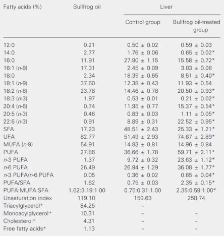
bullfrog oil administration (Table 1). The proportion of SFA was decreased by 48%, with a significant reduction in the relative abundance of 14:0 (63%), 16:0 (44%), and 18:0 (54%; P < 0.05). Conversely, the pro-portion of PUFA was increased 62% (P < 0.05) and the PUFA/SFA ratio was increased 213% (P < 0.05). The most altered PUFA class was n-3 PUFA, which increased 143% (P < 0.05). The significant (P < 0.05) in-crease of n-6 PUFA was much smaller (34%). The n-3 PUFA/n-6 PUFA ratio was also increased 81% (P < 0.05). Among the n-3
Table 1. Fatty acid composition of bullfrog oil and of the liver of bullfrog oil-treated mice.
Fatty acids (%) Bullfrog oil Liver
Control group Bullfrog oil-treated group
12:0 0.21 0.50 ± 0.02 0.59 ± 0.03
14:0 2.77 1.76 ± 0.06 0.65 ± 0.02*
16:0 11.91 27.90 ± 1.15 15.58 ± 0.72*
16:1 (n-9) 17.31 2.45 ± 0.09 3.03 ± 0.08
18:0 2.34 18.35 ± 0.65 8.51 ± 0.40*
18:1 (n-9) 37.60 12.38 ± 0.43 11.93 ± 0.54
18:2 (n-6) 23.78 14.46 ± 0.78 20.50 ± 0.93*
18:3 (n-3) 1.97 0.53 ± 0.01 0.21 ± 0.02*
20:4 (n-6) 0.74 11.95 ± 0.77 15.37 ± 0.54*
20:5 (n-3) 0.46 0.83 ± 0.03 1.11 ± 0.05*
22:6 (n-3) 0.91 8.89 ± 0.31 22.52 ± 0.95*
SFA 17.23 48.51 ± 2.43 25.33 ± 1.21*
UFA 82.77 51.49 ± 2.93 74.67 ± 2.89*
MUFA (n-9) 54.91 14.83 ± 0.81 14.96 ± 0.84
PUFA 27.86 36.66 ± 1.78 59.71 ± 2.11*
n-3 PUFA 1.37 9.72 ± 0.32 23.63 ± 1.12*
n-6 PUFA 26.49 26.94 ± 1.29 36.08 ± 1.77*
n-3 PUFA/n-6 PUFA 0.05 0.36 ± 0.02 0.65 ± 0.04*
PUFA/SFA 1.62 0.75 ± 0.03 2.35 ± 0.15*
PUFA:MUFA:SFA 1.62:3.19:1.00 0.75:0.31:1.00 2.35:0.59:1.00*
Unsaturation index 119.10 150.63 258.74
Triacylglycerol+ 84.25
-Monoacylglycerol+ 10.31 -
-Cholesterol+ 4.31 -
-Free fatty acids+ 1.13 -
-Mice (25-30 g) received the oil (0.4% of body weight) by gavage daily for 4 weeks. Data are reported as the mean ± SEM of percent recovered fatty acids by weight for three determinations in each of the 6 mice in each group. SFA = sum of saturated fatty acids (FA); UFA = sum of unsaturated FA; MUFA = sum of monounsaturated FA; PUFA: sum of polyunsaturated FA; n-3 PUFA = sum of n-3 PUFA; n-6 PUFA = sum of
n-6 PUFA. +Lipid class composition (%).
PUFA, the liver of bullfrog oil-treated ani-mals had a higher proportion (P < 0.05) of 22:6n-3 (153%) and 20:5n-3 (34%) and a decreased proportion of 18:3n-3 (60%). A 29 and 42% increase of 20:4n-6 and 18:2n-6 was observed in n-6 PUFA, respectively (P < 0.05). No significant differences were observed in the proportion of MUFA between groups.
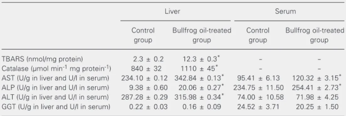
TBARS content (Table 2) in the liver of bullfrog oil-treated animals changed from 2.3 ± 0.2 to 12.3 ± 0.3 nmol TBA-MDA formed per mg protein (435%). Catalase ac-tivity (Table 2) was increased by bullfrog oil administration from 840 ± 32 to 1110 ± 45 µmol reduced H2O2 min-1 mg protein-1 (32%).
AST, ALT, and ALP activities were in-creased by 46, 10, and 114%, respectively, in the liver of bullfrog oil-treated animals com-pared to the control (P < 0.05). There was a statistically significant 26 and 8% increase in AST and ALP activities, respectively, in the serum of bullfrog oil-treated mice (Table 2), but no significant effect on ALT activity in serum, or on GGT activity in liver and serum (Table 2).
As previously mentioned, bullfrog oil is a MUFA-rich animal-derived oil. MUFA (n-9 and n-7) account for 55% of total FA,
con-sisting of 38% oleic acid (18:1n-9) and 17% palmitoleic acid (16:1n-7). Linoleic acid (18:2n-6) was the second most abundant FA (24%) and accounted for almost 100% of n -6 FA in bullfrog oil. Bullfrog oil was poor in linolenic acid (18:3n-3) and in FA contain-ing 20 or more carbons (Table 1). Although bullfrog oil is similar to canola oil in terms of total PUFA and MUFA, the former contains almost 100% more oleic acid and 350% more linolenic acid (ALA), as well as about 200% less total SFA than the latter.
The influence of dietary lipids on liver FA composition has been demonstrated in exper-imental animal studies using different oils (1,14,15). In the present study, intragastric administration of bullfrog oil caused an in-crease in long-chain PUFA (20:4n-6, 20:5n-3, and 22:6n-3) and a decrease in SFA (14:0, 16:0, and 18:0) in the liver. Bullfrog oil led to an increase in total liver PUFA, n-3 PUFA, and n-6 PUFA, concomitant with a decrease in total SFA. Total MUFA content did not differ between groups, although bullfrog oil-treated liver had a higher relative content of these FA. Arachidonic acid (AA), eicosapentaenoic acid (EPA), and docosahexaenoic acid (DHA) are PUFA obtained from the diet or
Table 2. Effect of intragastric bullfrog oil administration on TBARS, catalase, alkaline phosphatase, and aminotransferase activities of mouse liver and serum. Bullfrog oil administration is described in the legend to Table 1.
Liver Serum
Control Bullfrog oil-treated Control Bullfrog oil-treated
group group group group
TBARS (nmol/mg protein) 2.3 ± 0.2 12.3 ± 0.3* -
-Catalase (µmol min-1 mg protein-1) 840 ± 32 1110 ± 45* -
-AST (U/g in liver and U/l in serum) 234.10 ± 0.12 342.84 ± 0.13* 95.41 ± 6.13 120.32 ± 3.15*
ALP (U/g in liver and U/l in serum) 9.38 ± 0.60 20.06 ± 0.27* 234.75 ± 11.50 254.41 ± 2.73*
ALT (U/g in liver and U/l in serum) 287.28 ± 0.29 315.98 ± 0.34* 74.00 ± 10.58 71.98 ± 4.25
GGT (U/g in liver and U/l in serum) 0.22 ± 0.03 0.16 ± 0.09 24.52 ± 3.71 20.25 ± 1.50
Data are reported as the mean ± SEM of three determinations in each of the 6 mice in each group. AST = aspartate aminotransferase; ALT = alanine aminotransferase; ALP = alkaline phosphatase; GGT = gamma-glutamyltransferase; TBARS = thiobarbituric acid reactive substances. One unit of the aminotransferases and ALP is defined as the amount of enzyme required to produce 1 µmol of NAD or p-nitrophenol, respectively, per minute under the conditions of the assay.
synthesized from their direct precursors, linoleic acid for AA and ALA for EPA and DHA, in a process involving elongase/de-saturase enzymes. Therefore, the relative in-crease of AA in the liver of bullfrog oil-treated animals may be explained by direct incorporation and/or by formation from its precursor, since it is present in relatively high amounts in bullfrog oil compared to other oils. Similarly, the increase in EPA and DHA may be explained by direct incorpora-tion and/or synthesis from ALA present in bullfrog oil. An increase in the proportions of total n-3 PUFA in brain and liver (includ-ing EPA and DHA) has been observed in a number of studies involving feeding of olive oil, which contains a low proportion of n-3 PUFA and its precursors (15,16). Interest-ingly, in the present study the incorporation of n-9 FA was very low. Weber et al. (17), studying the metabolism of mead acid (20:3n -9), observed an accumulation of n-3 and n-6 products such as DHA, EPA, and AA, as also observed in the present study. These investigators proposed that a feedback mech-anism might down-regulate the n-9 pathway in the liver, preventing further conversion of dietary precursors and/or direct incorpora-tion. In this case, n-9 FA catabolism might be stimulated. Sprecher et al.(18) studying
n-6 FA, also suggested an inverse relation-ship between rates of peroxisomal ß-oxida-tion and FA esterificaß-oxida-tion.
Increased catabolism via peroxisomal ß-oxidation leads to an in vivo increase of hydrogen peroxide production that is poten-tially toxic to the cell. Catalase is predomi-nantly found in the peroxisomes of most cells and converts hydrogen peroxide to oxy-gen and water, preventing oxidative stress and playing an antioxidant defense role (19). Supporting the postulated increase in per-oxisomal ß-oxidation in bullfrog oil-treated animals, there was an increase in liver cata-lase activity. The increase in the activity of this enzyme is perhaps an attempt to protect against hydrogen peroxide toxicity.
An increase in the proportion of PUFA in the membranes and of the unsaturation in-dex was directly associated with the eleva-tion of membrane lipid peroxidaeleva-tion. Lipid peroxidation is a highly destructive phenom-enon that propagates free radicals and leads to the release of a wide variety of relatively stable breakdown products known to be toxic. Our study showed an elevated TBARS con-tent in the liver of bullfrog oil-treated ani-mals, which was associated with the increase in the PUFA/SFA ratio (Table 1). These results agree with previous studies indicat-ing that a high PUFA/SFA ratio in the diet is partially responsible for the increase of TBARS content leading to histological and functional damage in the liver (20).
AST, ALT, GGT, and ALP activities serve as biomarkers of liver injury (20). It is gener-ally assumed that an increase of these en-zyme activities reflects active inflammation and necrosis of hepatic cells, whereas a de-crease indicates a decline of hepatic inflam-mation and may lead to morphological im-provement (20). Our results indicate that the increase in lipid peroxidation in the liver of bullfrog oil-treated animals promoted dam-age to liver cells, which led to an increase of AST, ALT, and ALP activities in the liver. However, in spite of this, an increase (P < 0.05) of the AST and ALP activities, but not of ALT, was observed in blood serum. These results suggest that bullfrog oil supplemen-tation can promote damage to the liver. This small change, however, did not lead to mor-phological changes or leukocyte infiltration, as observed by light microscopy of the liver (data not shown).
Acknowledgments
We thank Rander Ltda. (Brasília, DF, Brazil) for providing the bullfrog oil used in
this study, and Laboratório Sabin (Brasília) for determination of the enzyme activities using the ADVIA 1650 system.
References
1. Pompeia C, Freitas JJS, Kim JS, Zyngier SB & Curi R (2002). Arachi-donic acid cytotoxicity in leukocytes: implications of oxidative stress and eicosanoid synthesis. Biology of the Cell, 94: 251-265. 2. Yaqoob P (1998). Monounsaturated fat and immune function.
Pro-ceedings of the Nutrition Society, 57: 511-520.
3. Pereira B, Costa-Rosa LFBP, Bechara EJH, Newsholme P & Curi R (1998). Changes in the TBARs content and superoxide dismutase, catalase and glutathione peroxidase activities in the lymphoid or-gans and skeletal muscles of adrenodemedullated rats. Brazilian Journal of Medical and Biological Research, 31: 827-833.
4. Pompeia C, Lopes LR, Miyasaka CK, Procopio J, Sannomiya P & Curi R (2000). Effect of fatty acids on leukocyte function. Brazilian Journal of Medical and Biological Research, 33: 1255-1268. 5. Albuquerque KT, Ramalho RA, Soares AG & Tavares-do-Carmo MG
(1998). Effects of ethanol intake on retinol concentration in the milk of lactating rats. Brazilian Journal of Medical and Biological Re-search, 31: 929-932.
6. Maggi-Capeyron MF, Cases J, Badia E, Cristol JP, Rouanet JM, Besançon P, Leger CL & Descomps B (2002). A diet high in choles-terol and deficient in vitamin E induces lipid peroxidation but does not enhance antioxidant enzyme expression in rat liver. Journal of Nutritional Biochemistry, 13: 296-301.
7. Tran TN, Retterstol K & Christophersen BO (2001). Differences in the conversion of the polyunsaturated fatty acids [1-14C]22:4(n-6)
and [1-14C]22:5(n-3) to [14C]22:5(n-6) and [114C]22:6(n-3) in isolated
rat hepatocytes. Biochimica et Biophysica Acta, 1532: 137-147. 8. Kushiro M, Masaoka T, Hageshita S, Takahashi Y, Ide T & Sugano M
(2002). Comparative effect of sesamin and episesamin on the activ-ity and gene expression of enzymes in fatty acid oxidation and synthesis in rat liver. Journal of Nutritional Biochemistry, 13: 289-295.
9. Abushufa R, Reed P & Weinkove C (1994). Fatty acids in erythro-cytes measured by isocratic HPLC. Clinical Chemistry, 40: 1707-1712.
10. Miyasaka CK, Mendonça JR, Nishiyama-Naruke A, Alves de Souza JA, Pires de Melo M, Pithon-Curi TC & Curi R (2001). Comparative effects of fish oil by gavage and fish oil-enriched diet on leukocytes.
Life Sciences, 69: 1739-1751.
11. Winterbourn CC, Gutteridge JM & Halliwell B (1985). Doxorubicin dependent lipid peroxidation at low pressures of O2. Free Radical Biology and Medicine, 1: 43-49.
12. Beutler E (1975). Red Cell Metabolism: A Manual of Biochemical Methods. Grune & Stratton, New York, 89-90.
13. Bradford MA (1976). A rapid and sensitive method for the quantifi-cation of microgram quantities of protein utilizing the principle of protein-dye binding. Analytical Biochemistry, 72: 248-254. 14. Jeffcoat R (1979). The biosynthesis of unsaturated fatty acids and
its control in mammalian liver. Essays in Biochemistry, 15: 1-36. 15. Weber N & Mukherjee KD (1998). Steep rise of docosahexaenoic
acid in phosphatidylethanolamines of heart and liver of rats fed native olive oil or rapeseed oil. Nutrition Research, 18: 851-861. 16. Brenner RR, Ayala S & Garda HA (2001). Effect of dexamethasone
on the fatty acid composition of total liver microsomal lipids and phosphatidylcholine molecular species. Lipids,36: 1337-1345. 17. Weber N, Vosmann K, Bruhl L & Mukherjee KD (1997). Metabolism
of dietary petroselinic acid: A dead-end metabolite of desaturation/ chain elongation reactions. Nutrition Research, 17: 89-97. 18. Sprecher H, Luthria DL, Mohammed BS & Baykousheva SP (1995).
Reevaluation of the pathway for the biosynthesis of polyunsatu-rated fatty acids. Journal of Lipid Research, 36: 2471-2477. 19. Miyasaka CK, de Souza JAA, Torres RP, Mancini J, Lajolo FM & Curi
R (1998). Effect of the administration of fish oil by gavage on activities of antioxidant enzymes of rat lymphoid organs. General Pharmacology, 30: 759-762.