CLONING AND EXPRESSION OF CELLULASE XF-818
OF
Xylella fastidiosa
IN
Escherichia Coli
Nelson Arno Wulff1,2; Helaine Carrer3; Sérgio Florentino Pascholati1*
1
USP/ESALQ Depto. de Entomologia, Fitopatologia e Zoologia Agrícola, C.P. 9 13418900 Piracicaba, SP Brasil.
2
Fundecitrus, Av. Dr. Adhemar P. de Barros, 201 - 14801-970 - Araraquara, SP - Brasil.
3
USP/ESALQ - Depto. de Ciências Biológicas. *Corresponding author <[email protected]>
ABSTRACT: Xylella fastidiosa’s genome was the first of a plant pathogen to be completely sequenced. Through comparative sequence analysis many genes were identified and, among them, several potentially involved in plant-pathogen interaction. However, the biological role of each gene should be assigned experimentally. On this regard, heterologous protein expression is a powerful tool to produce proteins from such genes, allowing their characterization. X. fastidiosa lives inside xylem vessels and eventually would degrade pit membranes from xylem cells to move radialy into the host. The identification of several putative plant cell wall degrading enzymes on X. fastidiosa genome prompted the assession of the function of such proteins. The open reading frame (ORF) Xf-818 was cloned into expression vector pET20b and E. coli cells harboring such plasmid exhibited cellulase activity. Using IPTG at 0.4 mmol L-1 with a 12 h incubation at
32°C are the best conditions to produce higher amounts of heterologous protein. The enzyme degrades cellulose confirming the endoglucanase activity of Xf-818.
Key words: citrus variegated chlorosis, cellulases, cloning and expresssion
CLONAGEM E EXPRESSÃO DA CELULASE XF-818 DE
Xylella Fastidiosa
EM
Escherichia Coli
RESUMO: Xylella fastidiosa foi a primeira bactéria fitopatogênica que teve seu genoma completamente seqüenciado. A identificação de diversos genes, através de similaridade de seqüências, indicou os possíveis mecanismos de patogenicidade da bactéria. Entretanto, a determinação da função de um gene requer a confirmação experimental e, neste aspecto, a expressão heteróloga é uma poderosa ferramenta. X. fastidiosa coloniza somente o xilema das plantas hospedeiras e a identificação putativa de diversos genes semelhantes a enzimas que degradam a parede celular vegetal, estimularam o presente estudo de catacterização destas enzimas. A clonagem da ORF Xf-818 de X. fastidiosa no vetor de expressão pET20b possibilitou a produção da proteína heterologamente em E. coli. O emprego de IPTG a 0,4 mmol L-1 com 12 h a 32°C, possibilitou as melhores condições para E. coli produzir a proteína heteróloga. Clones de E. coli que expressam Xf-818, apresentam atividade celulásica, degradando eficientemente a celulose. A identificação de Xf-818 como uma endoglicanase foi assim confirmada.
Palavras-chave: clorose variegada dos citros, celulases, clonagem e expressão
INTRODUCTION
Xylella fastidiosa Wells is a xylem restricted
pathogen of several plants and important crops, causing different symptoms according to the host (Hopkins, 1989). Citrus variegated chlorosis (CVC) was first ob-served in the states of São Paulo and Minas Gerais (Bra-zil) in 1987 (Rossetti & De Negri, 1990), and X. fastidiosa
was identified as the etiological agent of CVC (Rossetti et al., 1990; Chang et al., 1993). The bacterium spreads among citrus trees by means of leafhopper sharpshooter vectors and infected budwoods (Rossetti et al., 1995; Lopes, 1996). CVC spread fast from 1987 to 1992 on cit-rus growing areas of the São Paulo State, and the main
cause was the use of graft-infected seedlings with X .
fastidiosa (Tubelis et al., 1993). In grapevines, this
patogencauses the Pierce’s Disease (PD), and most of the
knowledge about the bacterium is derived from studies on this host.
X. fastidiosa is a major concern regarding losses
on citrus plantations. The Fundecitrus estimates that in 2001 about 36 % of the orange trees in São Paulo were infected, and that the highest incidence was in the most important region for citrus production, Bebedouro (20º56’58"S, 48º28’45"W) and Barretos (20º33’26"S, 48º34’04"W) (www.fundecitrus.com.br). Currently, pro-ducers deal with CVC through cultural practices.
A recent scientific advance was achieved by
unravelling the genome of X. fastidiosa from citrus
poten-tially pathogenic or virulence associated factors and, among them, there is a streaking similarity with virulence
mechanisms in Xanthomonas campestris (Simpson et al.,
2000; Lambais et al., 2000; Dow & Daniels, 2000). A search on candidate genes revealed an almost complete similar xantham gum operon (Silva et al., 2001), several putative plant cell wall degrading enzymes, regulatory
components such as rpf gene family members (Dow &
Daniels, 2000), genes potentially involved in antibiotic, siderophores, and even toxin synthesis. Leite et al. (2002) described a potential mechanism driving adhesion to plant cells and aggregation, a hypothesis supported by several
adhesion and pili genes putatively found in the X .
fastidiosa genome (Simpson et al., 2000).
Among such diverse, but complementary, puta-tive factors, the plant cell wall degrading enzymes deserve especial attention. The presence of a cellobiohydrolase, three endoglucanases, three xylanases, one
polygalactu-ronase, one pectate lyase and the β-glucosidase and β
-xylosidase (Table 1) provides the bacterium with a plant-cell-degrading enzyme arsenal.
Hopkins (1985) did not detect cellulolytic and
pectinolytic activity on culture medium of X. fastidiosa
from grapevine. Fry et al. (1994) detected proteases
pro-duced by X. fastidiosa-PD but there was no correlation
between enzyme production and virulence. Purcell & Hopkins (1996) report that the movement of xylem lim-ited bacteria between vessels would suggest the produc-tion of enzymes capable to degrade pit membranes. Ad-jacent middle layer and primary cell walls form the pit membrane, which are composed by pectic components such as pectin and pectate, cellulose and hemicelluloses. As cellulose and hemicellulose are structural components of primary walls, the degradation of such polymers would
enable X. fastidiosa to move through pit membranes,
moving from one cell to another, on radial spread into the host.
As cellulases and xylanases were found in the
X. fastidiosa genome, this study worked on the
hypoth-esis that these enzymes posses such hydrolytic activity. To test this hypothesis, a putative cellulase gene Xf-818
was cloned, introduced and expressed into E. coli to
pro-duce and purify the protein, allowing enzyme charac-terization.
E. coli expression system has several advantages:
E. coli grows faster than X. fastidiosa and in a cheaper
and simpler medium. Also, as E. coli does not have
endoglucanase, all endoglucanase activity is derived from
the cloned gene. As X. fastidiosa has three putative
cel-lulases, expressing each one separately in E. coli make
possible to circumvent co-elution of cellulases during purification steps. Cloned genes can also be engineered to produce proteins containing fusion tags designed to help in purification steps, such as hexahistidine tag,
mal-tose binding protein and protein A (Nilsson et al., 1997).
The optimization of the expression of the X. fastidiosa
gene Xf-818 in E. coli, and the cellulase activity (EC
3.2.1.4 b-1,4 endoglucanase) of its protein product is herein described.
MATERIAL AND METHODS
Similarity searches between the translated open reading frame (ORF) sequence from Xf-818 and
previ-ously described proteins were made with Blastp (Altschul
et al., 1990) using the swissprot protein database at NCBI (http://www.ncbi.nlm.nih.gov/blast/Blast.cgi?). Prediction of localization of the signal peptide was achieved with SignalP V1.1 (Nielsen et al., 1997; http://www.cbs.dtu.dk/ services/SignalP/).
Reagents and culture medium
Oligonucleotides for both PCR and DNA se-quencing were purchased from Operon and Life Tech-nologies. Restriction enzymes and amplification reagents were from Gibco BRL. Sequencing reagents were
pur-chased from Perkin Elmer. E. coli was grown on LB
me-dium (Sambrock et al., 1989); solid meme-dium contained agar at 1.8% (w/v) and appropriate antibiotics were
supplemented to this medium (50 µg kanamycin mL-1 and
100 µg ampicillin mL-1).
Plasmid, cosmid and E. coli strains
Expression vector pET20b(+) (Novagen), Cosmid
07H04 containing ORF Xf-818 from X. fastidiosa 9a5c
employed during sequencing project (ONSA/FAPESP),
the E. coli DH5α cloning strain and strain BL21(λDE3)
(Studier & Moffatt, 1986; Novagen) were obtained from other institutes. Plasmids and cosmid were extracted through alkaline lysis (Sambrock et al., 1989) and were quantified on 0.8% agarose gels (w/v).
Subcloning of Xf-818 into expression vector pET20b(+)
The coding sequence of Xf-818 (Simpson et al., 2000) was amplified from cosmid 07H04 by PCR.
For-ward primer containing an engineered NdeI restriction
site (underlined) at the beginning of the ORF (F592Sl 5’CCGGTCGACATATGTCGTTTTCCAAACAC)
and reverse primer containing a engineered HindIII
re-striction site spanning the stop codon (R592Hd 5’GGAAAATAAGCTTCAATAGTTTGAAC) were em-ployed. Around 50 ng of cosmidial DNA, 0.2 pmoles of
each primer, 20 mmol L-1 Tris-HCl pH 8.4, 50 mmol L-1
KCl, 1.5 mmol L-1 magnesium chloride, 200 µM of each
dNTP and 1.5 U of Taq DNA polymerase were
em-ployed on PCR amplification in a 40 µL volume.
Am-plification was preceeded by 3 min at 94°C, followed by 35 cycles of 30 s at 94°C, 60 s at 60°C, and 120 s at
72°C. At the end of 35th cycle, the reaction was kept for
PCR-amplified DNA was cut with NdeI
and HindIII and linked to pET20b cut with the same
enzymes. Electroporated E. coli DH5α clones containing
the plasmids were screened by restriction digestion. The coding region of Xf-818 was sequenced to assure that no mutation was introduced during PCR amplifica-tion. The primers described above, primers T7 promoter (5’TAATACGACTCACTATAGGG) and T7 terminator (5’TGCTAGTTATTGCTCAGCGGT), and two internal primers that anneal inside Xf-818 (818intN – 5’GCGTCATCGGCTTGG and 818intC 5’CGCGCACGTGATTCC) were used. Sequence reads were analyzed on Phred + Phrap + Consed package (Gor-don et al., 1998). Plasmid pNAW3, containing Xf-818 cloned into pET20b, was electroporated into competent
E. coli BL21(λDE3).
Expression of Xf-818 protein inE. coli
Seven single colonies resulting from
transforma-tion of pNAW3 into BL21(λDE3) were selected and
grown on liquid LB medium. After overnight growth (37°C at 220 rpm), an aliquot of 1 mL was centrifuged
(1 min; 10,000 g), the pellet was suspended in the same
medium and a 100 µL aliquot was stored at -20°C for
electrophoresis. The remaining culture was induced with
IPTG at 0.4 mmol L-1. Cells were kept at 37°C, 200 rpm
for 3.5 h. A drop of 50 µL of each culture medium with
cells of E. coli was deposited on the surface of a plate
containing solid LB with carboxymethyl cellulose (CMC) at 0.1% (w/v). Plates were stored overnight at 37°C and revealed for cellulase as described below. From the cul-ture supernants, endoglucanase was evaluated through spectrophotometric assay and cell pellet was used to SDS-PAGE.
Another assay was performed to determine the better temperature and time of harvesting after induc-tion. An overnight culture of the single clone selected on the previous assay (electrophoresis, plate and spec-trophotometric assay) was inoculated on 1.5 L of LB medium and grown for 3 h at 37°C/220 rpm. The
me-dium was induced with 0.4 mmol L-1 IPTG, divided into
portions and stored at 28, 31 or 37°C under agitation. One mL aliquots were withdrawn at 0, 2, 4, 8 and 19 h after induction. Each sample was centrifuged (2 min;
12,000 g) and the pellet was used on electrophoresis as
described above. The supernatant was used to evaluate endoglucanase activity.
For time course endoglucanase activity, 300 mL of LB medium were inoculated with 5 mL of an over-night culture of BL21(DE3) harboring pNAW3 clone pre-viously selected. The culture was grown for 3 h, IPTG
was added at 0.4 mmol L-1 final concentration and after
8 h (37°C /280 rpm) cells were pelleted by
centrifuga-tion (10 min; 5,000 g). The pellet was suspended into
buffer (50 mmol L-1 Tris-HCl pH 7.5, 300 mmol L-1 NaCl,
10% glycerol and 1 mmol L-1 PMSF). Triton X-100 was
added to a final concentration of 0.1%. After three cycles
of freezing and thawing, MgCl2 (10 mmol L-1) and
DNaseI (10 µg mL-1) were added. All reagents were
ho-mogenized and standed at room temperature for 10 min.
This lysate was centrifuged to 5,000 g for 25 min at 4°C.
The resulting supernatant was centrifuged again at 40,000
g for 30 min at 4°C. We used ammonium sulfate at 20
and 80 % saturation and EDTA was added to 1 mmol L-1
before protein precipitation. Each fraction was dialyzed against distilled water and the desalted solution was
cen-trifuged (20,000 g; 10 min; 4°C). Proteins pelleted at 80
% saturation were used on endoglucanase assays to con-firm the cellulase activity of heterologous protein.
SDS-PAGE and SDS-PAGE zymograms
The same cell cultures employed on the plate test were used for electrophoresis to visualize the amount of
recombinant protein. A 100-µL aliquot of each culture,
both before and after induction with 0.4 mmol L-1 IPTG,
were centrifuged (2 min; 12,000 g) and the pellet was
dis-solved with 100 µL buffer (1 volume of TE buffer and 1
volume of electrophoresis sample buffer). Lysozyme was
added (10 mg µL-1) and kept for 15 min at 30°C. This
lysate was heated in a water bath at 100°C for 5 min,
cooled and centrifuged (2 min; 12,000 g). A 10-µL
ali-quot of each sample was applied on the top of a 4.5% stacking gel. Resolving gel was at 10%. SDS-PAGE was run according to Laemmli (1970). After electrophoresis run at 4°C, the gel was stained with Coomassie blue.
To detect CMCase activity after SDS-PAGE, 0.1% CMC was included in the polyacrylamide solution before gel was cast. After electrophoresis, the gel was
washed and stored at 37°C into 20 mmol L-1 Tris-HCl
buffer, pH 7.2. After two buffer exchanges (1.5 h each wash) the gel was immersed into the same buffer with 0.1% (v/v) Triton X-100. This buffer was exchanged twice and the gel was stored overnight at 37°C in buffer without Triton X-100. The gel was stained with Congo red and destained with 1 M NaCl washes (Park et al., 1998; Wood et al., 1988).
Endoglucanase assay
Plates with cultures had the cells washed off with water and killed with 2% sodium hipochloride. The me-dium was covered with Congo Red 0.1% (w/v) for 1 h, washed with water and several times with NaCl 1 M. Cel-lulase positive clones give a yellow halo against a red background on this cellulase plate assay (Wood et al., 1988).
An indirect assay for endoglucanase was made by
incubation of a 50-µL aliquot of supernatant (from
ex-pression assay above) with 200 µL of 1% CMC (w/v) into
50 mmol L-1 sodium acetate buffer, pH 5.2. After 2 h at
Lever procedure (Lever, 1972), having glucose as a stan-dard. The amount of protein present in the supernatant was measured by the Bradford procedure (Bradford, 1976), having BSA as standard. One unit of
endoglucanase (EC 3.2.1.4 cellulase, CMCase or β
-1,4-endoglucanase) is the amount of protein in milligrams
enough to produce 1 µmol of reducing sugars (an amount
of 0.18 µg of glucose as determined according to Lever,
1972) from the substrate by min (Foong & Doi, 1992).
RESULTS AND DISCUSSION
On recent years, high-throughput genome analy-sis enabled several complete genomes to be unravelled. Although such information is invaluable, genomic data needs functional confirmation in order to allow biologi-cal roles of genes to be confirmed. Although a putative function can be assigned to an open reading frame (ORF) by sequence comparison, such information needs to be confirmed through analysis of the protein product of the putative gene. Also, characterization of the product is es-sential in case of enzymes to determine the specificity to-wards different substrates, stability and other properties (Hough & Danson, 1999).
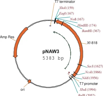
The coding sequence of 1779 bp from Xf-818 was sucessfully cloned into pET20b, creating pNAW3 (Figure 1). The Xf-818 gene is under the control of the inducible promoter T7, and remains translatable from its original start codon.
Simpson et al. (2000) have assigned putative
functions to around 50 % of ORFs from X. fastidiosa
9a5c, and among the genes described there are several potentially involved in plant-pathogen interaction, spe-cially in plant cell wall degradation (Table 1). Using Blast (Altschul et al., 1990) we have analyzed the best matches of protein Xf-818 (Table 2) and the highest similarities were among bacterial endoglucanases. It has a better
ho-mology with EngXCA from Xanthomonas campestris pv.
campestris, an endoglucanase with minor role on initial
stages of pathogenic process in radish and turnip (Gough et al., 1988; 1990). All matches of endoglucanases Xf-818 belong to the family 5 of glycoside hydrolases (Table 2) (Henrissat, 1991; Henrissat & Bairoch, 1993). In fact,
all three endoglucanases putatively found on X. fastidiosa
(Xf-810, Xf-818 and Xf-2708) belong to family 5 (Simpson et al., 2000).
Protein Xf-818 has two domain structures, with a N-terminal domain composed of a catalytic domain of family 5, interspersed by a lynker sequence to a type II cellulose binding domain (CBD) (Figure 2B). Cellulase activity was confirmed on CMC plates, a diagnostic test that shows the catalytic behavior of cellulases over cel-lulose chains (Figure 3). Congo red dye complexes with cellulose polymer but not with oligomers formed after its cleavage. Therefore, alterations associated with dye
bind-ing can be used to monitor hydrolytic activity on endoglucanases, but not cellobiohydrolases, another kind of cellulases acting over cellulose (Wood et al., 1988). Cellulase activity was also assessed by the formation of reducing sugars during hydrolysis of CMC (Figure 4). Confirmation of Xf-818 protein as a endoglucanase,
sup-Figure 1 - Restriction map of the plasmid pNAW3. Gene Xf-818 was cloned between NdeI and HindIII sites, under control of the T7 promoter. It has T7 terminator at its end, after the HindIII site. Important features of the plasmid are shown.
pNAW3
5 3 83 b p
Xf-818 Amp Res
T7 promoter
ori
T7 terminator
BamHI (367)
BglII (2052)
EagI (167)
HindIII (174)
NcoI (1866)
NdeI (1956) NotI (167)
SacI (1627)
XbaI (1994)
XhoI (159)
Figure 2 - (A) Annealing positions of primers on the Xf-818 gene cloned into pNAW3. NdeI and HindIII sites are shown as well as flanking structures of the pET20b plasmid; (B) Structure of Xf-818 protein (NCBI AAF83628/ SwissProt Q9PF60), as determined from the sequence derived from ORF identified on X. fastidiosa 9a5c (Simpson et al., 2000). Relevant features of Xf-818 protein domains are shown, with numbered positions referring to beginning and end of domains or specific structures.
+1 ATG
T7 promoter
T7 promoter primerF592Sl primer 818intN primer 818intC primer R592Hd primer T7 terminator T7 terminator primer 515 - 529 1061 - 1075
Nde I Hind III
Xf - 818
His6 tag
1779 TGA
pNAW3 5383 bp
A
592 aa
+1
Signal Catalytic domain GH 5 Lynker CBD II
peptide
Xf - 818
1 20 31 348 373 505 512 588
Domain involved in cellulose binding. It has five conserved trytoplan molecules flanqued by cysteines Sequence
composed of 70 % glycine and 29 % serine This domains has two
conserved amino acids E181 e E303 potentially
localized in the β-1.4 glycosidic bond cleavage Clivage at
LSA AP
port annotation that was performed during a sequencing project (Simpson et al., 2000), a fact that elicits assum-ing that other putative plant cell wall degradassum-ing enzymes are in fact true enzymes that can act over the plant cell wall (Table 1).
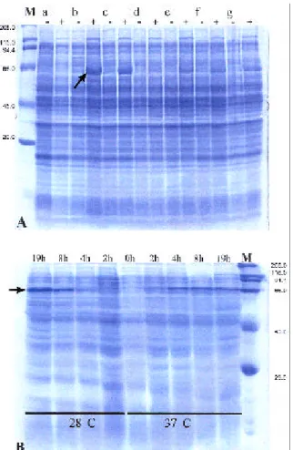
To optimize protein production, an E. coli clone
that produces high amount of heterologous protein was selected, and a few parameters that can influence protein production and accumulation were evaluated. The clone
of E. coli harboring pNAW3 and named clone b is the
best Xf-818 protein producer to be employed when scal-ing up the process of heterologous protein expression (Table 3). Such analysis was confirmed by means of elec-trophoretic analysis of total protein (Figure 5A).
The next step was to analyze the accumulation
of cellulase in the selected clone of E. coli after
induc-tion. Protein accumulation increased positively a few hours from induction, reaching the highest rate 19 h af-ter induction (Figure 5B). The rate of protein accumula-tion was apparently dependent of temperature during
ex-Gene1 Putative function2 Size (bp)* Protein (aa)* kDa* e-value*
Xf-1267 Cellobiohydrolase 2052 683 70.9 e-142
Xf-810 Endoglucanase 1698 565 61.6 3e-50
Xf-818 Endoglucanase 1779 592 60 1e-145
Xf-2708 Endoglucanase 1071 356 39.3 2e-85
Xf-878 Endoxylanase 774 257 28.6 9e-04
Xf-2395 Acetylxylan esterase 861 286 32.2 1e-13
Xf-2779 Endoxylanase 2712 903 101.1 8e-15
Xf-2466 Polygalacturonase 1636 544 59.8 e-112
Xf-2359 Pectate lyase 711 236 26.1 1e-10
Xf-439 β-glucosidase 2304 767 83.2 e-137
Xf-845 β-xylosidase 2649 882 95.0 2e-80
Table 1 - Putative genes of Xylella fastidiosa related to plant cell wall degrading enzymes (PCWDE).
*bp=base pairs, aa=amino acids, kDa=kilodalton, e-value= expect value
1Gene number, size in bp and amino acid number are from Simpson et al. (2000) found at http://aeg.lbi.ic.unicamp.br/xf/.
2Gene function was assigned according to the similarity found with other genes deposited at GenBank and the highest similarity is shown
on the last column.
Acession Number Cellulase Organism Probability Score
P19487 Endo-1,4-β-glucanase [engXCA] X. c. pv. campestris e-137 487
P23548 Endo-1,4-β-glucanase Bacillus polymyxa 4e-77 287
P54583 Endo-1,4-β-glucanase E1 Acidothermus cellulolyticus 4e-64 244
P10474 cellobiohydrolaseEndo-1,4-β-glucanase / 1,4 -β- Caldicellulosiruptor
saccharolyticus 2e
-42 172
Q05332 Endo-1,4-β-glucanase G Clostridium thermocellum 1e-41 169
P50400 Endo-1,4-β-glucanase D Celullomonas fimi 5e-33 140
P04956 Endo-1,4-β-glucanase B Clostridium thermocellum 1e-30 133
P27033 Endo-1,4-β-glucanase C Pseudomonas fluorescens 3e-18 92
Table 2 - Homologs of Xf-818 protein as found on protein-protein Blast search at NCBI (http://www.ncbi.nlm.nih.gov/ BLAST/)1.
1swissprot protein database
Clone1 µg glucose produced2 SA3 Plate assay4
pNAW3 a 3.41 ± 0.56 2.65 Positive pNAW3 b** 7.02 ± 1.48 5.52 Positive pNAW3 c 1.67 ± 0.27 1.41 Positive pNAW3 d 3.64 ± 0.17 2.94 Positive pNAW3 e 3.96 ± 0.94 3.26 Positive pNAW3 f 3.95 ± 1.2 3.30 Positive pNAW3 g 1.55 ± 0.53 1.14 Positive pET20 b - 1.43 ± 0.64 nd Negative Table 3 - Cellulase activity of selected clones of E. coli
BL21(DE3) harboring pNAW3 after 0.4 mmol L- 1
IPTG induction.
1Double asterisk indicates selected clone for cellulase production; 2Mean of 3 samples ± standard deviation;
3SA = Specific activity (µg glucose per mg of protein on sample
per min); nd = not determined;
4Carboxymethyl cellulose degradation on plate assay (LB medium
pression. At 37°C protein accumulated faster, but it reached a slightly higher level at the lower temperature. A suitable option is to keep the culture medium at 32°C after IPTG induction and then perform protein extraction after 12 h.
Confirmation that Xf-818 has cellulase activity, considered as an endoglucanase by the Congo red assay is by far the most important fact. Endoglucanase Xf-818 produces increasing amounts of reducing sugars in rela-tion to incubarela-tion time (Figure 4), an indicarela-tion of high activity and stability. An interesting feature of many car-bohydrate hydrolysing enzymes is the ability to bind to the substrate, a characteristic also found for cellulases, that is mediated by a domain called cellulose binding do-main, which is independent of the catalytic domain (Fig-ure 2B). Although Hopkins (1985) did not detected
cel-lulase activity on the culture medium of X. fastidiosa PD,
Scarpari (2001) was able to detect the presence of mRNA of Xf-818 on the culture medium, an indicator of
cellu-lase production by X. fastidiosa 9a5c.
The ability to knockout X. fastidiosa genes
(Monteiro et al., 2001; Silva Neto et al., 2002) opens the
way to analyze functionally X. fastidiosa genes, such as
Xf-818 and addresses the role of this protein during plant-pathogen interaction. Cellulases have minor roles on the beginning of symptom development as assessed by
cel-lulase mutants of E. carotovora subsp. carotovora (Mäe
et al., 1995), Ralstonia solanacearum (Roberts et al.,
1988) and X. c. pv. campestris (Gough et al., 1990).
How-ever, this pattern is different with Clavibacter
michiganensis ssp. michiganensis (Jahr et al., 2000) and
C . m. ssp. sepedonicus (Laine et al., 2000), since
endoglucanases are essential for symptom development. The next step is to produce and purify high amounts of Xf-818 to characterize its substrate specific-ity, raise antibodies and evaluate its expression during
cit-rus colonization by X. fastidiosa. The recent sequencing
of other X. fastiosa strains, such as Dixon strain from
al-mond, Ann1 from oleander (Bhattacharyya et al., 2002) and a grapevine strain (Van-Sluys et al., 2003), opens the possibility to study the diversity of pathogenicity factor among strains of the same bacterium, but with different host range.
Figure 4 - Reducing sugars produced by Xf-818 during hydrolysis of CMC at 40°C. Measurements of reducing sugars were performed at indicated time intervals. Mean of 3 replicates ± standard deviation.
Time (h)
0 2 4 6 8 10 12 14 16 18
u
g glucose
0 20 40 60 80 100 120 140
Figure 3 - Cellulase plate assay showing cellulose degradation by the action of Xf-818 expressed in E. coli BL21(DE3) by pNAW3. Untransformed E. coli was taken as control. Yellow-white halos indicate cellulose degradation.
ACKNOWLEDGMENTS
This work was supported by grant from FAPESP (Fundação de Amparo à Pesquisa do Estado de São Paulo). First author was supported by a scholarship from FAPESP, and the third author was fellow of CNPq (Conselho Nacional de Desenvolvimento Científico e Tecnológico). Authors also thank Dra. M. Fiore (USP/CENA), Dr. J. Ferro (UNESP/Jaboticabal) and Dra. M.L. Targon (IAC/ CCSM) for providing essential material for this research.
REFERENCES
ALTSCHUL, S.F.; GISH, W.; MILLER, W.; MYERS, E.W.; LIPMAN, D.J. Basic local alignment search tool. Journal of Molecular Biology, v.215,
p.403-410, 1990.
BHAT TACHARYYA, A.; STILWAGEN, S.; IVANOVA, N. et al., Whole-genome comparative analysis of three phytopathogenic Xylella fastidiosa strains. Proceeding of the National Academy of Sciences of USA, v.99,
p.12403-12408, 2002.
BRADFORD, M.M. A rapid and sensitive method for the quantitation of microgram quantities of protein utilizing the principles of protein-dye binding. Analytical Biochemistry, v.72, p.248-254, 1976.
CHANG, C.J.; GARNIER, M.; ZREIK, L.; ROSSETTI, V.; BOVÉ, J.M. Culture and serological detection of the xylem-limited bacterium causing citrus variegated chlorosis and its identification as a strain of Xylella
fastidiosa. Current Microbiology, v.27, p.137-142, 1993.
DOW, J.M.; DANIELS, M.J. Xylella fastidiosa genomics and bacterial pathogenicity to plants. Yeast, v.17, p.263-271, 2000.
FOONG, F.C.-F.; DOI, R.H. Characterization and comparison of Clostridium
cellulovorans endoglucanases-xylanases EngB and EngD hyperexpressed
in Escherichia coli. Journal of Bacteriology, v.174, p.1403-1409, 1992.
FRY, S.M.; HUANG, J.S.; MILHOLLAND, R.D. Isolation and preliminary characterization of extracellular proteases produced by strains of Xylella
fastidiosa from grapevines. Phytopathology, v.84, p.357-363, 1994.
GORDON, D.; ABAJIAN, C.; GREEEN, P. Consed: a graphical tool for sequence finishing. Genome Research, v.8, p.195-202, 1998.
GOUGH, C.L.; DOW, J.M.; BARBER, C.E.; DANIELS, M.J. Cloning of two endoglucanases genes of Xanthomonas campestris pv. campestris: analysis of the role of the major endoglucanase in pathogenicity.
Molecular Plant Microbe Interactions, v.1, p.275-281, 1988.
GOUGH, C.L.; DOW, J.M.; KEEN, J.; HENRISSAT, B.; DANIELS, M.J. Nucleotide sequence of the engXCA gene encoding the major endoglucanase
of Xanthomonas campestris pv. campestris. Gene, v.89, p.53-59, 1990.
HENRISSAT, B. A classification of glycosyl hydrolases based on amino-acid sequence similarities. Biochemical Journal, v.280, p.309-316, 1991.
HENRISSAT, B.; BAIROCH, A. New families in the classification of glycosyl hydrolases based on amino-acid sequence similarities.
Biochemical Journal, v.293, p.781-788, 1993.
HOPKINS, D.L. Physiological and pathological characteristics of virulent an avirulent strains of the bacterium that causes Pierce’s disease of grapevine. Phytopathology, v.75, p.713-717, 1985.
HOPKINS, D.L. Xylella fastidiosa: xylem-limited bacterial pathogen of plants. Annual Review of Phytopathology, v.27, p.271-290, 1989.
HOUGH, D.W.; DANSON, M.J. Extremozymes. Current Opinion i n Chemical Biology, v.3, p.39-46, 1999.
JAHR, H.; DREIER, J.; MELETZUS, D.; BAHRO, R.; EICHENLAUB, R. The endo-β-1,4-glucanase CelA of Clavibacter michiganensis subsp.
michiganensis is a pathogenicity determinant required for induction of
bacterial wilt of tomato. Molecular Plant Microbe Interactions, v.13,
p.703-714, 2000.
LAEMMLI, U.K. Cleavage of structural proteins during the assembly of the head of bacteriophage T4. Nature, v.227, p.680-685, 1970.
LAINE, M.J.; HAAPALAINEN, M.; WAHLROSS, T.; KANKARE, K.; NISSINEN, R.; KASSUWI, S.; METZLER, M.C. The cellulase encoded by the native plasmid of Clavibacter michiganensis ssp. sepedonicus plays a role in virulence and contains an expansin-like domain.
Physiological and Molecular Plant Pathology, v.57, p.221-233, 2000.
LAMBAIS, M.R.; GOLDMAN, M.H.S.; CAMARGO, L.E.A.; GOLDMAN, G.H. A genomic approach to the understanding of Xylella fastidiosa pathogenicity. Current Opinion in Microbiology, v.3, p.459-462, 2000.
LEITE, B.; ISHIDA, M.L.; ALVES, E.; CARRER, H.; PASCHOLATI, S.F.; KITAJIMA, E.W. Genomics and X-ray microanalysis indicates that Ca2+
and thiols mediate the aggregation and adhesion of X. fastidiosa. Brazilian Journal of Medical and Biological Research, v.35, p.645-650, 2002. LEVER, M. A new reaction for colorimetric determination of carbohydrates.
Analytical Biochemistry, v.47, p.273-279, 1972.
LOPES, J.R.S. Estudos com vetores de Xylella fastidiosa e implicações no manejo da clorose variegada dos citros. Laranja, v.20, p.329-344, 1996. MÄE, A.; HEIKINHEIMO, R.; PALVA, E.T. Structure and regulation of
Erwinia carotovora subsp. carotovora SCC3193 Cellulase gene celV1
and the role of cellulase in phytopathogenicity. Molecular and General Genetics, v.247, p.17-26, 1995.
MONTEIRO, P.B.; TEIXEIRA, D.C.; P ALMA, R.R.; GARNIER, M.; BOVE, J.M.; RENAUDIN, J. Stable transformation of the Xylella
fastidiosa citrus variegated chlorosis strain with oriC plasmids. Applied
and Environmental Microbiology, v.67, p.2263-2269, 2001.
NIELSEN, H.; ENGELBRECHT, J.; BRUNAK, S.; VON HEIJNE, G. Identification of prokaryotic and eukaryotic signal peptides and prediction of their cleavage sites. Protein Engineering, v.10, p.1-6, 1997.
NILSSON, J.; STAHL, S.; LUNDEBERG, J.; UHLÉN, M.; NYGREN, P.-A. Affinity fusion strategies for detection, purification, and immobilization of recombinant proteins. Protein Expression and Purification, v.11, p.1-16, 1997.
PARK, Y.W.; LIM, S.T.; YUN, H.D. Cloning and sequencing of the celA gene encoding CMCase of Erwinia carotovora subsp. carotovora LY34.
Molecules and Cells, v.8, p.27-35, 1998.
PURCELL, A.H.; HOPKINS, D.L. Fastidious xylem-limited bacterial plant pathogens. Annual Review of Phytopathology, v.34, p.131-151, 1996.
ROBERTS, D.P.; DENNY, T.P.; SCHELL, M.A. Cloning of the egl gene of
P s e u d o m o n a s s o l a n a c e a r u m and analysis of its role in
phytopathogenicity. Journal of Bacteriology, v.170, p.1445-1451, 1988. ROSSETTI, V.; DE NEGRI. D. Clorose variegada dos citros: revisão.
Laranja, v.11, p.1-14, 1990.
ROSSETTI, V.; GARNIER, M.; BOVÉ, J.M.; BERETTA, M.J.G.; TEIXEIRA, A.R.R.; QUAGGIO, J.A.; DE NEGRI, J.D. Présence de bactéries dans le xylème d’oranges atteints de chlorose variégée, une nouvelle maladie des agrumes au Brésil. Comptus Rendus de Academie Sciences Paris, v.310, p.345-349, 1990.
ROSSETTI, V.; CARVALHO, M.L.V.; CHAGAS, C.M. Ensaios de transmissão de clorose variegada dos citros (CVC) em campo.
Fitopatologia Brasileira, v.20, p.67, 1995. Suplemento.
SAMBROCK, J.; FRITISCH, E.F.; MANIATIS, J. Molecular cloning. 2.ed.
Plainview: Cold Spring Harbor Laboratory. Press, 1989.
SCARPARI, L.M. Modulação da expressão de genes de patogenicidade putativos em Xylella fastidiosa sob condições de baixa e alta densidade celular. Piracicaba: USP/ESALQ, 2001. 85p. (Dissertação - Mestrado) SILVA, F.R. DA; VETORE, A.L.; EDSON, L.K.; LEITE, A.; ARRUDA, P. Fastidian gum: the Xylella fastidiosa exopolysaccharide possibly involved in bacterial pathogenicity. FEMS Microbiology Letters, v.203, p.165-171, 2001.
SILVA NET O, J.F.; KOIDE, T.; GOMES, S.L.; MARQUES, M.V. Site-directed gene disruption in Xylella fastidiosa. FEMS Microbiology Letters, v.210, p.105-110, 2002.
SIMPSON, A.J.G.; REINACH, F.C.; ARRUDA, P. et al. The genome sequence of the plant pathogen Xylella fastidiosa. Nature, v.406, p.151-159, 2000. STUDIER, F.W.; MOFFAT T, B.A. Use of bacteriophage-T7 RNA-polymerase to direct selective high-level expression of cloned genes.
Journal of Molecular Biology, v.189, p.113-130, 1986.
TUBELIS, A.; BARROS, J.C.; LEITE, R.M.V.B. Difusão da clorose variegada dos citros em pomares comerciais de laranja doce no Brasil.
Laranja, v.14, p.239-254, 1993.
VAN-SLUYS, M.A.; OLIVEIRA, M.C.; MONTEIRO-VITORELLO, C.B. et al., Comparative analyses of the complete genome sequences of Pierce’s disease and citrus variegated chlorosis strains of Xylella
fastidiosa. Journal of Bacteriology, v.185, p.1018-1026, 2003.
WOOD, P.J.; ERFLE, J.D.; TEATHER, R.M. Use of complex formation between congo red and polysaccharide in detection and assay of polysaccharide hydrolases. Methods in Enzymology, 160, p.59-74, 1988.