(Annals of the Brazilian Academy of Sciences) ISSN 0001-3765
www.scielo.br/aabc
Cloning, expression, and analysis of the group 2 allergen
from
Dermatophagoides farinae
from China
YU-BAO CUI, YING ZHOU, WEIHONG SHI, GUIFANG MA, LI YANG and YUNGANG WANG
Department of Laboratory Medicine, Yancheng Health Vocational & Technical College, Jiangsu Yancheng 224006, P.R. China
Manuscript received on March 3, 2010; accepted for publication on July 6, 2010
ABSTRACT
To obtain the recombinant group 2 allergen product ofDermatophagoides farinae(Der f 2), the Der f 2 gene was
synthesized by RT-PCR. The full-length cDNA comprised 441 nucleotides and was 99.3% identical to the reference sequence (GenBankAB195580). The cDNA was bound to vector pET28a to construct plasmid pET28a(+)-Der f 2, which was transformed intoE. coliBL21 and induced by IPTG. SDS-PAGE showed a specific band of about 14kDa in the whole cell lysate. As estimated by chromatography, about 3.86 mg of the recombinant product was obtained, which conjugated with serum IgE from asthmatic children. The protein had a signal peptide of 17 amino acids. Its secondary structure comprised an alpha helix (19.86%), an extended strand (30.82%), and a random coil (49.32%). The subcellular localization of this allergen was predicted to be at mitochondria. Furthermore, its function was shown to be associated with an MD-2-related lipid-recognition (ML) domain. The results of this study provide a solid foundation for large-scale production of the allergen for clinical diagnosis and treatment of allergic disorders.
Key words: Dermatophagoides farinae, house-dust-mite allergy, mite allergens, Der f 2, allergen engineering,
im-munotherapy, bioinformatics.
INTRODUCTION
Dust mites have been recognized to be associated with the rising incidence of asthma and other allergic dis-eases (Milián and Díaz 2004, Nadchatram 2005). At present, mite extract is used in clinical diagnosis and treatment of allergic diseases, especially in developing countries. However, the extract is a complex mixture of allergens, non-allergic or toxic proteins, and some en-zymes, which have been reported to be associated with flushing, swelling, induration, necrosis and other local reaction, shock, laryngeal edema, bronchospasm, urti-caria, angioedema, generalized erythema, and other sys-temic side effects (Mauro et al. 2006, Zhang et al. 2009). Over the past 10 years, some groups of mite allergens have been cloned, sequenced, expressed and purified.
Correspondence to: Yu-bao Cui E-mail: [email protected]
Present address: Department of Physics, University of Texas at San Antonio, One UTSA Circle, San Antonio, Texas 78249 USA.
The three-dimensional structures of some allergens were determined, and B and T-cell epitopes were mapped (Jeong et al. 2006, Mothes et al. 2006). The recom-binant allergens were produced in bacteria, yeasts and insect cell expression systems, and have been demon-strated to have good IgE binding activity similar to their natural counterparts. They have also shown good effects in skin prick tests and other allergen diagnosis methods (Jeong et al. 2006, Mothes et al. 2006). These recombinant allergens are intended to replace natural al-lergens and to allow the development of new immuno-therapy projects, including the use of hypoallergens, al-lergens coupled to IgE suppressive adjuvants, and pep-tide-based therapies (Dinakar and Portnoy 2004, Linhart and Valenta 2005).
preval-ent allergens in differpreval-ent regions (Linhart and Valpreval-enta 2005). In our previous study, we obtained the gene encoding the group 2 allergen fromDermatophagoides farinae(Der f 2). An additional region of 87 bp (from 77 to 163 bp) was present in our strain that was absent from the reference sequence (GenBankAB195580) (Cui
et al. 2007). We attempted to express this gene in Es-cherichia coli(E. coli) with no success. We, then, iso-lated total RNA fromDermatophagoides farinaeagain, and synthesized the cDNA encoding Der f 2 by RT-PCR. We obtained a Der f 2 gene that was identical in length to the reference sequence, and successfully ex-pressed it inE. coli.
MATERIALS AND METHODS
PATIENTSERA
Five asthmatic children were involved in this study. They were demonstrated to be allergic to dust mites by the skin prick test with Dermatophagoides farinae extract provided by the Department of Parasitology, Medical College of Fudan University. Blood (5 ml) was extracted from each child and centrifuged for serum. We mixed five sera samples and treated the mixture as the positive control in western-blotting. On the other hand, umbili-cal serum samples were taken from five children whose mothers had no atopic history, centrifuged, mixed, and used as negative controls.
PREPARATION OF CDNAANDPOLYMERASECHAIN
REACTION(PCR)
According to our previous report (Cui et al. 2007, 2008), house dust mites were cultured and isolated. Total RNA was obtained using RNA isolator (TaKaRa Biotech Co. Led, Dalian, China, Code No. D312) and stored at – 80◦C. For PCR amplification of the cDNA encoding Der f 2, two primers were designed and synthesized based on the GenBank sequence AB195580. The
for-ward primer sequence was (5′ GGATCCATGATTTCC-AAAATCTTGTGCC3′), and the reverse was (5′ CTC-GAGTTAATCACGGATTTTACCATGG3′). These two primers had a BamHI site and a XhoI site at their 5’ ends (underlined), respectively. Reverse transcription (RT) was performed using the total RNA isolated from mites with High Fidelity PrimeScriptTM RT-PCR Kit (TaKaRa Biotech Co. Led, Code No. DR027A) in the
PCR Thermal Cycler Dice (TaKaRa Biotech Co. Led, Code No. TP 600). The reaction mixture for RT had total RNA (2µl), 20µM of the reverse primer (1µl),
10 mM of dNTP Mixture (1µl), 20µM of Random
6-mers (1µl), and RNase Free H2O (5µl), which was set
at 65◦
C for 5 min, followed by ice-bath for 2 min. Then, 5×PrimeScript RT Buffer (4µl), 40 U/µL of RNase In-hibitor (0.5µl), PrimeScript RTase (0.5µl), and RNase
Free dH2O (5µl) were added in. 20µl of the final re-action mixture was incubated at 30◦C for 10 min, 42◦C for 30 min and 95◦C for 5 min. The RT product was used as a template for PCR in the same thermal Cycler Dice with PrimeSTARrHS DNA Polymerase (TaKaRa Biotech Co. Led, Code No. DR010A). The total reaction mixture had RT products (2µl), 5×PrimeSTAR PCR Buffer (10µl), 2.5 mM of dNTP Mixture (4 µl), 20 µM of the forward primer (1µl), 20µM of the reverse
primer (1µl), 2.5 U/µL of PrimeSTAR HS DNA
Poly-merase (0.5µl), and dH2O (31.5µl). PCR conditions used here included an initial incubation for 3 min at 94◦C, followed by 30 cycles of 10 sec at 98◦C, 15 sec at 55◦C, and 30 sec at 72◦C. After a final incubation for 10 min at 72◦C, the amplicons were analyzed by agarose gel electrophoresis (1.0%) and visualized with ImageMasterrVDS.
CLONING ANDDNA SEQUENCING
The PCR-amplified DNA was recovered from the gel with Agarose Gel DNA Purification Kit Ver. 2.0 (TaKaRa Biotech Co. Led, Code No. DV805). A poly-A tail was added with DNA A-Tailing Kit (TaKaRa Biotech Co. Led, Code No. D404), and the products were bound to pMD19-T simple vector (TaKaRa Biotech Co. Led, Code No. D104). E. coliJM109 (TaKaRa Biotech Co. Led, Code No. D9052) was, then, transformed with the recombinant plasmids; positive clones were selected by blue/white screening on Luria-Bertani (LB) plates con-taining 100µg/ml ampicillin, and confirmed by
restric-tion enzyme analysis and automatic DNA sequencing.
CONSTRUCTION OFEXPRESSIONPLASMIDS PET28A(+)-DER F2
re-action mixture had pMD19-T-Der f 2 10µl, 10 U/µl of
BamH I (5µl), 10 U/µl XhoI (5 µl), 10×K Buffer (10µl), and dH2O (70µl), which was set at water-bath
for four hours at 37◦C. The digested fragment was sub-jected to agarose gel electrophoresis and recovered from the gel using Agarose Gel DNA Purification Kit Ver. 2.0 (TaKaRa Biotech Co. Led, Code No. DV805). The fragment was, then, subcloned into expression vector pET28a(+) (Novagen, Germany, Kit Lot No. N72770) to create pET28a(+)-Der f 2 using the DNA Ligation Kit (TaKaRa Biotech Co. Led, Code No. D6023). E. coliCompetent Cells JM109 (TaKaRa Biotech Co. Led, Code No. D9052) were transformed with pET28a(+)-Der f 2 plasmids; positive clones were selected by blue/ white screening and verified by restriction enzyme ana-lysis withBamH I andXhoI.
EXPRESSION OF RDER F2INE. coliBL21 (DE3)
5µl of pET28a (+)-Der f 2 plasmid were purified using
the MiniBEST Plasmid Purification Kit Ver. 2.0 (TaKa-Ra Biotech Co. Led, Code No. DV801A) and trans-formed into 100µl ofE. coliBL21 (DE3, Stratagene, USA).E. coliBL21 carrying pET28a (+)-Der f 2 was grown on LB plates containing 50µg/ml of
kanamy-cin at 37◦C overnight. A single colony was inoculated into 2 ml LB plates at 37◦C overnight, 100
µl of which
was taken and added into a glass tube containing 5 ml LB fluid containing kanamycin and, then, cultured at 37◦C. 100 mM of
isopropyl-β-D-thiogalactopyranoside
(IPTG, 50µl, final 1 mM) was added to induce thelac promoter. TheE. colicells were harvested by centrifu-gation, and PBS Buffer (200µl/tube) was added in.
Af-ter centrifugation and resuspension, ultrasonic disrup-tion was used until the obtendisrup-tion of a transparent fluid, 50µl of which was taken as the whole cell lysate. The
lysate was centrifuged and the supernatant and pellet were regarded as the soluble and insoluble protein sam-ples, respectively. The whole cell lysate (10µl), soluble
and insoluble protein samples were taken individually, and 2.5µl of 5×SDS sample buffer were added into each of them. The samples were heated for 10 min at 95◦C and subjected to sodium dodecylsulfate-polyacryl-amide gel electrophoresis (SDS-PAGE) with 12.5% of polyacrylamide gel and CBB-R250 staining.
ISOLATION, PURIFICATION,ANDRENATURATION OF
RECOMBINANT RDER F2 PROTEINS
E. coliBL21 carrying pET28a (+)-Der f 2 was grown on LB plates containing 50 µg/ml of kanamycin at
37◦
C overnight. A single colony inoculated into 5 ml of LB fluid with shaking at 250 rpm overnight at 37◦C was treated as the seed fluid for fermentation. 50 ml of the seed fluid were inoculated into 2000 ml LB fluid containing 50µg/ml of kanamycin at 30◦C shaking at 250 rpm for fermentation. 100 mM IPTG were added until the final concentration reached 1 mmol/l. The in-duced cells were collected, suspended in PBS (pH 7.4), and broken by ultrasonic waves in an ice bath until the fluid became transparent. This fluid with broken cells was centrifuged at 7000 rpm at 4◦C for 15 min to col-lect rough inclusion bodies, which were then washed in PBS buffer containing 2 mol/l of urea for the purified product. The purified inclusion bodies were resuspended in PBS buffer (pH 8.0) containing 8 mol/L of urea and 5 mmol/l of DTT, stirred slightly at 4◦C overnight, and centrifuged at 1000 rpm for 10 min. The supernatant was filtered by microfiltration membrane with a pore size of 0.22µm. This protein product was further purified by
nickel-affinity chromatography at a speed of 4.0 ml/min, eluted with 25, 50, 75, 100, 125, and 150 mmol/L of imi-dazole solution at a speed of 6 ml/min. The elution peak at each stage was collected and determined for molecu-lar weight and purity by SDS-PAGE. All of the elution fractions were freeze-dried for protein power. Lastly, the product was re-natured by gradient dialysis with a renat-uration solution containing 2 mol/l to 0.2 mol/L of urea, and 0.2 mol/l of EDTA continuously.
WESTERNBLOTTING
For western blotting analysis, proteins were transferred to a PVDF membrane (TIANGEN, Biotech Co. Led, Beijing, China) and incubated with primary antibodies kept in the sera from asthmatic children allergic to dust mites at 4◦
NUCLEOTIDESEQUENCE, INFERREDAMINO
ACIDSEQUENCE, STRUCTURAL ANDFUNCTION
ANALYSIS, ALIGNMENT ANDPHYLOGENY
Sequences were edited to remove the vector sequence and the extra restriction sites. The open reading fragment (ORF) was obtained using the ORF finder in the NCBI (National Center for Biotechnology Information) web-site. The amino acid sequence of Der f 2 was deduced using Translate Tools, and its physiochemical proper-ties were predicted by ProtParam Tools in ExPaSy web server. The signal peptide of Der f 2 was analyzed by the SignalP 3.0 server. The functional site was searched for in the InterPro database in EBI (European Bioin-formatics Institute) website. The subcellular localiza-tion for Der f 2 was predicted by CELLO v.2.5, and its secondary structure was analyzed by GOR4.0 software. The published sequences that matched our sequenced cDNA were obtained from GenBank using BLAST. The published sequences of ticks and mites were chosen for alignment using ClustalW2 at the EBI website, and com-puted for similarity by VECTOR NTI 9.0 software (IBI, New Haven, CT, USA). A phylogenetic tree was con-structed with maximum parsimony methods in Molec-ular Evolutionary Genetics Analysis (MEGA) software version 4.0 software.
RESULTS
CLONING ANDSEQUENCING OF CDNA ENCODING
DER F2
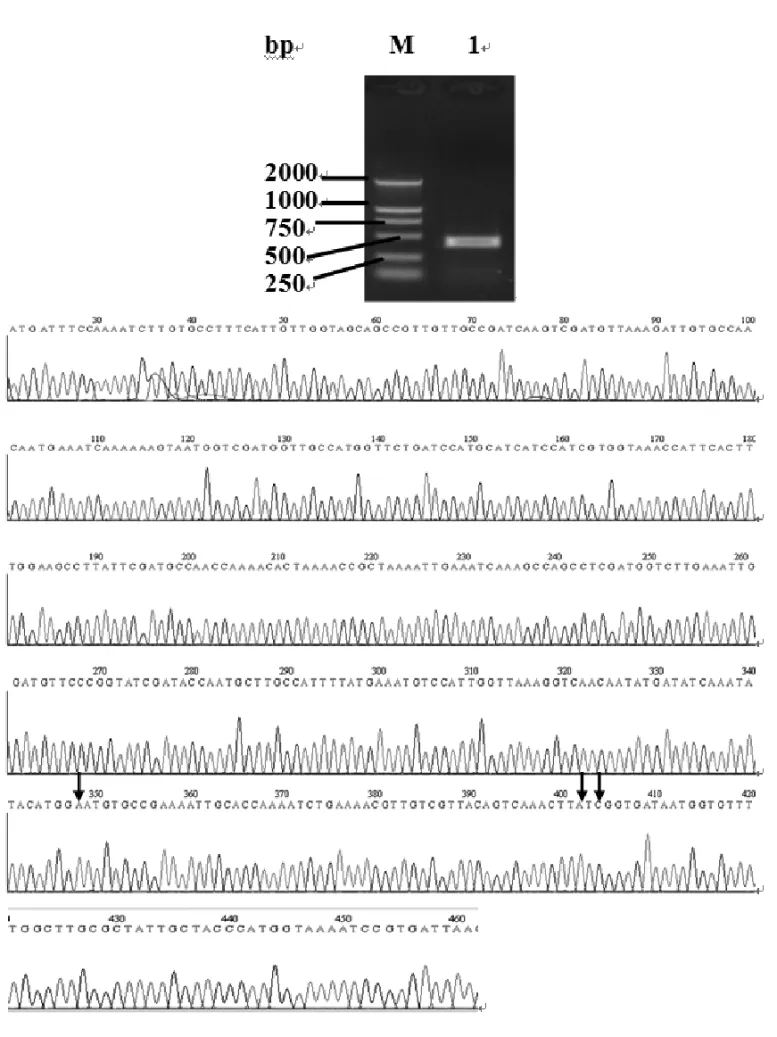
Total RNA was isolated from adult mites, and Der f 2 cDNA fragments were amplified by RT-PCR, obtaining a 441 bp product (Fig. 1A). The recovered PCR prod-uct was cloned into vector pMD19-T and transformed intoE. colicompetent cells (JM109) and positive clones. The product was confirmed by restriction enzyme diges-tion and automatic DNA sequencing (Fig. 1B, the vector sequence and the added restriction sites were removed from the sequencing results). The sequencing results showed that our Der f 2 cDNA is 99.3% identical to the previously published sequence (GenBank AB195580).
There is one mutation at position 328 (G>A) resulting
in amino acid mutation from Aspartate to Asparagine, and the other two mutations at positions 382 (G>A)
and 384 (T>C) resulting in amino acid mutation from
Valine to Isoleucine. Using the ORF Finder, a complete
ORF was found within the Der f 2 cDNA; its length is 441 bp from the start codon ATG to the stop codon TGA (Fig. 1B).
EXPRESSION ANDPURIFICATION OFRECOMBINANT
PROTEINDER F2
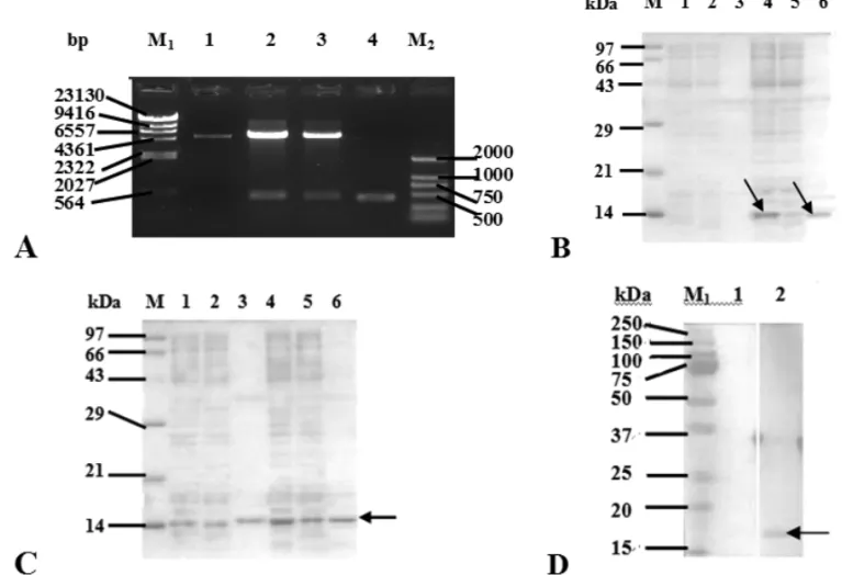
The cDNA encoding Der f 2 allergen was excised from recombinant plasmid pMD19-T-Der f 2, sub-cloned into expression vector of pET28a (+) and confirmed by re-striction analysis (Fig. 2A). E. coli BL21 was trans-formed with plasmid pET28a (+)-Der f 2, and protein expression was induced with IPTG. We observed a sin-gle band of about 14 kDa on SDS-PAGE, which agreed with the predicted molecular weight of 14.076 kDa for the Der f 2 protein (Fig. 2B). E. coli BL21 carrying pET28a (+)-Der f 2 was recovered from a 2000 ml of fermentation solution, collected, ultrasonically dis-rupted, and centrifuged. The precipitate was washed, dissolved and filtered. The supernatant was purified by nickel-affinity chromatography and eluted with imida-zole solution, and the pure protein presented in 25, 50, 75, 100, and 125 mmol/l of elution fluid was shown by SDS-PAGE (Fig. 2C). These elution fractions were col-lected and freeze-dried, and 3.86 mg of protein power was obtained. The recombinant protein can bind the IgE antibody in asthmatic children by western-blotting (Fig. 2D), which demonstrated that the expression prod-uct has good allergenicity.
BIOINFORMATICS FORAMINOACIDCOMPOSITION, PHYSIOCHEMICALPROPERTIES, SECONDARY
STRUCTURE, SUBCELLULARLOCALIZATION,AND
FUNCTIONALSITE OF THERECOMBINANT
PROTEIN RDER F2
Fig. 2 – Expression and purification of the recombinant protein Der f 2. (A) Confirmation of pET28a (+)-Der f 2 plasmid by restriction enzyme digestion. After subcloning and blue/white screening, the positive pET28a (+)-Der f 2 plasmids were confirmed by restriction enzyme digestion withBamH I andXhoI and, then, separated on a 1% agarose gel containing ethidium bromide. Lane M1,λ-Hind III DNA Marker;Lane M2, DNA Marker DL2,000;Lane 1, pET28a(+) Vector;Lane 2, 3, pET28a(+)-Der f 2 plasmids digested withBamH I andXhoI;Lane 4, the gene fragment encoding for Der f 2 allergen. (B) SDS-PAGE analysis of expression rDer f 2 inE. coliBL21 cells. Lane M, TaKaRa Protein Marker (Broad);Lane 1, the whole cell lysate ofE. coliBL21 cells containing pET28a;Lane 2, the supernatant of cells containing pET28a;Lane 3, the pellet of cells containing pET28a;Lane 4, the whole cell lysate ofE. coliBL21 cells containing pET28a (+)-Der f 2;Lane 5, the supernatant of cells containing pET28a (+)-Der f 2;Lane 6, the pellet of cells containing pET28a (+)-Der f 2. Arrows indicate the rDer f 2 band. (C) SDS-PAGE analysis of purified rDer f 2 protein fromE. coliBL21 cells.Lane M: TaKaRa Protein Marker (Broad);Lane 1, 2, 3, 4, 5, 6, eluted fraction with 25, 50, 75, 100, 125 and 150 mmol/L imidazole elution buffer, respectively. (D) Western blotting analysis of rDer f 2 inE. coliBL21 cells.Lane 1, the reaction result between rDer f 2 and negative serum;Lane 2, the reaction result between rDer f 2 and positive serum;Lane M1, Precision Plus Protein Standards;Lane M2, perfect protein marker.
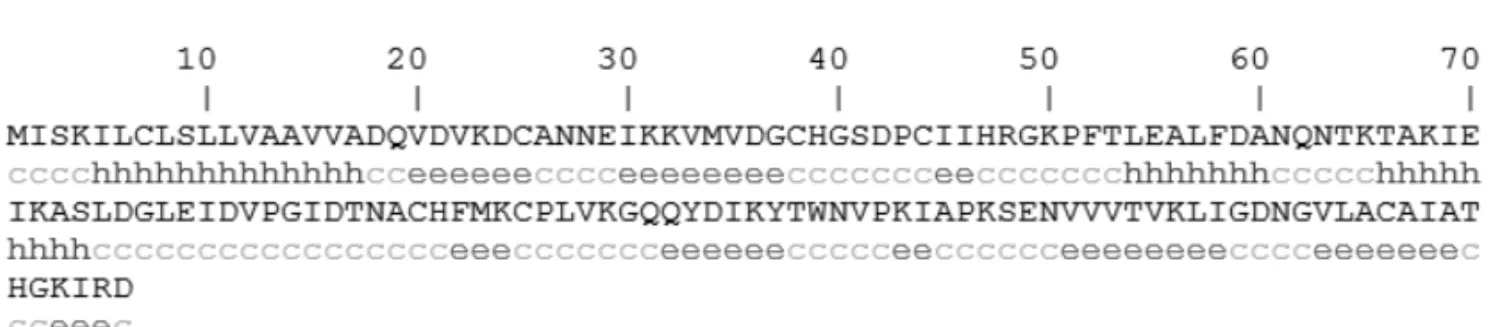
of 0.105 by ProtParam Tools, which indicated that it should be hydrophilic. With regard to Der f 2 secondary structure, GOR4 predicted that 19.86% (29 aa) of the protein were in alpha helices, 30.82% (45 aa) in extended strands, and 49.32% (72 aa) in random coils (Fig. 3). Using CELLO v.2.5, the subcellular localization of Der f 2 from China was concluded to be mitochondrial (Table I). A MD-2-related lipid-recognition (ML) do-main from residue 22 to 141 was found in the Der f 2 pro-protein by InterProScan.
AMINOACIDSEQUENCEHOMOLOGYANALYSIS,
ALIGNMENT,ANDPHYLOGENY
Fig. 3 – The secondary structure of the Der f 2 pro-protein (GOR 4.0). Based on the deduced amino acid sequence of the Der f 2 allergen, its secondary structure was predicted as described in “Materials and Methods”. Alpha helix (Hh): 29 (19.86%); extended strand (Ee): 45 (30.82%); and random coil (Cc): 72 (49.32%).
TABLE I
Protein subcellular localization for Der f 2 by CELLO v.2.5. Support Vector Machine Localization Reliability Amino Acid Comp. Nuclear 0.345 N-peptide Comp. Mitochondrial 0.514 Partitioned seq. Comp. Plasma Membrane 0.368 Physicochemical Comp. Mitochondrial 0.446 Neighboring seq. Comp. Mitochondrial 0.883 CELLO Prediction:
Mitochondrial 2.044
Nuclear 1.057
Chloroplast 0.537 Plasma Membrane 0.452 Cytoplasmic 0.389 Extracellular 0.379 Peroxisomal 0.035
Lysosomal 0.030
Endoplasmic Reticulum 0.026 Cytoskeletal 0.020
Vacuole 0.017
Golgi 0.016
ticks and mites, i.e.Dermatophagoides siboney (Gen-Bank ABC96702), Dermatophagoides pteronyssinus (GenBankCAK22338),Suidasia medanensis(GenBank
AAS75831),Aleuroglyphus ovatus(GenBank ABU97-461), Blomia tropicalis (GenBank ABG76185), Pso-roptes ovis(GenBankQ965E2),Lepidoglyphus destruc-tor(GenBankP80384),Tyrophagus putrescentiae (Gen-BankABU97478), Ixodes ricinus (GenBank ABL61-513), Euroglyphus maynei (GenBank Q9TZZ2) and
Acarus siro (GenBank ABU97459) were chosen.
Af-ter sequences coding for signal peptides were deleted, they were aligned using the ClustalW2 (Fig. 4), and a phylogenetic tree was constructed using Mega 4.0 soft-ware. The similarity between Der f 2 and Der p 2,
Der s 2 and Eur m 2 were 88%, 96% and 83% (Table II), and these four mites were clustered with 100% of Bootstrap values (Fig. 5).
DISCUSSION
Fig. 4 – Alignment between Der f 2 allergen and its homologous protein fractions from some other species of allergenic mites and ticks by ClustalW2 (http://www.ebi.ac.uk/Tools/clustalw2/). Notes: “∗” means that the residues or nucleotides in that column are identical in all sequences
in the alignment, “:” indicates conserved substitutions, “∙” indicates semi-conserved substitutions. The group 2 allergens ofDermatophagoides
farinae,Dermatophagoides siboney,Dermatophagoides pteronyssinus,Suidasia medanensis,Aleuroglyphus ovatus,Blomia tropicalis,Psoroptes ovis,Lepidoglyphus destructor,Tyrophagus putrescentiae,Ixodes ricinus,Euroglyphus mayneiandAcarus siroare abbreviated as Der_f, Der_s, Der_p, Sui_m, Ale_o, Blo_t, Pso_o, Lep_d, Tyr_p, Ixo_r, Eur_m and Aca_s.
pET28a (+)-Der f 2 was transformed intoE. coliBL21, the obvious bands of about 14 kDa were observed in the whole cell lysate and the pellet of cells on SDS-PAGE, which agreed with the predicted result and sug-gest that the prokaryotic expression system for Der f 2 was successfully developed, as well as the product pre-sented in the inclusion bodies. The expression vector PET28a(+), with a total length of 5369bp, expressed the recombinant protein containing the six-histidine tail, which bound to the nickel resin and could be eluted
with a chelating agent after other proteins were removed with the appropriate buffers. The single specific reac-tion band of about 14 kDa was present in the 25, 50, 75, 100, and 125 mmol/l of imidazole elution fractions on SDS-PAGE. Furthermore, the purified recombinant al-lergen could bind to IgE in sera from asthmatic children, which showed that the recombinant protein had good allergenicity and was a good foundation for the produc-tion of genetic allergens.
TABLE II
Analyses of similarity between the allergen Der f 2 and its homologous protein fractions from some other species of allergenic mites and ticks by VCETOR NTI 9.0 software.
Aca_s Ale_o Sui_m Blo_t Lep_d Der_f Der_s Der_p Eur_m Ixo_r Pso_o Tyr_p
Aca_s 56 56 52 52 43 42 43 43 33 47 42
Ale_o 19 66 62 56 44 42 42 44 34 49 48
Sui_m 20 14 58 60 46 45 44 45 30 50 44
Blo_t 16 14 15 54 42 42 42 43 30 48 42
Lep_d 20 17 16 15 42 42 38 40 33 47 46
Der_f 18 18 17 21 17 96 88 83 27 38 35
Der_s 20 20 18 19 17 3 89 82 27 38 34
Der_p 17 21 18 22 20 7 7 85 26 39 36
Eur_m 17 18 16 20 18 9 9 9 28 38 32
Ixo_r 20 20 22 24 20 22 22 23 24 34 26
Pso_o 20 22 19 17 22 26 26 25 24 23 45
Tyr_p 24 19 24 21 22 22 22 20 22 20 18
Note: The similarity is above the diagonal line, and the divergence is below. The group 2 allergens ofDermatophagoides farinae, Dermatophagoides siboney,Dermatophagoides pteronyssinus,Suidasia medanensis,Aleuroglyphus ovatus,Blomia tropicalis,Psoroptes ovis,Lepidoglyphus destructor,Tyrophagus putrescentiae,Ixodes ricinus,Euroglyphus maynei, andAcarus siroare abbreviated as Der_f, Der_s, Der_p, Sui_m, Ale_o, Blo_t, Pso_o, Lep_d, Tyr_p, Ixo_r, Eur_m, and Aca_s.
Fig. 5 – Tree of the amino acid sequence homology constructed for Der f 2 allergen and other homologous allergens from some species of allergenic mites and ticks with maximum parsimony methods in Mega 4.0. The group 2 allergens ofDermatophagoides farinae,Dermatophagoides siboney, Dermatophagoides pteronyssinus, Suidasia medanensis, Aleuroglyphus ovatus,Blomia tropicalis,Psoroptes ovis,Lepidoglyphus destructor, Tyrophagus putrescentiae,Ixodes ricinus,Euroglyphus mayneiandAcarus siroare abbreviated as Der_f, Der_s, Der_p, Sui_m, Ale_o, Blo_t, Pso_o, Lep_d, Tyr_p, Ixo_r, Eur_m and Aca_s.
into an amino acid sequence, and the signal peptide se-quence was deduced at position 1 to 17. After the re-moval of the signal peptide, the mature protein was predicted to be a hydrophilic protein of 129 residues and 14.076 kDa of molecular weight. Interestingly, the
domain, which is associated with lipid recognition, par-ticularly with the recognition of pathogen-related prod-ucts. The ML domain has an immunoglobulin-like beta-sandwich fold similar to that of E-set Ig domains.
Many studies have been published concerning house dust mites, which belong to the family Pyroglyphi-dae, especially Dermatophagoides pteronyssinus, Der-matophagoides farinae, andEuroglyphus maynei. Other mite species, referred to as “storage mites” because of their consumption of stored food, are also considered allergenic, although their study is more limited (Fernán-dez-Caldas et al. 2007). We used the deduced amino acid sequence of Der f 2 from China to search the pro-tein databases at NCBI using BLASTp. Similar amino acid sequences of other mite species were identified, in-cludingD. siboney,D. pteronyssinus andEuroglyphus mayneilisted in Pyroglyphidae;Lepidoglyphus destruc-tor andBlomia tropicalis in Glycyphagidae; Suidasia medanensis,Aleuroglyphus ovatus,Tyrophagus putres-centiae andAcarus siroin Acaridae. Besides Derma-tophagoidesspp. other mite-species, especiallyBlomia tropicalisdue to its abundance in tropical and subtrop-ical regions, were previously considered to be impor-tant from economic and sanitary perspectives, but are now being recognized as important contributors to the allergen content in house dust. They can cause tional respiratory allergies in farmers and other occupa-tionally exposed individuals (Nadchatram 2005). Many of these allergens have shown sequence homology and biological functional similarity to those described in Dermatophagoidesspp. The amino acid sequence sim-ilarity between the group 2 allergen of D. farinae and that ofD. pteronyssinus,D. siboneyandE. mayneiwere calculated at 88%, 96%, and 83%, respectively, in this paper. In the phylogenetic tree constructed by Mega 4.0, D. farinae, D. pteronyssinus, D. siboney and E. maynei were clustered with 100% of bootstrap values, which agreed with the present taxonomy. D. farinae and D. siboneywere clustered with 59% of bootstrap values, and D. pteronyssinus andEuroglyphus maynei with 63%. Generally, if the bootstrap value is below 70%, the relationship in the phylogenetic tree will be out of accord with the real taxonomy. In our previ-ous report (Cui et al. 2008), Der p1 shared more than 87% identity in an amino acid sequence with Eur m1,
but only 80% with Der f 1. D. pteronyssinuswas evo-lutionarily closer toE. mayneithan toD. farinae, even thoughD. pteronyssinus andD. farinae belong to the same genus (Dermatophagoides) in the phylogenetic tree constructed for the group 1 allergens from differ-ent mite-species. Therefore, the relationship among the common mite-species should be investigated using much more molecular data.
ACKNOWLEDGMENTS
This work was supported by National Sciences Founda-tion of China (NSFC 30060166), and Health Department of Jiangsu Province in China (Grant Number: Z200914 and J200907).
RESUMO
Com a finalidade de obter o produto recombinante do aler-geno grupo 2 doDermatophagoides farinae(Der f2), o gene Der f 2foi sintetizado por RT-PCR. O cDNA continha 441
nucleotídeos e era idêntico em 99,3% à sequência de refe-rência (GenBank AB195580). O cDNA foi ligado ao vetor pET28a para construir o plasmídeo pET28a(+)-Der f2, o qual foi introduzido por transformação emE. coliBL21 e induzido por IPTG. Em SDS-PAGE foi vista uma banda específica de 14 kDa no lisado celular. Conforme estimado por cromato-grafia, cerca de 3,86 mg do produto recombinante foi obtido, que reagia com IgE sérica de crianças asmáticas. A proteína continha um peptídeo sinal de 17 amino ácidos. Sua estrutu-ra secundária consistia de uma alfa hélice (19,86%), uma fita estendida (30,82%), e uma sequência randômica (49,32%). A localização subcelular desse alergeno foi predita ocorrer nas mitocôndrias. Sua função foi associada com o domínio de reconhecimento lipídico (ML) relacionado a MD-2. Os resul-tados desse estudo permitem a produção em larga escala do alergeno para o diagnóstico clínico e tratamento das doenças alérgicas.
Palavras-chave: Dermatophagoides farinae, alergia a ácaro
de poeira doméstica, alergenos acarianos, Der f 2, alergeno recombinante, imunoterapia, bioinformática.
REFERENCES
CUIYB, PENGJL, ZHOUP, PENGMANDQIANSY. 2007. Bioinformatic studies on the group 2 allergens of Der-matophagoides farinaefrom China. Asian Pac J Allergy
CUIYB, ZHOUP, PENGJL, PENG M, ZHOUYANDLIN
YZ. 2008. Cloning, sequence analysis, and expression of cDNA coding for the major house dust mite allergen, Der f 1, inEscherichia coli. Braz J Med Biol Res 41:
380–388.
DINAKARCANDPORTNOYJM. 2004. Allergen Immuno-therapy in the prevention asthma. Curr Opin Allergy Clin Immunol 4: 131–136.
FERNÁNDEZ-CALDASE, IRAOLAVANDCARNÉSJ. 2007. Molecular and biochemical properties of storage mites (exceptBlomiaspecies). Protein Peptide Lett 14: 954–
959.
JEONGKY, HONGBCSANDYONGTS. 2006. Recombinant allergens for diagnosis and immunotherapy of allergic dis-orders, with emphasis on cockroach allergy. Curr Protein Pept Sci 7: 57–71.
LINHART BANDVALENTAR. 2005. Molecular design of allergy vaccines. Curr Opin Immunol 17: 646–655. MAUROM, RUSSELLOM, ALESINAR, SILLANOV, ALES
-SANDRINIA, DAMAA, PASSALACQUAGANDSENNA
G. 2006. Safety and pharmacoeconomics of a cluster ad-ministration of mite immunotherapy compared to the tra-ditional one. Eur Ann Allergy Clin Immunol 38: 31–34.
MILIÁN E AND DÍAZ AM. 2004. Allergy to house dust mites and asthma. P R Health Sci J 23: 47–57.
MOTHESN, VALENTARANDSPITZAUERS. 2006. Allergy testing: the role of recombinant allergens. Clin Chem Lab Med 44: 125–132.
NADCHATRAM M. 2005. House dust mites, our intimate associates. Trop Biomed 22: 23–37.
THOMAS WR, SMITH WA, HALES BJ, MILLS KL AND
O’BRIEN RM. 2002. Characterization and immuno-biology of house dust mite allergens. Int Arch Allergy Immunol 129: 1–18.
THOMAS WR, SMITHWA ANDHALESBJ. 2004. The
al-lergenic specificities of the house dust mite. Chang Gung Med J 27: 563–569.
ZHANG L, WANG C, HAN D, WANG X, ZHAO Y AND
LIUJ. 2009. Comparative study of cluster and conven-tional immunotherapy schedules withDermatophagoides pteronyssinusin the treatment of persistent allergic