ORIGINAL
ARTICLE
An overview of a diagnostic and epidemiologic
reappraisal of cutaneous leishmaniasis in Iran
Authors
Mahin Farahmand1
Hossein Nahrevanian2
Hasti Atashi Shirazi3
Sabah Naeimi3
Zahra Farzanehnejad3
1Academic Member,
Department of Parasitology, Pasteur Institute of Iran, Tehran, Iran
2Associate Professor and
Chief of Parasitology Laboratory, Department of Parasitology, Pasteur Institute of Iran, Tehran, Iran
3Department of
Parasitology, Pasteur Institute of Iran, Tehran, Iran
Submitted on: 04/29/2010 Approved on: 06/06/2010
Correspondence to:
H Nahrevanian Department of Parasitology, Pasteur Institute of Iran, 69 Pasteur Avenue, Tehran
1316943551, Iran Phone/Fax: (0098-21) 66968855 [email protected]; [email protected]
We declare no confl ict of interest.
ABSTRACT
Cutaneous leishmaniasis (CL) is a widespread tropical infection which has a high incidence rate in Iran. Leishmania tropica, the causative agent of anthroponotic cutaneous leishmaniasis (ACL), and Leishmania major, which causes zoonotic cutaneous leishmaniasis (ZCL), are endemic in various parts of Iran with a high incidence rate. The aim of this study was to evaluate the reappraisal of the diagnosis and epidemiology of CL in Iran, by different clinical, parasitological and molecular as-says among patients suspected of CL referred to the Department of Parasitology, at the Pasteur Institute of Iran during 2006-2009. Two hundred samples from patients with ulcerative skin lesions were collected, clinical analyses were applied, data questionnaire was completed and samples were examined for CL by using both direct microscopic and culture methods. Moreover, PCR assay was applied for detection of Leishmania species in CL isolates resulting from parasitological assay. Clini-cal observation revealed that the majority (58%) of lesions was single; double lesions were observed in 22% of patients, and only 20% of CL had multiple lesions. Out of 200 patients, Leishman body was observed in 77 samples (38.5%) by direct smear and 40% by cultivation assay. Most patients (21.3%) had a travel history to the Isfahan province, one of the most important endemic areas of CL located in center of Iran. PCR assay by kDNA indicated 32 and 18 out of 50 isolates respectively had similar patterns with standard L. major and L. tropica. In conclusion, clinical manifestations and an appropriate diagnostic assay with a parallel molecular characterization of CL may lead to a screening evaluation of disease, prognosis, treatment and control strategies.
Keywords: parasitological analysis; molecular biology; leishmaniasis; cutaneous; Iran; epidemiology.
[Braz J Infect Dis 2011;15(1):17-21]©Elsevier Editora Ltda.
INTRODUCTION
Leishmaniasis is endemic in 88 countries throughout Africa, Asia, Europe, and North and South America. There are estimated 12 million cases worldwide, with onemillion new cases each year.1,2 At least 21 species and
subspecies of Leishmania have been recorded as being infective to humans, many of which cause extensive morbidity and are responsible for a wide spectrum of clinical symptoms. Cu-taneous leishmaniasis (CL) is a common skin disease in the Middle East region affecting all ages and both sexes.3
Both CL and visceral leishmaniasis (VL) occur in different parts of Iran.4 Leishmania
(L.) tropica, the causative agent of
anthro-ponotic cutaneous leishmaniasis (ACL), and L.
major, which causes zoonotic cutaneous
leish-maniasis (ZCL), are endemic in various regions
of Iran with a high incidence rate.5,6 CL is still
considered an important health problem in many areas of the world, especially the Eastern Mediterranean region, and almost all countries of the Middle East, including Iran.7,8 In Iran the
disease prevalence is high in some foci, includ-ing Isfahan,9 Shiraz,10 Khorasan,11 Khuzestan
and Kerman12 provinces.
Correct diagnosis and characterization of the particular parasite is important for prog-nostic evaluation and for prescribing appropri-ate treatment.13 According to routine assays,
diagnosis is based primarily on clinical symp-toms, microscopic observation of parasites in stained smears, and/or cultivation of promas-tigotes.14 A timely and defi nitive diagnosis of
cases associated with this method underscores its defi ciency. The only rapid method for the diagnosis of CL is the PCR; however, it is not yet available outside the research settings and remains expensive for fi eld deployment. Diagnosis of CL by cultivation in liquid media has several advantages, mainly the possibility of examining the entire sample collected in a closed system for the emergence of a few motile promas-tigotes. For decades, blood-based biphasic media of various formulations have been used for this purpose with variable degrees of success. The culture often turns out positive when inoculated with lesion aspirates from CL suspected patients having a large number of amastigotes. Also, a prolonged incubation is often required for a positive result. Effi cient culture techniques for diagnosis of leishmaniasis require an
in vitro environment for rapid conversion of a small number of amastigotes into a population of motile promastigotes visible by microscopy.15
This study was performed to evaluate the reappraisal of the diagnosis and epidemiology of CL in Iran, by different clinical, parasitological and molecular assays among CL pa-tients suspected referred to the Department of Parasitology, at the Pasteur Institute of Iran, Tehran, from 2006 to 2009. The parasitological techniques (microscopy and culture) will be applied as gold standard for CL diagnosis; PCR assay was also used for detection of CL species in collected samples.
MATERIALS AND METHODS
Study population
This descriptive study was carried out with patients clinical-ly suspected of having CL, who were referred to the Depart-ment of Parasitology, at the Pasteur Institute of Iran, Tehran, for laboratory confi rmation. The diagnosis of CL was based on clinical observation, positive smear and culture assay. For each case having cutaneous lesions, a questionnaire was completed to record the necessary information including name, age, gender, address, location of ulcer on the body, data and place of acquiring the disease, previous travel his-tory and work address. A total number of 200 patients with skin lesions, suspected of CL were examined during the pe-riod of 2006-2009.
Samples and smears
Generally, samples were obtained from those ulcers which showed the most indurate margins. The lesions were cleaned from debris with normal saline. Purulent or necrotic ulcers were treated with particular care and debris removed before sampling. According to clinical history, none of the patients had received any antileishmanial chemotherapy prior to diagnosis. Samples for parasitological diagnosis were dermal scraping of the active indurate margins of lesions or dermal scrap-ing of the bottoms of the ulcer. Skin scrapscrap-ings from the le-sion were obtained and smears prepared on a slide, stained
with Giemsa and examined microscopically for presence of amastigotes. Bacterial contamination of Leishmania cultures was minimized by cleaning lesions with 70% methanol and local debridement before obtaining specimens. At least two Giemsa-stained slides for each patient were prepared for mi-croscopic examination.
Culture method
The samples were aspirated from the edge of the skin le-sions and cultured in liquid phase (normal saline) of Novy Macneal Nicolle (NNN) media. The culture was incubated at 25°C and checked for growth of Leishmania promastig-otes and supervised every day using an inverted microscope for 28 days. Penicillin-G and Streptomycin were added to the phosphate buffered saline (PBS) solution utilized in the NNN media culture.16-18
DNA extraction
DNA extraction and PCR assay were performed on promas-tigotes isolated from NNN media. The mass cultured pro-mastigotes were harvested by centrifugation (3,000 rpm) at 4°C for 10 min and washed three times in cold sterile PBS (pH 7.2). DNA was extracted by DNG-plus extraction Kit (Cinnagen Co., Iran) according to the manufacturer’s manual. Briefl y, the pellet was mixed in 400 µL DNG-plus solution and vortexed for 15-20 s. Three hundred microlit-ers of isopropanol were added and mixed by vortexing, and the specimen was centrifuged at 12,000 rpm for 10 min. The tube was decanted by gently inverting and placed on tissue paper for 2-3 s downward. One milliliter of 75% ethanol was added to the pellet, mixed by 3-5 s vortexing and centri-fuged at 12,000 rpm tor 5 min. The ethanol was poured off completely, and the pellet dried at 65°C for 5 min. The DNA pellet was dissolved in 50 µL of sterile distilled water and incubated in a water bath at 65°C for 5 min and stored at 4°C until use.19
PCR assay
RESULTS
Frequency of cutaneous leishmaniasis according to age groups and gender
Leishmania amastigotes were detected by microscopic observa-tion in 77 cases (38.5%) out of 200 patients; however the NNN culture led to the growth of promastigotes in 80 samples (40%). Association of CL infection and gender was observed in 63.8% (51) males and 36.2% (29) females. Although the highest rate (31.3%) of infection was recorded in 21-30 years age group, the lowest rate (10%) was represented by the 31-40 years age group.
Frequency of CL lesion number
Number of lesions varied with single lesions being ob-served in the majority of patients (58%), appearing as a
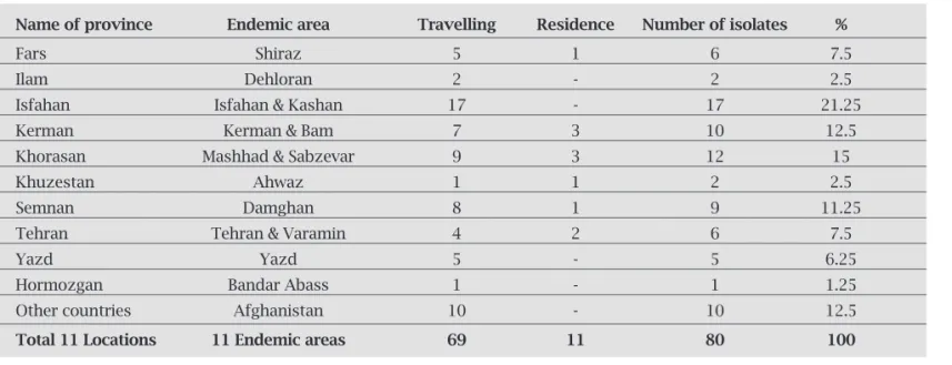
Table 1. Frequency of CL by place of residence and travel history
Name of province Endemic area Travelling Residence Number of isolates %
Fars Shiraz 5 1 6 7.5
Ilam Dehloran 2 - 2 2.5
Isfahan Isfahan & Kashan 17 - 17 21.25
Kerman Kerman & Bam 7 3 10 12.5
Khorasan Mashhad & Sabzevar 9 3 12 15
Khuzestan Ahwaz 1 1 2 2.5
Semnan Damghan 8 1 9 11.25
Tehran Tehran & Varamin 4 2 6 7.5
Yazd Yazd 5 - 5 6.25
Hormozgan Bandar Abass 1 - 1 1.25
Other countries Afghanistan 10 - 10 12.5
Total 11 Locations 11 Endemic areas 69 11 80 100
Figure 1: Single and multiple lesions in patients with CL in Iran.
round papular plaque with a 4-80 mm diameter. In ad-dition, double lesions were seen in 22% of cases; finally, 20% of patients presented multiple lesions between 3-7 mm (Figure 1).
Frequency of CL by place of residence and travel history
CL lesion location in body
The samples were taken from different sites of suspected lesions. Hand and arm were the most commonly affected sites (40.0%), while other major sites of lesion location were face and neck (37.5%). Detailed location of lesions is shown in Table 2.
DISCUSSION
Findings of this research revealed that Leishmania para-site was less detected by microscopic observation (38.5%) than by culture method (40%). This is in agreement with authors’ previous report,18 but not with Kumar et al. who
indicated that direct microscopy or parasite culture alone detected respectively 65.5% and 48.2% of the positive sam-ples.20
CL infection was more prevalent among males (63.8%) than females (36.2%). Previous reports con-firmed the same results indicating that males are more commonly infected than females, most likely because of their exposure, possibly as a result of occupational contact with the outdoor sand fl y vectors.18-21 Moreover, the highest
proportion of infection (31.3%) was recorded in 21-30 years age group, and the lowest (10%) was in the 31-40 years age group, which is in agreement with previous reports indi-cating more exposure as a result of educational and oc-cupational situations.18-21
According to the number of lesions in the patients, sin-gle lesions were more common (58%) than both double (22%) or multiple lesions (20%), which is consistent with a previous report by Talari et al.7 that showed 69.7% had
only one lesion.22 Multiple biting habits by the sand fl y
could explain the fi nding of about 42% of patients having more than one skin lesion.
Knowing the geographical location of infection acquisi-tion is important in order to focus on epidemiological pat-tern of disease. The majority of patients were resident or at least had a travel history to Isfahan and Kashan (21.25%), central Iran, while a few patients were traveling or living in Hormozgan (1.5%) and Khuzestan provinces (2.5%), south Iran or in Ilam province (2.5%), west part Iran. There is an agreement between our fi ndings with previous reports which confi rm disease dissemination for many years in the rural areas of Kashan, specially in Abouzeidabad. This might be due to the high transmission rate maintained by the pres-ence of both vector (Phlebotomus papatasi) and reservoir (Rhombomys opimus) in those areas.7,18 However some
stud-ies have indicated different results.23 Two species of
Leish-mania are involved in CL infections in Iran. L. major for ZCL and L. tropica for ACL. ACL has a disperse frequency whereas ZCL is found in many rural foci in the north, east and south of Iran.7,24
Hand and arm were the most commonly affected sites of suspected lesions (40%), followed by face and neck (37.5%), legs (20%) and other parts of the body (2.5%). Due to sand fl y attack to the exposed body areas to have a blood meal, the most of the lesions appear in the hands, face, and legs, and upper parts of the body, which may be due to the outdoor sleeping on the terraces or yards with-out using bed net during the summer. Previous report by Talari et al.7 confi rms this fi nding.22
Table 2. Body location of lesions among cutaneous leishmaniasis suspected patients in Iran
% Number of patients Lesion location
37.5 30 Face and neck
40 32 Hand and arm
20 16 Legs
2.5 2 Other parts of body
100 80 Total
Electrophoretic patterns of PCR products from genomic DNA
Electrophoresis patterns from each isolates were compared with reference strains of Iranian L. tropica and L. major. In this study, a single 620 bp band for identifi cation of L. ma-jor and an 800 bp band for detection of L. tropica were evi-denced. Thirty and twenty samples out of 50 were identifi ed as
L. major (64%) and L. tropica (36%), respectively (Figure 2).
Figure 2: Electrophoretic patterns of PCR products obtained from crude parasite genomic DNAs for Leishmania species detection 1; L. tropica, MHOM/IR/09/Mash-F 2; L. major
MRHO/IR/75/ER, M; marker.
2 M 1
620 bp
According to PCR fi ndings, two strains of L. tropica and
L. major are the causative agents of CL in Iran. Most of iso-lates collected from patients infected in the Mashhad area and the majority of isolates from Kashan were character-ized as L. tropica. These fi ndings lend support to previous publications about Leishmania species in Iran.12,24,25 Most of
the isolates characterized as L. major species were collected from patients with a travel history to endemic foci in Shi-raz, Chabahar and Sabzevar, as previously shown. Razmjou
et al. found a 23.2% prevalence rate among 1,000
inhab-itants of the three villages of Shiraz, with L. major being identifi ed in the majority of cases in rural areas of Shiraz.26
However, these fi ndings have not been confi rmed by other researchers. In the study by Rahbarian et al. all isolates de-tected by PCR method in Gonbad-e Qavus county, north Iran were of L. major.27 Parvizi et al (2010) also reported
L. major in Natanz, Isfahan province in centre of Iran, in
a rural ZCL focus.28 Conclusively, there are indication that
Isfahan has been a major endemic focus of ZCL in Iran for several decades.24
ACKNOWLEDGMENTS
The authors thank Dr. M Assmar and Dr. S Naddaf academic members of the Department of Parasitology, Pasteur Insti-tute of Iran for their assistance during this study.
REFERENCES
1. WHO Expert Committee, Geneva. Control of leishmaniasis. WHO Technical Report Series 1990; 793:9-10.
2. Desjeux P. Leishmaniasis: Current situation and new perspec-tives. Com Immunol Microbiol Infect Dis. 2004; 27:305. 3. Omidian M,Khosravi A, Nazari M, Rashidi A. The comparison
of histopathological fi ndings and polymerase chain reaction in lesions with primary clinical diagnosis of cutaneous leishma-niasis with negative smear. Pak J Med Sci 2008; 24: 96-99. 4. Nadim A, Javadian E, Seyedi-Rashti MA. Epidemiology of
leishmaniasis in Iran. In: Leishmania parasites and
leishmani-ases. Ardehali S, Rezai H, Nadim A. (Authors). 1994 2nd ed, Tehran University Press, Iran, pp. 178-80.
5. Yaghoobi-Ershadi MR, Hanafi -Bojd AA, Javadian E, Jafari R, Zahraei-Ramazani AR. A new focus of cutaneous
leish-maniasis caused by Leishmania tropica. Saudi Med J. 2002;
23:291-294.
6. Yaghoobi-Ershadi MR, Jafari R, Hanafi -Bojd AA. New epi-demics focus of zoonotic cutaneous leishmaniasis in central Iran. Ann Saudi Med. 2004; 24:98-101.
7. Talari SA, Rezvan Talaei R, Shajari GR. Childhood cutaneous leishmaniasis: report of 117 cases from Iran. Kor J Parasitol. 2006; 44(4):355-360.
8. Momeni A. Clinical picture of CL in Isfahan, Iran. Int J Der-matol. 1994; 33(4):260-265.
9. Salimi MA. Clinical and epidemiological comparison on the cutaneous leishmaniasis in the city and villages of Isfahan. Irn J Pub Health 2000; 2(4):214-219.
10. Moaddeb, Gettner S, Ardehali S. Studies on the causative agent of cutaneous leishmaniasis in Shiraz Iran. Irn J Med Sci 1993; 18:28-33.
11. Javadian E, Nadim A, Tahvildari A, Assefi V. Epidemiology of cutaneous leishmaniasis in Khorassan Iran. Bull Soc Path Exot. 1967; 69:140-143.
12. Nadim A, Seyedi-Rashti MA. A brief review of the epidemi-ology of various types of leishmaniasis in Iran. Acta Medica Iranica 1971; XIV:99-106.
13. Blum J, Desjeux P, Schwartz E, Beck B, Hatz C. Treatment of cutaneous leishmaniasis among travellers. J Antimicrob Chemother. 2004; 53:158-166.
14. Magill AJ. Cutaneous leishmaniasis in the returning traveler. Infect Dis Clin N Am. 2005; 19:241-266.
15. Allahverdiev AM, Uzun S, Bagirova M, Durdu M, Memisoglu HRA. Sensitive new micro culture method for diagnosis of cutaneous leishmaniasis. Am J Trop Med Hyg. 2004; 70(3): 294-297.
16. Ramirez JR, Agudelo S, Muskus C. Diagnosis of cutaneous leishmaniasis in Colombia: the sampling site within lesions in-fl uences the sensitivity of parasitological diagnosis. J Clin Mic. 2000; 38 (10):3768-3773.
17. Romero SA, Sampaio RN. Sensitivity of a vacuum aspiratory culture technique for diagnosis of localized CL in an endemic
area of L. braziliensis transmission. Mem Inst Oswaldo Cruz
1999; 94(4):505-508.
18. Farahmand M, Assmar M, Nahrevanian H, Farzanehnejad Z Piazak. Cutaneous leishmaniasis in patients referred to the Pasteur Institute of Iran during 2003-2006. Internet J Parasit Dis. 2008; 3(2):1-5.
19. Shahbazi F, Shahabi S, Kazemi B, Mohebali M, Abadi A, Zare Z. Evaluation of PCR assay in diagnosis and identifi cation of cutaneous leishmaniasis: a comparison with the parasito-logical methods. Parasitol Res. 2008; 103:1159-1162.
20. Kumar R, Bumb RA, Ansari N, Metha R. Cutaneous
leish-maniasis caused by leishmania tropica in Bikaner, India:
Parasite identification and characterization using molecu-lar and immunologic tools. Am J Trop Med Hyg. 2007; 76(5):896-901.
21. Hsia RY, Halpern J, FACEP D. Leishmaniasis. eMedicine Spe-cialties. Updated: Apr 20, 2010.
22. Talari SA, Shajari G, Talaei R. Clinical fi nding of Cutaneous Leishmaniasis as a new focus of Iran. Int J Infec Dis. 2006; 5(2):1-5.
23. Motazedian H, Noamanpoor B, Ardehali S. Characterization of Leishmania parasites isolated from provinces of the Islamic Republic of Iran. East Mediterr Health J. 2002; 8(2-3):338-44. 24. Nadim A, Faghih MA. The epidemiology of CL in Isfahan
province of Iran, I, the reservoir, II, the human disease. Trans R Soc Trop Med Hyg. 1968; 62:534-542.
25. Tashakori M, Ajdary S, Kariminia A, Mahboudi F,
Alimoham-madian MH. Characterization of Leishmania species and
L. major strains in different endemic areas of Cutaneous Leish-maniasis in Iran. Irn Biomed J. 2003; 7(2):43-50.
26. Razmjou SH, Hejazy H, Motazedian MH, Baghaei M, Emamy M, Kalantary M. A new focus of zoonotic cutaneous leishma-niasis in Shiraz, Iran. Trans Royal Soc Trop Med Hyg. 2009; 103:727-230.
27. Rahbarian N, Mesgarian A, Mahmoudi Rad M, et al.
Identi-fi cation of Leishmania Species isolated from human
cutane-ous leishmaniasis using PCR method. J Res Health Sci. 2009; 9(2):48-51.
28. Parvizi P, Baghban N, Alaee Novin E, Absavaran A.
Detec-tion, identification and molecular typing of Leishmania