Immunological effects of donor
lymphocyte infusion in patients
with chronic myelogenous leukemia
relapsing after bone marrow
transplantation
1Departamento de Análises Clínicas, Toxicológicas e Bromatológicas,
Faculdade de Ciências Farmacêuticas de Ribeirão Preto, Universidade de São Paulo, Ribeirão Preto, SP, Brasil
2Fundação Hemocentro de Ribeirão Preto, Ribeirão Preto, SP, Brasil
3Departamento de Clínica Médica, Faculdade de Medicina de Ribeirão Preto, Universidade de São Paulo, Ribeirão Preto, SP, Brasil
F.A. Castro1,2, P.V.B. Palma2, F.R. Morais2 and J.C. Voltarelli2,3
Abstract
Allogeneic bone marrow transplantation (alloBMT) is the only cura-tive therapy for chronic myelogenous leukemia (CML). This success is explained by the delivery of high doses of antineoplastic agents followed by the rescue of marrow function and the induction of graft-versus-leukemia reaction mediated by allogeneic lymphocytes against host tumor cells. This reaction can also be induced by donor lympho-cyte infusion (DLI) producing remission in most patients with CML who relapse after alloBMT. The immunological mechanisms involved in DLI therapy are poorly understood. We studied five CML patients in the chronic phase, who received DLI after relapsing from an HLA-identical BMT. Using flow cytometry we evaluated cellular activation and apoptosis, NK cytotoxicity, lymphocytes producing cytokines (IL-2, IL-4 and IFN-γ), and unstimulated (in vivo) lymphocyte prolif-eration. In three CML patients who achieved hematological and/or cytogenetic remission after DLI we observed an increase of the percent of activation markers on T and NK cells (CD3/DR, CD3/ CD25 and CD56/DR), of lymphocytes producing IL-2 and IFN-γ, of NK activity, and of in vivo lymphocyte proliferation. These changes
were not observed consistently in two of the five patients who did not achieve complete remission with DLI. The percent of apoptotic mark-ers (Fas, FasL and Bcl-2) on lymphocytes and CD34-positive cells did not change after DLI throughout the different study periods. Taken together, these preliminary results suggest that the therapeutic effect of DLI in the chronic phase of CML is mediated by classic cytotoxic and proliferative events involving T and NK cells but not by the Fas pathway of apoptosis.
Correspondence
J.C. Voltarelli
Fundação Hemocentro de Ribeirão Preto (FUNDHERP)
Av. Bandeirantes, 3900 14051-140 Ribeirão Preto, SP Brasil
Fax: +55-16-3963-9309 E-mail: jcvoltar@fmrp.usp.br
Research supported by the Center for Research in Cell-Based Therapy (CTC/FAPESP/No. 9814247-6), FUNDHERP, FAEPA-HCFMRPUSP and CNPq (No. 351860/92-4).
Received February 20, 2003 Accepted September 2, 2003
Key words
•Chronic myelogenous
leukemia
•Donor lymphocyteinfusion •Immune system
•Bone marrow
Chronic myelogenous leukemia (CML) is a myeloproliferative disorder characterized by the presence of a cytogenetic marker, the Philadelphia (Ph) chromosome, which derives from a reciprocal translocation be-tween chromosomes 9 and 22, i.e., t (9;22) (q34;q11), and the resultant production of constitutively activated bcr-abl tyrosine ki-nase in pluripotent hematopoietic progenitor cells (1). The disease evolves along three clinical phases of increasing severity: chronic phase, accelerated phase and blastic phase (1). Therapies currently available for CML in the chronic phase are chemotherapy (hy-droxyurea), interferon-α (IFN-α), tyrosine kinase inhibitors (STI571; Gleevec), alloge-neic bone marrow transplantation (alloBMT) and donor lymphocyte infusion (DLI) (1). Over the past 35 years, alloBMT has emerged as an effective and potentially curative thera-py of a variety of lethal hematological malig-nancies including CML. In the chronic phase the disease-free survival at 5 years reaches 70% for allografted patients in most centers (2).
The curative potential of alloBMT could be partially explained by delivery of very high doses of antineoplastic agents followed by the rescue of marrow function and the occurrence of the graft-versus-leukemia (GVL) effect (3), which is mediated by im-munocompetent donor cells contained in the graft. These cells exert antileukemic effects in a number of ways (3), but donor T cells could also induce graft-versus-host disease (GVHD), which is a major cause of trans-plant-related mortality (3).
Definite evidence for the GVL effect was provided when infusion of allogeneic lym-phocytes (DLI) from the BMT donor alone induced complete remission in CML pa-tients who relapsed after BMT (4). Clinical experience with BMT, DLI and experimen-tal tests in vitro indicates that CML is
par-ticularly susceptible to immune response regulation (5-7). There are few in vivo longi-tudinal studies investigating the
immuno-logical system of CML patients treated with DLI. In one of these studies, after CD4+ lymphocyte infusion T cells from four CML patients expressed a restricted T cell recep-tor Vß repertoire, which persisted for three months and coincided with the time of the cytogenetic response (8). In another study, Verfuerth et al. (9) showed an expansion of a few numbers of T cell clonotypes after DLI and thus a restricted T cell repertoire diver-sity. These two studies suggest that an oligo-clonal T cell response could be associated with the GVL effect after DLI in CML pa-tients.
The mechanisms underlying the antileu-kemic activity of DLI in CML need to be better understood to improve the current results of CML treatment and help the de-sign of more efficient and less toxic proto-cols of immunotherapy (10).
The aim of this study was to investigate effects of DLI on several immunological phenotypes and functions in CML patients who relapsed after HLA-identical related BMT. We evaluated immunological param-eters pre- and post-DLI (+11, +30, +60, +90 and +365 days) in the peripheral blood mono-nuclear cells (PBMC) of five CML patients in the chronic phase, three men and two women with a median age of 37.8 years (range 36-42 years). The chronology and characteristics of the relapse and of DLI, including number of infusions, dose of T cells infused, GVHD occurrence, and chi-merism studies are described in Table 1.
Patients were on regular follow-up at the Bone Marrow Transplantation Unit of the University Hospital of the School of Medi-cine of Ribeirão Preto, State of São Paulo, Brazil or of the Federal University of Rio Grande do Sul.
CML relapse was confirmed by the de-tection of the Ph chromosome in the bone marrow by cytogenetics and the bcr-abl
re-arrangement by RT-PCR.
Fa-culdade de Medicina de Ribeirão Preto, Uni-versidade de São Paulo. After signing the informed consent form, 20 ml heparinized blood and 5 ml EDTA-anticoagulated blood were collected, PBMC were isolated by den-sity gradient centrifugation on Ficoll-Hypaque (Sigma, St. Louis, MO, USA) (11) and the following tests were performed: natu-ral killer (NK) cell activity (12), apoptotic and activation markers on T lymphocytes, NK cells and stem cells, percentage of lym-phocytes producing cytokines (IL-2, IL-4 and IFN-γ) (13), and in vivo lymphocyte
proliferation (14).
For cell immunophenotyping, surface la-beling was performed by a direct fluores-cence technique using monoclonal antibodies (Becton-Dickinson, San Jose, CA, USA), against human CD3, CD8, CD19, CD56, CD34, CD3/CD25, CD3/DR, CD3/DQ, CD56/DR, CD19/DR, CD3/CD95 (Fas-R), CD56/CD95, CD19/CD95, CD34/CD95, CD8/CD95L (FasL), CD56/CD95L, CD34/ CD95L, and CD34/Bcl-2. To detect intracel-lular Bcl-2 protein, mononuclear cells were permeabilized with FACS permeabilizing solution (Becton-Dickinson). Flow cytom-etry analyses were carried out with a FACSort equipment (Becton-Dickinson) using the Cellquest software. The results are reported
as the percent of stained cells calculated from 10,000 events for all immunopheno-types and 50,000 cells for the CD34+ cell quantification, subtracted from the test back-ground percentage.
NK activity against K562 target cells was assessed by a flow cytometry assay (12) using the DIO membrane dye (Molecular Probes, Eugene, OR, USA), to stain live K562 cells and propidium iodide (Sigma) nuclear dye to stain dead cells. The percent of specific lysis was calculated by the for-mula:
% dead target cells x 100
100 - % (debris and fragments)
NK activity is reported as 40% lytic units per 107 cells.
The percent of lymphocytes producing IFN-γ, IL-2 and IL-4 was determined on PBMC stimulated with 25 ng/ml phorbol-myristate-acetate and 1 µg/ml ionomycin in the presence or absence of 10 µg/ml brefel-dine-A. All reagents were purchased from Sigma. For intracellular IL-2 and IFN-γ/IL-4 detection, cells were incubated for 24 and 4 h, respectively. After incubation, cells were fixed, permeabilized and labeled with spe-cific antibodies (Becton-Dickinson).
Analy-Table 1. Chronology and characteristics of donor lymphocyte infusions for the treatment of chronic myelogenous leukemia.
Patient BMT Relapse post-BMT DLI Infused T cells GVHD post-DLI Chimerism after last DLI
1 10/8/1996 7/18/1997 1st 9/12/1997 1st 1.0 x 10⁸/kg No
2nd 9/28/1997 2nd 1.7 x 10⁸/kg* No 86% XY-H
2 4/25/1995 7/14/1998 1st 7/27/1998 1st 1.0 x 10⁷/kg No
2nd 11/14/1998 2nd 1.8 x 10⁸/kg No 100% Ph negative
3rd 3/30/1999 3rd 6.7 x 10⁸/kg Day +100
3 11/1/1995 12/26/1996 1/7/1997 2.8 x 10⁸/kg No 100% XY-D (CNS relapse)
4 5/5/2000 9/1/2000 1/31/2001 2.1 x 10⁸/kg No Not done
5 3/10/1997 9/9/1999 1st 11/17/1999 1st 2.0 x 10⁷/kg No
2nd 2/14/2000 2nd 3.8 x 10⁸/kg No 100% XY-D
3rd 4/30/2000 3rd 5.3 x 10⁸/kg Day +130
Dates = month/day/year; BMT = bone marrow transplantation; CNS = central nervous system; D = donor; DLI = donor lymphocyte infusion; GVHD = graft-versus-host disease; H = host; Ph = Philadelphia chromosome.
ses were performed with a FACSort cytom-eter using the Cellquest software and the results are reported as percent of stained cells per 10,000 events and subtracted from the test background percentage.
In vivo mixed lymphocyte culture assay
was performed by a modification of the stan-dard mixed lymphocyte culture method. Briefly, 1 x 10⁵ mononuclear cells obtained from patients were cultured without stimula-tion in triplicate in trays for 6 days in a humidified atmosphere with 5% CO2. On the fifth day, cultures were pulsed with 1 µCi of tritiated thymidine and further incubated for 20 h at 37ºC in a humidified atmosphere with 5% CO2. After harvesting the cells, total isotope incorporation was determined by scintillation counting in a ß-counter.
Over the study period, three patients (Nos. 2, 4 and 5) achieved hematological remis-sion (defined as a white blood cell count <10 x 109/l, platelets <450 x 109/l, <5% circulat-ing immature cells, and absence of organo-megaly) and/or cytogenetic remission (de-fined as absence or 35-65% of Ph-positive metaphases by cytogenetic analysis), but the other two patients (Nos. 1 and 3) did not
achieve disease remission and died on days +83 and +98 post-DLI, respectively.
Clinical responses were associated with the infusion of larger cell doses and the occurrence of GVHD. However, immuno-logical changes were observed earlier than GVHD. Chimerism after DLI was studied cytogenetically and showed a predominance of donor cells in one patient (No. 5) who responded and of host cells in one patient (No. 1) who did not respond. One patient (No. 3) with central nervous system relapse showed donor cells in the marrow.
Statistical differences among the five study periods (pre-DLI, +11, +30, +60 and +90) in the groups of patients who achieved hematological and/or cytogenetic remission (patients 2, 4 and 5) and patients without remission (patients 1 and 3) were analyzed by the Friedman test followed by the Dunn test for multiple comparisons. A P value <0.05 was taken as significant. All analyses were performed using Prism 2.01 software. In the present study, we evaluated the effects of DLI as a therapeutic intervention on the immune system of five CML patients in the chronic phase, who relapsed after
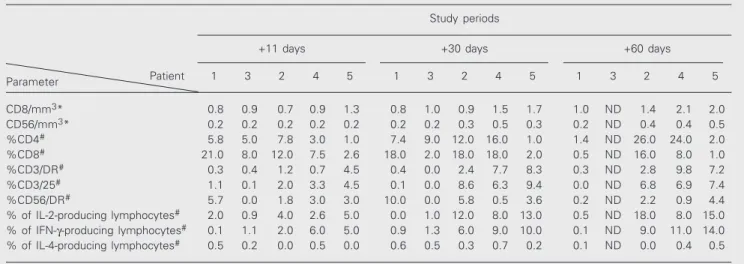
Table 2. Immunological evaluation of chronic myelogenous leukemia patients after donor lymphocyte infusion treatment.
Study periods
+11 days +30 days +60 days
Parameter Patient 1 3 2 4 5 1 3 2 4 5 1 3 2 4 5
CD8/mm3* 0.8 0.9 0.7 0.9 1.3 0.8 1.0 0.9 1.5 1.7 1.0 ND 1.4 2.1 2.0
CD56/mm3* 0.2 0.2 0.2 0.2 0.2 0.2 0.2 0.3 0.5 0.3 0.2 ND 0.4 0.4 0.5
%CD4# 5.8 5.0 7.8 3.0 1.0 7.4 9.0 12.0 16.0 1.0 1.4 ND 26.0 24.0 2.0
%CD8# 21.0 8.0 12.0 7.5 2.6 18.0 2.0 18.0 18.0 2.0 0.5 ND 16.0 8.0 1.0
%CD3/DR# 0.3 0.4 1.2 0.7 4.5 0.4 0.0 2.4 7.7 8.3 0.3 ND 2.8 9.8 7.2
%CD3/25# 1.1 0.1 2.0 3.3 4.5 0.1 0.0 8.6 6.3 9.4 0.0 ND 6.8 6.9 7.4
%CD56/DR# 5.7 0.0 1.8 3.0 3.0 10.0 0.0 5.8 0.5 3.6 0.2 ND 2.2 0.9 4.4
% of IL-2-producing lymphocytes# 2.0 0.9 4.0 2.6 5.0 0.0 1.0 12.0 8.0 13.0 0.5 ND 18.0 8.0 15.0
% of IFN-γ-producing lymphocytes# 0.1 1.1 2.0 6.0 5.0 0.9 1.3 6.0 9.0 10.0 0.1 ND 9.0 11.0 14.0
% of IL-4-producing lymphocytes# 0.5 0.2 0.0 0.5 0.0 0.6 0.5 0.3 0.7 0.2 0.1 ND 0.0 0.4 0.5
*Absolute number x 103. #Results are reported as percent of positive cells subtracted from the values obtained pre-donor lymphocyte infusion.
BMT. The immune system was investigated immunophenotypically (by determination of activation and apoptotic markers on T, B and NK cells) and functionally (by NK activity, production of cytokines and lymphocyte pro-liferation). We also monitored the expres-sion of Bcl-2, Fas and FasL antigens on CD34+ stem cells. The results were associ-ated with the achievement of a hematologi-cal and cytogenetic response.
The percent of activation markers on T and NK cells (CD3/DR, CD3/CD25 and CD56/DR, Table 2) showed a significant increase in patients who achieved remission post-DLI but not in patients who did not achieve remission. In the entire group of patients the percent values of cells positive for CD3/Fas, CD56/Fas, CD34/Fas, CD56/ FasL, CD8/FasL, CD34/Fas, CD34/DR, CD34/DQ, CD34/Bcl-2, CD19/DR, CD19/ Fas did not change after DLI therapy through-out the different study periods. On the basis of these results, apoptosis, through the Fas-FasL pathway, seems not to be involved in the antileukemic effect of DLI. In fact, Deininger et al. (15) and Ravandi et al. (16) reported that Ph-positive progenitor cells are resistant to apoptosis (Fas-FasL pathway), providing a growth advantage to leukemic cells over normal cells. Interestingly, all pa-tients presented a reversed CD4/CD8 cell ratio on day +11 post-DLI suggesting an involvement of CD8 T cells in the response against relapsing CML (Table 2).
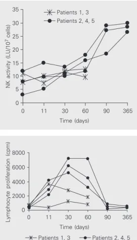
NK activity tended to increase in all pa-tients, but was statistically significant only in patients who achieved remission (Figure 1). These results agree with those reported by Pawelec et al.(17), who demonstrated a correlation between NK cell activation and elimination of bcr-abl-positive cells in vitro.
Therefore, it seems that NK cell cytotoxicity plays a role in the antileukemic effector mech-anism in CML, contributing to hematologi-cal and cytogenetic remission. Thus, the re-lationship between NK activity and the GVL effect mediated by DLI needs to be better
investigated.
Patients who presented a response to DLI treatment showed a statistically significant increase in the percent of lymphocytes ca-pable of producing IL-2 and IFN-γ, in con-trast to patients who maintained relapse (Table 2). These cytokines are involved in classic Th1 cell-mediated functions such as clonal expansion of cytotoxic T lympho-cytes, macrophage activation, up-regulation of co-stimulatory and major histocom-patibility complex molecules on antigen-pre-senting cells, T cell proliferation, and NK activation. IL-2 and IFN-γ improve the inter-action between host and donor cells and prevent the escape of leukemic cells from the immune system (18). In contrast, the percent of lymphocytes capable of produc-ing IL-4, a Th2 cytokine, did not change over the study periods in any patient (Table 2).
In vivo lymphocyte proliferation tended to
Figure 1. Natural killer (NK) cell activity in chronic myelogenous leukemia patients after donor lymphocyte infusion (DLI). NK activity is reported as 40% lytic units (LU) per 107 cells. NK
cyto-toxicity increased significantly (P < 0.0009) in patients who achieved remission (numbers 2, 4 and 5) after DLI, but remained unchanged (P > 0.05) in patients 1 and 3 who did not respond to DLI (Friedman test followed by Dunn test for multiple compari-sons). Note that the abscissa scale is not linear.
Figure 2. Lymphocyte prolifera-tion in chronic myelogenous leu-kemia patients after donor lym-phocyte infusion (DLI). Prolifera-tion is expressed as incorpo-rated tritiated thymidine (cpm). Lymphocyte proliferation in-creased significantly (P = 0.0028) in patients who achieved remis-sion (numbers 2, 4 and 5) after DLI treatment, but remained un-changed (P > 0.05) in patients 1 and 3 who did not respond to DLI (Friedman test followed by Dunn test for multiple compari-sons). Note that the abscissa scale is not linear
Lymphocyte proliferation (cpm)
8000
6000
4000
2000
0
0 11 30 60 90 365
Time (days)
Patients 2, 4, 5 Patients 1, 3
NK activity (LU/10
7 cells)
35
30
25
20
15
10
5
0
0 11 30 60 90 365
increase in all patients between days +11 and +60, but this increase in proliferation was statistically significant only in patients 2, 4 and 5 who achieved remission after DLI (Figure 2). This result fits the clinical observations that allogeneic T cells are capable of generating tumor-specific immunity and could be used as immunotherapy for CML (19). In fact, the efficacy of allogeneic donor lymphocytes based on histocompatibility differences between do-nors and recipients is essential to obtain remis-sion in CML patients (20).
Taken together, these results suggest that DLI could induce a GVL effect and up-regulate the immune response in CML pa-tients. Our preliminary observations also agree with previous clinical studies indicat-ing that an allogeneic immune response
con-tributes to the control of CML after BMT relapse and support the further use of DLI associated with other CML therapies such as IFN-γ, tyrosine-kinase inhibitors and alloBMT. If confirmed in a study with more patients, these data will provide important information to design effective immunothera-pies for other malignancies such as multiple myeloma, lymphoma and acute leukemias.
Acknowledgments
We are grateful to the Bone Marrow Trans-plantation Unit of the Federal University of Rio Grande do Sul, specifically to Dr. Henrique Bittencourt, for sending samples and data for one patient included in the study.
References
1. Sawyers CL (1999). Chronic myelogenous leukemia. New England Journal of Medicine, 340: 1330-1340.
2. Clift RA & Anasetti C (1997). Allografting for chronic myeloid leu-kaemia. Baillieres Clinical Haematology, 10: 319-336.
3. Weiden PL, Flournoy N, Thomas ED, Prentice R, Fefer A, Buckner CD & Storb E (1979). Antileukemic effects of graft versus host disease in human recipients of allogeneic marrow grafts. New Eng-land Journal of Medicine, 300: 1068-1073.
4. Kolb JH, Mittermuller J, Clemm C, Ledderooe G, Brehm G, Heim M & Wilmans W (1990). Donor leukocyte transfusions for treatment of recurrent chronic myelogenous leukemia in marrow transplant pa-tients. Blood, 76: 2462-2465.
5. Cullis JO, Jiang YZ, Schwarer AP, Hughes TP, Barrett AJ & Goldman JM (1992). Donor leukocyte infusions for chronic myeloid leukemia in relapse after allogeneic bone marrow transplantation. Blood, 79: 1379-1381.
6. Jiang YZ, Barrett AJ, Goldman JM & Mavroudis DA (1997). Associa-tion of natural killer cell immune recovery with a graft-versus-leuke-mia effect independent of graft-versus-host disease following allo-geneic bone marrow transplantation. Annals of Hematology, 14: 1-6.
7. Guilhot F & Lacotte-Thierry L (1998) Interferon-α: mechanisms of action in chronic myelogenous leukemia in chronic phase. Hematol-ogy and Cell Therapy, 40: 237-239.
8. Claret EJ, Alyea EP, Orsini E, Pickett CC, Collins H, Wang Y, Neuberg D, Soiffer RJ & Ritz J (1997). Characterization of T cell repertoire in patients with graft-versus-leukemia after donor lymphocyte infu-sion. Journal of Clinical Investigation, 100: 855-866.
9. Verfuerth S, Peggs K, Vyas P, Barnett L, O’Reilly RJ & Mackinnon S (2000) Longitudinal monitoring of immune reconstitution by CDR3 size spectratyping after T-cell-depleted allogeneic bone marrow transplant and the effect of donor lymphocyte infusions on T-cell repertoire. Blood, 95: 3990-3995.
10. Luznik L & Fuchs EJ (2002). Donor lymphocyte infusions to treat hematologic malignancies in relapse after allogeneic blood or
mar-row transplantation. Cancer Control, 9: 123-135.
11. Boyum A (1977). Isolation of lymphocytes, granulocytes and macro-phages. Scandinavian Journal of Immunology, 5 (Suppl): 9-15. 12. Chang L, Gusewitch AG, Chritton DBW, Folz JC, Lebeck LK &
Cannarella SLN (1993). Rapid flow cytometric assay for the assess-ment of natural killer cell activity. Journal of Immunological Meth-ods, 166: 45-52.
13. Jung T, Schauer U, Heusser C, Neumann C & Rieger C (1993). Detection of intracellular cytokines by flow cytometry. Journal of Immunological Methods, 159: 197-207.
14. Bain B, Vas MR & Lowenstein L (1964). The development of large immature mononuclear cells in mixed leukocyte cultures. Blood, 23: 108-116.
15. Deininger MWN, Goldman JM & Melo JV (2000). The molecular biology of chronic myeloid leukemia. Blood, 96: 3343-3356. 16. Ravandi F, Kantarjian HM, Talpaz M et al. (2001). Expression of
apoptosis proteins in chronic myelogenous leukemia: associations and significance. Cancer, 91: 1964-1972.
17. Pawelec G, da Silva P, Max H, Kalbacher H, Schmidt H, Bruserud O, Zugel U, Baier W, Rehbein A & Pohla H (1995). Relative roles of natural killer- and T cell-mediated anti-leukemia effects in chronic myelogenous leukemia patients treated with interferon-alpha. Leu-kemia and Lymphoma, 18: 471-478.
18. Murray JS (1998). How the MHC selects Th1-Th2 immunity. Immu-nology Today, 19: 157-162.
19. Smit WM, Rijnbeek M, van Bergen CAM, Willemze R & Falkenburg JHF (1998). Generation of leukemia-reactive cytotoxic T lympho-cytes from HLA-identical donors of patients with chronic myeloid leukemia using modifications of a limiting dilution assay. Bone Marrow Transplantation, 21: 553-560.