*Correspondence: C. L. Vituri. Departamento de Análises Clínicas, Centro de Ciências da Saúde – CCS, Universidade Federal de Santa Catarina, 88010-970 - Florianópolis - SC, Brasil. E-mail: [email protected]
A
rti
Pharmaceutical Sciences vol. 47, n. 2, apr./jun., 2011
Assessment of ibrosis and vascularization of bone marrow
stroma of Chronic Myeloid Leukemia patients treated with imatinib
mesylate and their relationship with the cytogenetic response
Caroline Regina de Jesus
1, Lee I-Ching
1, Teresinha de Jesus Carvalho Neiva
2,
Cidônia de Lourdes Vituri
2,*1Laboratory of Pathology, Oncology Research Center (CEPON), Florianópolis, Brazil, 2Center for Health Sciences, Department of Clinical Analyses, Universidade Federal de Santa Catarina
Chronic Myeloid Leukemia (CML) is a myeloproliferative disease characterized by the presence of the Philadelphia chromosome (translocation between chromosomes 9 and 22), resulting in the formation of the hybrid BCR-ABL protein. Currently, the treatment of CML patients is performed with imatinib mesylate (IM), which promotes the elimination of leukemic cells by inhibiting the kinase activity of BCR-ABL. This study evaluated the effectiveness of IM by monitoring 22 CML patients in a chronic phase treated at the CEPON/SC with IM for a minimum follow-up period of two years. Cytogenetic Response (CR) and bone marrow biopsies (BMB) were evaluated before and after IM treatment. BMB were evaluated by detection of reticulin degree and vascularization. The results were correlated to the CR. Mean time to achieve CR was 9 months and was attained by 77.27% of the patients. The results from the initial BMB analysis showed that 59.09% presented reticulin of between 2+ and 4+ whereas after treatment, only 27.17% presented this degree. With regard to vascularization of the initial sample, 90.91% were graded between II and IV, whereas after treatment, 40.91% had this degree. The results suggest a positive correlation of degree of reticulin and vascularization with CR.
Uniterms: Chronic myeloid leukemia/treatment. Imatinib mesylate/effectiveness/leukemia treatment. Bone marrow/biopsies. Fibrosis. Vascularization.
A Leucemia Mielóide Crônica (LMC) é uma doença mieloproliferativa caracterizada pela presença do cromossomo Filadélia (translocação entre os cromossomos 9 e 22), que resulta na formação da proteína híbrida BCR-ABL. Atualmente o tratamento de pacientes com LMC é realizado com mesilato de imatinibe (MI), o qual promove a eliminação das células leucêmicas pela inibição da atividade quinase de BCR-ABL. O presente estudo avaliou a eicácia do MI por meio do acompanhamento de pacientes portadores de LMC em fase crônica, atendidos no CEPON/SC tratados com MI pelo tempo mínimo de dois anos. Foram avaliadas a Resposta Citogenética (RC) e as biópsias de medula óssea (BMO) antes e após o tratamento com MI. As BMO foram avaliadas quanto ao grau de reticulina e vascularização. Os resultados correlacionaram-se com a RC cujo tempo médio para obtenção da RC foi de 9 meses, sendo atingida por 77.27% dos pacientes. Na primeira BMO, 59.09% dos pacientes apresentaram grau de reticulina entre 2+ e 4+ e após o tratamento, apenas 27.17% apresentaram esta graduação. Quanto à vascularização da primeira amostra, 90.91% foram graduadas entre II e IV e após o tratamento, 40.91% apresentaram esta graduação. Os resultados sugerem uma correlação diretamente proporcional entre os graus de reticulina e vascularização com a RC.
INTRODUCTION
Chronic Myeloid Leukemia (CML) is a clonal dise-ase characterized by the presence of the Philadelphia (Ph1)
chromosome, which results from a reciprocal and balanced translocation between chromosomes 9 and 22 (Sawyers, 1999). The molecular consequence of this translocation is the generation of a hybrid protein called BCR-ABL, which presents increased tyrosine kinase activity, compromising the regulation of the cell cycle (Faderl et al., 1999; Sattler, Grifin, 2003). The diagnostic presumptive of CML usu-ally arises with the results obtained in blood cell counts. The total leukocyte count is elevated and is invariably greater than 25,000/µL. The total leukocyte count rises progressively in untreated patients. Granulocytes at all stages of development are present in the blood and are generally morphologically normal. Blast cell prevalence is approximately 3% but can range from 0% to 10%. The platelet count is elevated in approximately 50% of pa-tients and largely normal in the rest. Deinitive diagnosis is obtained by evaluation of bone marrow biopsy (BMB), myelogram and cytogenetic examination (Lichtman et al., 2006; Brasil, Ministério da Saúde, 2001; Geary, 2000).
The hematopoietic microenvironment of bone mar-row (BM) is also changed in CML. This microenvironment is composed of stromal cells, accessory cells and their pro-ducts (extracellular matrix proteins and cytokines), which inluence the self-renewal, proliferation and differentiation of hematopoietic stem and progenitor cells (Vituri et al., 2000; Vituri et al., 2006). Collagen type III, visualized by silver staining, is generally increased at diagnosis (reticulin ibrosis). In almost half of patients, the reticulin is remarkably increased in BM, and this increase shows a correlation with the proportion of megakaryocytes (Licht-man et al., 2006; Guimarães et al., 2006). The BM of CML patients have a two-fold increase in microvessel density compared to healthy controls while in CML there is more angiogenesis in BM than is found in other forms of leuke-mia. The concentration of vessels, or the BM microvessel density (MVD), can easily be investigated by immunohis-tochemistry in hematological disorders because the BM is a primary site of disease activity and readily accessible tissue. This measurement is also useful for evaluating the prognosis of patients (Panteli et al., 2004).
Regarding treatment of CML, imatinib mesylate (IM) is currently the irst-line drug of choice. This drug, chemically known as 2-phenylaminopyrimidine, entered clinical trials in 1998 as STI571. The drug is a potent and speciic inhibitor of all ABL related kinases, Platelet Deri-ved Growth Factor (PDGF) and c-kit. As the hybrid protein BCR-ABL is responsible for the occurrence of events
related to the LMC, its inhibition leads to the elimination of leukemic cells (Savage, Antman, 2002).
This study examined the BM stromal of CML patients treated with IM correlating to other laboratory parameters. The effectiveness of the drug was analyzed over a two-year period of monitoring based on observa-tion of the time needed for patients to achieve CR. The BMB were evaluated for degree of reticulin (ibrosis) and degree of vascularization. Finally, the results obtained from the analysis of BMB collected after treatment, were correlated to CR.
PATIENTS AND METHODS
Firstly, a survey was carried out involving all regis-tered patients with CML treated using IM at the Oncology Research Center Units of Santa Catarina, in Florianópo-lis, Santa Catarina (CEPON/SC) in the period spanning from August 2005 to August 2007. During the period, 67 patients were enrolled, all from Santa Catarina. Only the 22 patients that presented at least two reports of anatomi-cal and pathologianatomi-cal bone marrow, one of diagnosis and another after 6±2 months of treatment with IM, were con-sidered for the study. Therapy with IM, at a daily dosage of 400 mg was given throughout the study, and no other cytotoxic or supportive agents were combined to IM.
Inclusion criteria
Patients with CML at a chronic phase, Ph1
chromoso-me positive, treated with IM, submitted to two BMB, and able to perform histological sections, were included. The determination of the stage of disease was done according to the laboratory peripheral blood indings (Table I) and indings on BMB (Table 2) (Sawyers, 1999). All patients included were previously treated with IFN-a before star-ting the use of IM. Patients were informed in accordance with the guidelines of the local ethics committee (CONEP/ ANVS). The demographics and baseline characteristics of the patients included in the study are shown in Table III.
TABLE I - Laboratory findings of peripheral blood of CML
patients, at a chronic phase
Peripheral Blood
Leukocytosis (> 25,000/mm3)
Platelets: Increased or normal Basophilia
Reduced activity of leukocyte alkaline phosphatase
Cytogenetic response
The analysis of Cytogenetic Response(CR) was done by examining the medical records of patients. CR was acknowledged when patients achieved Major Cytoge-netic Response, according to current treatment guidelines for CML in Brazil (Table IV). The tests were evaluated in-dividually and were used to analyze a possible correlation
between CR and degrees of reticulin and vascularization. The data used to construct the graph considered the per-centage of chromosome Ph1 positive cells and the grading
of the second BMB, related to the degree of reticulin and vascularization (Figures 6 and 7).
Gomori staining
BMB slides were prepared from parafin-embedded blocks. After deparaffinization, the BMB slides were immersed in potassium permanganate solution 0.5% (Quimex) for 2 minutes. The slides were then washed and immersed quickly in oxalic acid solution 5% (Ve-tec). Subsequently, these were immersed in alumen iron solution 5% (Reagen) for 2 minutes. After washing, the slides were immersed in silver ammonium solution - Gomori (Vetec) for 2 minutes. After immersion in distil-led water, the slides were quickly immersed in formalin TABLE II - Findings on BMB of CML patients, at a chronic phase
Bone Marrow Hypercellularity
Reduction of intramedullary fat Increased number of megakaryocytes Less than 10% of blasts and promyelocytes Increased myeloid to erythroid cell ratio
TABLE III - Patient demographics and baseline characteristics
Patient demographics and baseline characteristics Values
Nº patients 22
MaleSex, % 50
MedianAge (range) 42.36 (18 to 69y)
History of illness, usage of INF-α, number of patients (%)
Hematologic/cytogenetic resistance 12 (54.54)
Intolerance 7 (31.82)
No reason 3 (13.64)
INF-α therapy duration, number of patients (%)
> 1 year 15 (68.18)
< 1 year 7 (31.82)
Splenomegaly, number of patients (%) 10 (45.46)
Time diagnosed with disease (early treatment with imatinib), number of patients (%)
< 1 year 0 (0)
> 1 year 22 (100)
Hemoglobin level at diagnosis, number of patients (%)
< 12.0 g/dl 14 (63.64)
> 12.0 g/dl 8 (36.36)
Leukocytes count at diagnosis, number of patients (%)
< 10,000/mm3 0 (0)
10,000-50,000/ mm3 15 (68.18)
> 50,000/ mm3 7 (31.82)
Platelet count at diagnosis, number of patients (%)
< 450,000/ mm3 13 (59.09)
> 450,000/ mm3 9 (40.91)
solution 10% (Vetec) and washed under running water. Subsequently, the slides were immersed in chloride old solution 1% (Synth) and in sodium thiosulfate solution 5% (Vetec). Finally, the sections were coverslipped with a speciic ixation luid (Entellan, Merck) (Michalany J, 1980).
- Measurement of fibrosis degree (reticulin fiber network)
The histological sections of BMB were examined using a light microscope. The entire section was scanned at 100 x magniication. Positive and negative control sam-ples were evaluated concomitantly. The positive controls were from patients with Primary Myeloibrosis (Chronic Idiopathic Myeloibrosis) and the negative controls were from patients without hematological disease. The reticulin ibers were graded as 0 to 4+. The classiication used was based on the Bauermeister Graduation (Alves, 2009). The normal samples had a degree of reticulin 0, indicating ab-sence of reticulin, and served as negative controls (Figure 1A). Samples with the presence of thin and scattered ibers
or a focal network had degree 1+, also normal. The visuali-zation of a thin iber network in most of the fragment, and absence of thick ibers, represent degree 2+. Samples with a diffuse network of thin ibers with scattered thick ibers and absence of collagen were graded 3+. Samples with a diffuse network of coarse ibers and bundles of collagen were graded as 4+, similar to the positive controls (Figure 1B) (Alves, 2009; Bueso-Ramos et al., 2004).
Immunohistochemistry for CD34
Bone marrow microvessels were visualized by an immunohistochemical labelled for CD34. Although the CD34 antibody also stained myeloid progenitors, the number of cells stained was suficiently small as not to interfere with our analysis. Nevertheless, vessel speciicity the CD34-stained stroma considered for analysis was do-cumented thoroughly. In general, CD34 has been found to be a useful antigen for assessing intratumor angiogenesis (Kvasnicka et al, 2004).
Slides of the BMB were prepared from parafin-em-bedded blocks. After deparafinization, slides were steam pretreated in a solution containing 200 mL of methanol and 4 mL of hydrogen peroxide (Merck, concentration 30%) for 20 minutes, protected from the light. After wa-shing with buffer at pH 7.2, the slides were immersed in a citrate buffer, with pH 6.0 called Target Retrieval (Dako), for exposure to the antigen, for 45 minutes in a water bath at 85 °C. After cooling the solution for approximately 20 minutes, the slides were washed in buffer solution. Before incubating the slides, the tissue sections were demarcated with a hydrophobic pen. The primary antibody (CD34, TABLE IV - Criteria for classiication of CR (Brasil. Ministério
da Saúde, 2001)
Cytogenetic Response
Major Cytogenetic Response:
- Complete Cytogenetic Response: 0% Ph1 positive cells
- Partial Cytogenetic Response: 1% to < 35% Ph1 positive cells
Minor Cytogenetic Response: 35% to 90% Ph1 positive cells Lack of Cytogenetic Response: > 90% Ph1 positive cells
FIGURE 1A - Histological sections of normal BMB (degree
0) stained by Gomori silver solution, 100 x magniication. The ibers are stained in gray.
FIGURE 1B - Histological section of BMB stained by Gomori
Novocastra) was then incubated overnight in a 1:100 dilution at room temperature. The biotin-streptavidin kit (Novocastra) was used for antigen visualization and was incubated for 30 minutes at room temperature. Finally, Diaminobenzidine (DAB, Dako) was used as the substrate for the abovementioned marker enzyme. The sections were counterstained with hematoxylin (Merk) and coverslips were applied with a speciic ixation luid (Entellan, Merk) (Dabbs, 2006).
- Measurement of microvessel density
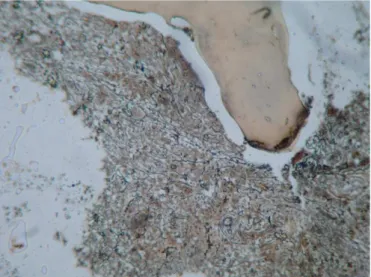
The BMB histological sections, stained immuno-histochemically for CD34, were examined using a light microscope in order to identify the areas with intense vascularization. The entire section was scanned at 100 x magniication. Three areas, with the highest number of microvessels, were chosen and deined as the hot spots. Microvessels were then counted at 400 x magniication in each of these hot spots.
The final MVD number was assigned by taking the average of the three separate visual counts. Micro-vessels were identiied as endothelial cells, either single or clustered in nets or tubes, clearly separated from one another, either with or without a lumen (Figure 2A). Vessels with muscular walls and vessels in the perios-teum and open sinusoids were excluded. Measurement also excluded areas occupied by fat. The vascularization was graded from I to IV degrees (Kvasnicka, 2004). Positive and negative control samples were evaluated concomitantly. The positive controls were obtained from patients with Multiple Myeloma whereas the negative controls were from patients without hematological disease (Figure 2B).
Data and statistical analysis
The percentage of patients that achieved CR in the two years of treatment and the degree of reticulin and vas-cularization of the BMB were evaluated. The irst biopsy was performed before IM treatment and the second sample was collected after at least six months of IM treatment.
The tests of correlation were also performed be-tween CR and the degree of vascularization and reticulin from the second BMB. The patients who responded were those with less than 35% of Ph1 chromosome positive cells
on the cytogenetic examinations. The evaluation of this correlation was veriied with the non-parametric test of Spearman, considering p< 0.05 as the minimum level of signiicance. Results were expressed in absolute values of correlation (r), where “0” indicates total absence of linear correlation and “1” a perfect linear relationship. Statistical analysis was performed using the INSTAT-2 program.
RESULTS
1- The study was conducted in 22 individuals tre-ated with IM that met the inclusion criteria. During the two years of monitoring, 77.27% patients achieved CR (Figure 3). By the sixth month, nearly 22% of patients had achieved CR and at the tenth month, 50% had attained this response. The last patient attained CR just in the fourteenth month. Beyond this timepoint, no other patients achieved CR throughout the rest of the monitoring. The average time taken to attain CR was 10 months.
FIGURE 2A - Histological section of normal (degree I) BMB
stained immunohistochemically for CD34, 100 x magniication.
F I G U R E 2 B - Histological section of BMB stained
2- Regarding evaluation of reticulin degree of the irst BMB, 59.09% of the samples showed signiicant i-brosis and were graded between 2+ and 3+ while 13.64% revealed myelofibrosis (degree +4). Only 9.09% of samples collected before treatment with IM were normal (degree 0) and 22.73% were graded as 1+. After treatment, less than half the samples were graded between 2+ and 3+ (22.73%), while no reticulin ibers (degree 0) were visualized in 45.46% of the BMB, and only 4.54% were graded as 4+ (Figure 4).
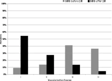
3-For the degree of vascularization of the irst BMB, 77.27% revealed a signiicant increase in the number of vessels, and were graded between III and IV. Only 9.09% of the pre-treatment samples were considered as having normal vascularity (degree I) and a small number of sam-ples (13.64%) were graded as II.
After IM treatment, the number of samples with a
degree of vascularization between III and IV was about 4 times lower compared to the number obtained before treatment (18.18%) and 27.27% of samples were graded at II. Most samples (54.54%) were considered to have normal vascularity and were graded as I (Figure 5).
4- The evaluation of reticulin degree of the BMB collected after IM was correlated to CR (Table V). Among patients who had their BMB graded at 0 reticulin, 90% achieved CR. The patients who presented 1+, 2+ and 3+ reticulin in the second sample, 80%, 75% and 50% of these achieved CR, respectively. None of the patients with second BMB graded as 4+ reticulin achieved CR. These data are presented in Figure 6. The results of cytogenetic tests were assessed from each patient record, observing the percentage of chromosome Ph1 positive cells and the
reticulin degree of the second BMB. Points below the line represent patients who achieved CR and points above the line represent patients who did not achieve CR. The value of “r” obtained by Spearman’s correlation test was 0.8082 and the value of P was < 0.0001.
5- The results obtained from the assessment of de-gree of vascularization of BMB collected after IM were correlated with CR (Table VI). No patients with BMB graded as IV achieved CR. Among patients with degrees I, II and III of vascularization, 91.67%, 83.33% and 33.33% achieved CR, respectively. These results show that the percentage of patients responding increases as the degree of vascularization decreases. In this situation, there is also an inverse correlation with CR. These data are also shown in graph form (Figure 7). For the construction of this graph, the results of cytogenetic tests from each patient were used, FIGURE 3 - Time required for patients to achieve CR. Patient
monitoring was performed for 2 years.
FIGURE 4 - BMB distribution of CML patients included in the
study, according to Fibrosis/Reticulin degree (Gomori stain).
FIGURE 5 - BMB distribution of CML patients included in the
noting the percentage of chromosome Ph1 positive cells and
vascularization degree of BMB collected after treatment. Points below the line represent patients who achieved CR and points above the line represent patients who did not achieve CR. The value of “r” obtained by Spearman’s cor-relation test was 0.8179 and the value of P was < 0.0001.
DISCUSSION
The therapy used to treat CML patients was extremely limited up until the late 1970s. This changed when IFN-α
came into use. The disadvantage of the use of IFN-α is that
the reversal of myeloibrosis occurs only at high doses of the drug and the side effects of this medication are signiicant. Attaining CR is the main goal of treatment, since this is TABLE V - Relationship between Reticulin degree of BMB from CML patients collected after IM treatment and CR
Reticulin degree of BMB (after 6 to 12
months of IM treatment) Distribution of BMB after IM by reticulin degree (%) % of patients that achieved CR
0 45.46 90
+1 27.27 80
+2 13.64 75
+3 9.09 50
+4 4.54 0
FIGURE 6 - Distribution of patients according to reticulin
degree on second BMB and percentage of Ph1 chromosome
positive cells obtained in first cytogenetic examination performed after treatment with IM. Points below the line represent patients who achieved CR, while points above the line represent non- responders. This graph was constructed assuming a cytogenetic response occurs when there is less than 35%
Ph1 chromosome positive cells on the cytogenetic test (Major
Cytogenetic Response).
TABLE VI - Relationship between Vascularization degree of BMB from CML patients collected after IM treatment and CR
Vascularization degree of BMB (after 6
to 12 months of IM treatment) Distribution of BMB after IM by vascularization degree (%) % of patients that achieved CR
I 54.54 91.67
II 27.27 83.33
III 13.64 33.33
IV 4.54 0
FIGURE 7 - Distribution of CML patients according to
vascularization degree on second BMB and percentage of
Ph1 chromosome positive cells obtained in first cytogenetic
examination performed after treatment with IM. Points below the line represent patients who achieved CR, while points above the line represent non-responders. This graph was constructed assuming a satisfactory response occurs when there is less than
directly related to patient survival. In addition, individuals who do not respond are called refractory and this event is associated to disease progression and worse prognosis (Gui-marães et al., 2006; Buesche et al., 2004; Sattler, Grifin, 2003; Goldman, Mello, 2003; Druker, Talpaz, Resta, 2002).
The results obtained showed that 77.27% of patients presented less than 35% of chromosome Ph1positive cells
in the cytogenetic tests during the monitoring period. The average time to achieve CR response was ten months. Although some patients proved to be refractory to IM, they continued the treatment for at least two years. The percentage of patients that achieved CR in this study exceeded the igure reported by another study in patients resistant to IFN-α, posteriorly treated with IM (Guilhot,
2004). The cited study, which has been used as a compa-rison, considered the stage of disease, and 60% of patients who were at a chronic phase achieved CR. In contrast, the CR attainment rates among patients who were at an accelerated phase and blast crisis were only 24% and 16%, respectively. The results of this study and those of other researchers have shown that patients who failed to attain CR within 2 years of treatment are probably refractory and present poor prognosis (Guilhot, 2004).
The evaluation of ibrosis by reticulin analysis was investigated because the BMB of CML patients generally exhibit a significant increase in reticulin fibers (Zago, Falcão, Pasquini, 2001). Moreover, myeloibrosis is asso-ciated historically with poor CML prognosis. The platelet-derived growth factor (PDGF) is a cytokine involved in myeloibrosis development in CML patients treated with IM (Kantarjian et al., 2005). Expression of this growth factor stimulates ibroblast activities including prolife-ration, migration and deposition of various extracellular matrix proteins, such as collagens, ibronectin, laminin and others (Anjos, Alvarez-Silva, Borelli, 2004). Some studies have shown that IM also acts on the BM stroma (Bueso-Ramos et al., 2004; Klion, 2004) .The drug acts not only by blocking the ATP link to the binding site of protein tyrosine kinase BCR-ABL but also acts by blocking the bind site of PDGF, present in ibroblasts. These cells are involved in the release of various elements that compose the extracellular matrix of BM, such as reticulin. IM has an independent anti-ibrotic effect on BM of CML patients, while IFN-α proves ineffective in patients with
myeloi-brosis at diagnosis (Kantarjianet al., 2005; Buescheet al., 2004; ; Mello, 2004;; Klion et al., 2004; Suetterlinet al., 2004; Hasselbach et al., 2003).
Regarding the reticulin analysis of BMB, 18.18% of samples collected before IM treatment presented a thin iber network in most fragments (degree 2+), 36.36% sho-wed a diffuse network of thin ibers with scattered thick
ibers (degree 3+) and 13.64% of samples showed coarse ibers and bundles of collagen (degree 4+), giving a total of 68.18% with signiicant changes in the reticulinic network. In contrast, 45.46% of BMB collected after the therapy revealed absence of reticulin (degree 0) and 27.27% sho-wed thin and scattered ibers or a focal network (degree 1+), 13.64% of samples were graded as 2+ and 9.09% as 3+. Interestingly, no samples revealed the highest degree of reticulin (4+). These results strengthen the hypothesis that the drug acts on the BM stroma and has the ability to reverse ibrosis. Several reports have shown signiicant reductions in myeloibrosis in patients with CML receiving IM. Bueso-Ramos et al. (2004) reported resolution of at least 2 degrees in 19 of their 31 patients (61%) and by at least 1 degree in 34 patients (85%). However, no correla-tion with cytogenetic response was found. Buesche et al (2007) also showed resolution of ibrosis, although during a follow-up period of up to 4.8 years, small foci with ab-normal iber increase emerged. In our study, patients were followed for only two years. In addition, IM is also being tested as a potent antiibrotic therapeutic agent in several diseases and beneicial results have been found that may not only halt further progression but also induce regression of preexisting dermal ibrosis (Akhmetshina et al., 2009). As in CML, there is a change in the distribution of elements present in the extracellular matrix of BM, and it has been suggested that the leukemic cells are protected or “hidden” by these elements. The increased fibrosis could hinder the performance of the drug, not allowing the apoptosis of Ph1 chromosome positive cells (Klion et al.,
2004). The study suggested that in samples where there was a reduction of ibrosis, the drug worked more easily on the leukemic cells.
myeloibrosis increases with age. In addition, our analysis included only 22 patients.
An increase in angiogenesis has been observed in pa-tients with hematological diseases, including CML (Lun-dberget al. 2000). One study investigated the role of IM on the BM angiogenesis in comparison with Hydroxyurea and IFN-α. The paper reported that, in vitro, the expression
of Vascular Endothelial Growth Factor (VEGF) reduced after treatment with IM. Also, the study suggested that the decrease in microvascularization presents an association with the reversal of myeloibrosis (Kvasnicka et al., 2004). The methods used in the present study to assess the vascu-larity of BM followed were the same as those adopted in the aforementioned study, which employed the technique of immunohistochemical expression of CD34 present in endothelial cells. Akin to ibrosis, vascularization is also altered in CML. Therefore, it has been suggested that the number of vessels also have an important role in disease progression and therapeutic response (Kvasnickaet al., 2004). And as a consequence, there may be a correlation with the prognosis of patients. Bone marrow study of angiogenesis was performed by using microvessel visual grading with antibody CD 34. The analysis showed that, of the irst samples collected prior to treatment with IM, degree IV occurred in 36.36% patients, degree III in 40.91%, degree II in 13.64%, and degree I in only 9.09%. However, in the analysis of vessels on the second biopsy after IM treatment, few patients showed degree III, degree II was evident in 27.27% and more than half the samples (54.54%) presented degree I. The results of our study corroborate those reported in the literature, showing that blood vessel density of patients decreased after 3 months on IM (Rumpel, Friedrich, Deininger, 2003).
Since some researchers have shown that IM acts on BM vascularization, we also performed correlation analysis of this parameter with CR. Our results showed a high correlation between the vascularization of BM samples collected after treatment and CR. Among the patients with degree I and II of vascularization on the se-cond BMB, 91.67% and 83.33% attained CR, respectively. Among patients whose samples were graded as III, 33.33% responded, while none of the patients with degree IV of vascularization achieved CR.
The present study strengthens the hypothesis that the stroma of patients with CML presents signiicant changes, and that IM is effective in normalizing this microenviron-ment, demonstrating correlation between stabilization and CR. In addition, in samples where the stroma is more disorganized, the drug appears to have greater dificulty acting. Consequently, the BM status of patients with CML has a direct correlation with prognosis.
REFERENCES
ALVES, A.C. Histologia de medula óssea. Rev. Bras. Hematol.
Hemoter., v.31, n.3, p.183-188, 2009.
AKHMETSHINA, A.; VENALIS, P.; DEES, C.; BUSCH, N.; ZWERINA, J.; SCHETT, G. Treatment with imatinib prevents ibrosis in different preclinical models of systemic sclerosis and induces regression of established fibrosis. Arthritis Rheum., v.60, n.1, p219-224, 2009.
ANJOS, A.R.; ALVAREZ-SILVA, M.; BORELLI, P. Interaction of leukemic cells with proteins of the extracellular matrix. Rev. Bras. Hematol. Hemoter., v.26, n.3, p.206-211, 2004.
BRASIL. Ministério da Saúde. Secretaria de Assistência à Saúde. Protocolo de Diretrizes Terapêuticas – Leucemia Mielóide Crônica do Adulto, 2001. Rio de Janeiro. p.1-4.
BUESCHE, G.; FREUND, M.; HEHLMANN, R.; GEORGII, A.; GANSER, A.; HECKER, H.; HEIMPEL, H.; FONATSCH, C.; HEINZE, B.; PFIRRMANN, M.; HOLGADO, S.; SCHMEIL, A.; TOBLER, A.; HASFORD, J.; BUHR, T.; KREIPE, H. H. Treatment intensity signiicantly inluencing ibrosis in bone marrow independently of the cytogenetic response: meta-analysis of the long-term results from two prospective controlled trials on chronic myeloid leukemia. Leukemia, v.18, n.9, p.1460-1467, 2004.
BUESO-RAMOS, C.E.; CORTES, J.; TALPAZ, M.; O’BRIEN, S.; GILES, F.; RIOS, M.B.; MEDEIROS, L.J.; KANTARJIAN, H. Imatinib mesylate reduces bone marrow ibrosis in patients with chronic myelogenous leukemia. Cancer, v.101, n.2, p.332-336, 2004.
DABBS, D. Diagnostic immunohistochemistry. 2.ed. Pittsburgh:
Elsevier, 2006. p. 1-37.
DRUKER, B.J.; TALPAZ, M.; RESTA, D.J. Eficacy and safety of a speciic inhibitor of the BCR-ABL tyrosine kinase in
chronic myeloid leukemia. N. Engl. J. Med., v.344, n.14,
p.1031-1037, 2002.
FADERL, S.; TALPAZ, M.; ESTROV, Z.; O’BRIEN, S.; KURZROCK, R.; KANTARJIAN, H.M. The biology of
chronic myeloid leukemia. N. Engl. J. Med, v.341, n.3,
p.164-172, 1999.
GEARY, C.G. The story of chronic myeloid leukemia. British
GOLDMAN, J.M.; MELLO, J.V. Chronic myeloid
leukemia-advances in biology and new approaches to treatment. N.
Engl. J. Med., v.349, n.15, p.1451-1464, 2003.
GUILHOT, F. Sustained durability of responses plus high rates of cytogenetic responses result in long-term benefit for newly diagnosed chronic-phase chronic myeloid leukemia (CML-CP) treated with imatinib (IM) therapy: update from
the IRIS Study (abstract). Blood, v.104, n.1, p.10, 2004.
GUIMARÃES, J.R. Q. Manual de oncologia. 2.ed.São Paulo:
BBS Editora, 2006. p.219-235.
HASSELBACH, H.C.; BJERRUM, O.W.; JENSEN, B.A.; CLAUSEN, N.T.; HANSEN, P.B.; BIRGENS, H.; THERKILDSEN, M.H.; RALFKJAER, E. Imatinib mesylate in idiopathic and postpolycythemic myeloibrosis. Am. J. Hematol., v.74, n.4, p.238-242, 2003.
KANTARJIAN, H.M.; BUESO-RAMOS, C.E.; TALPAZ, M.; O’BRIEN, S.; GILES, F.; FARDEL, S.; WIERDA, W.; RIOS, M.B.; SHAN, J.; CORTES, J. Signiicance of myeloibrosis in early chronic-phase myelogenous on imatinib mesylate
therapy. Cancer, v.104, n.4, p.777-780, 2005.
KLION, A. D.; ROBYN, J.; AKIN, C.; NOEL, P.; BROWN, M.; LAW, M.; METCALFE, D.D.; DUNBAR, C.; NUTMAN, T.B. Molecular remission and reversal of myeloibrosis in response to imatinib mesylate treatment in patients with the myeloproliferative variant of hypereosinophilic syndrome. Blood, v.103, n.2, p.473-478, 2004.
KVASNICKA, H. M.; THIELE, J.; STAIB, P.; SCHMITT-GRAEFF, A.; GRIESSHAMMER, M.; KLOSE, J.; ENGELS, K.; KRIENER, S. Reversal of bone marrow angiogenesis in chronic myeloid following imatinib mesylate
(STI571) therapy. Blood, v.103, n.9, p.3549-3551, 2004.
LICHTMAN, M.A.; BEUTLER, E.; KIPPS, T.J.; SELIGSOHN,
U.; KAUSHANSKY, K.; PRCHAL, J.T. Williams
Hematology. 7.ed. New York: McGraw-Hill Companies, 2006. p.1245-1263.
LUNDBERG, L.G.; LERNER, R.; SUNDELIN, P.; ROGERS, R.; FOLKMAN, J.; PALMBLAD, J. Bone marrow in polycytemia vera, chronic myelocytic leukemia, and
myeloibrosis has an increased vascularity. Am. J. Pathol.,
v.15, n.7, p.15-19, 2000.
MELLO, M. C.R. Avaliação da resposta clínica e citogenética em portadores de leucemia mielóide crônica, tratados com inibidor da tirosina quinase (imatinibe). São Paulo, 2004. p.1-12 [Graduated Thesis. Faculty of Medicine. University of São Paulo].
MICHALANY, J. Técnica histológica em anatomia patológica:
com instruções para o cirurgião, enfermeira e citotécnico. São Paulo: EPU, 1980. p.52-109.
PANTELI, K.; ZAGORIANAKOU, N.; BAI, M.; KATSARAKI, A.; AGNANTIS, N.J.; BOURANTAS, K. Angiogenesis in chronic myeloproliferative diseases detected by CD34
expression. Eur. J. Haematol., v.72, n.6, p.410-415, 2004.
RUMPEL, M.; FRIEDRICH, T.; DEININGER, M.W.N. Imatinib normalizes boné marrow vascularity in patients
with chronic myeloid leukemia in irst chronic phase. Blood,
v.101, n.11, p.4641-4643, 2003.
SATTLER, M.; GRIFFIN, J.D. Molecular mechanisms of
transformation by the BCR-ABL oncogene. Semin.
Hematol., v.40, n.2, p.4-10, 2003.
SAVAGE, D.G, ANTMAN, K.H. Imatinib- a new oral target
therapy. N. Engl. J. Med., v.346, n.9, p.683-693, 2002.
SAWYERS, C.L. Chronic myeloid leukemia. N. Engl. J. Med.,
v.340, n.17, p.1330-1338, 1999.
SUETTERLIN, R.; BASCHONG, W.; LAENG, R.; HUBERT, L.R. Immunofluorescence and confocal laser scanning microscopy of chronic myeloproliferative disorders on
archival formaldehyde-ixed bone marrow. J. Histochem.
Cytochem., v.52, n.3, p.347-354, 2004.
VITURI, C. L.; ALVAREZ-SILVA, M.; TRENTIN, A. G.; BORELLI, P. Alterations in proteins of bone marrow
extracellular matrix in undernourished mice. Braz. J. Med.
Biol. Res., v.33, n.8, p.889-895, 2000.
VITURI, C.L.; ALVAREZ-SILVA, M.; TRENTIN, A.G.; BORELLI, P. Capacidade da matriz extracelular da medula
óssea de induzir proliferação de células mielóides in vitro
no modelo de desnutrição protéica em camundongos. Rev.
Bras.Cienc. Farm., v.44, n.3, p.494-501, 2006.
ZAGO, M.A.; FALCÃO, R.P.; PASQUINI, R. Hematologia –
fundamentos e práticas. São Paulo: Editora Atheneu, 2001. 1081 p.
Received for publication on 25th May 2010.