ISSN 0100-879X
BIOMEDICAL SCIENCES
AND
CLINICAL INVESTIGATION
www.bjournal.com.br
www.bjournal.com.br
Volume 44 (7) 606-728 July 2011
Braz J Med Biol Res, July 2011, Volume 44(7) 666-670
doi: 10.1590/S0100-879X2011007500062
Dynamics of immunosuppression in hamsters with experimental
visceral leishmaniasis
C. Fazzani, P.A. Guedes, A. Sena, E.B. Souza, H. Goto and J.A.L. Lindoso
Faculdade de Medicina de Ribeirão Preto Campus
Ribeirão Preto
Institutional Sponsors
The Brazilian Journal of Medical and Biological Research is partially financed by
analiticaweb.com.br S C I E N T I F I C
Dynamics of immunosuppression in hamsters
with experimental visceral leishmaniasis
C. Fazzani
1,2, P.A. Guedes
1, A. Sena
1, E.B. Souza
1, H. Goto
1,3and J.A.L. Lindoso
1,41Laboratório de Soroepidemiologia e Imunobiologia (LIM-38), Instituto de Medicina Tropical de São Paulo, 2Departamento de Fisiopatologia Experimental, 3Departamento de Medicina Preventiva, Faculdade de Medicina,
Universidade de São Paulo, São Paulo, SP, Brasil
4Instituto de Infectologia Emilio Ribas, Secretaria Estadual da Saúde, São Paulo, SP, Brasil
Abstract
Immunosuppression has been reported to occur during active visceral leishmaniasis and some factors such as the cytokine
profile may be involved in this process. In the mouse model of cutaneous leishmaniasis using Leishmania (Leishmania) major, the Th1 response is related to protection while the Th2 response is related to disease progression. However, in hamsters, which are considered to be an excellent model for the study of visceral leishmaniasis, this dichotomy is not observed. Using outbred 45- to 60-day-old (140 to 150 g) male hamsters infected intraperitoneally with 2 x 10⁷L. (L.) chagasi amastigotes, we
evaluated the immune response of spleen cells and the production of cytokines. We used 3 to 7 hamsters per group evaluated. We detected a preserved response to concanavalin A measured by index of proliferation during all periods of infection studied, while a proliferative response to Leishmania antigen was detected only at 48 and 72 h post-infection. Messenger RNA from
cytokines type 1 (IL-2, TNF-α, IFN-γ) and type 2 (IL-4, IL-10 and TGF-β) detected by reverse transcriptase polymerase chain
reaction and produced by spleen cells showed no qualitative difference between control non-infected hamsters and infected hamsters during any period of infection evaluated. Cytokines were measured by the DNA band intensity on agarose gel using the Image Lab 1D L340 software with no differences observed. In conclusion, the present results showed an antigen-dependent
immunosuppression in hamsters with active visceral leishmaniasis that was not related to the cytokine profile.
Key words: Immunosuppression; Leishmania (Leishmania) chagasi; Hamster; Cytokine; T-cell proliferation
Introduction
Correspondence: J.A.L. Lindoso, Laboratório de Soroepidemiologia e Imunobiologia, IMT, USP, Av. Dr. Eneas C. Aguiar, 500, 05403-000 São Paulo, SP, Brasil. Fax:+55-11-3061-7023. E-mail: [email protected]
Received October 25, 2010. Accepted April 29, 2011. Available online May 13, 2011. Published July 25, 2011. Active visceral leishmaniasis (VL) is accompanied by
im-pairment of the T-cell response and can be fatal if not treated. During VL the suppression of Leishmania antigen- or mitogen-induced T cell-mediated immune responses is observed both in humans and in experimental animals (1-3). Leishmania-infected hamsters are considered to be an excellent experimental model for the evaluation of immunosuppression during VL since their disease is similar to the human disease with weight loss, he-patosplenomegaly and hypergammaglobulinemia (3-5). In this experimental model, immunosuppression is observed during active disease and a progressive impairment of Leishmania an-tigen- and concanavalin A (Con A)-induced lymphoproliferative response has been demonstrated. Some host-related factors may be involved in the development of immunosuppression, which is partially attributed to Leishmania antigen-specific
suppression, participation of non-adherent cells, possibly T lymphocytes (4), and reduced nitric oxide production by adherent cells from Leishmania-infected hamsters (5,6). Furthermore, an increased production of transforming growth
factor-β (TGF-β) by the spleen or peripheral blood mononuclear
cells of Leishmania-infected hamstersor Leishmania-infected mouseT cellshas beenrelated to immunosuppression (7-9) and to the impairment of protein kinase C activity or expan-sion of CD4+CD25+ cells (8,9). However, using reverse
tran-scriptase (RT)-PCR, dichotomy between the Th1 and Th2
cytokine profiles has not been observed during active VL in Leishmania donovani-infected hamsters (10). These results have been obtained during the late and intermediate phases of infection, but suppression is likely to occur during the initial phase of infection. Thus, in the present study, we evaluated
the immune response and cytokine profile of hamsters infected
with L. (L.) chagasi during the initial, intermediate and late phases of infection.
Material and Methods
Animals
Immunosuppression in the experimental visceral leishmaniasis 667
auratus) from the animal breeding facility of Faculdade de Medicina, Universidade de São Paulo, were maintained at the animal facility of Instituto de Medicina Tropical de São Paulo, Universidade de São Paulo, with free access to water and food throughout experiment.
Parasite
Leishmania (L.) chagasi (MHOM/BR/72/strain 46) were maintained in hamsters by successive inoculations with an infected spleen homogenate every 3 months. For the experi-ments, hamsters with VL after 2-3 months of infection were
sacrificed under anesthesia (20 mg/kg ketamine and 5 mg/
kg xylazine), the spleen was removed aseptically and the
amastigotes were purified by the method of Dwyer (11).
Experimental protocol
Three to 7 hamsters per experimental groupwere inocu-lated intraperitoneally with 2 x 10⁷purified amastigotes in
1.0 mL 199 medium (Cultilab, Brazil). Four control animals were injected with 1.0 mL 199 medium. The animals were
sacrificed and the spleens were weighed for analysis of
parasite load on spleen imprints. In addition, ex vivo spleen cells were used to measure the proliferative response and to analyze the qualitative and semi-quantitative cytokine
profile by RT-PCR.
In vitro spleen cell culture
Erythrocytes from the spleen homogenate were lysed with 2% ammonium chloride in distilled water for 2 min, the osmolarity was immediately adjusted with 10X phosphate-buffered salineand the preparation was washed twice in RPMI 1640 medium. The cell concentration was adjusted to 2 x 10⁷ cells/mL in RPMI 1640 medium supplemented with
10 mM HEPES, 100 IU/mL penicillin, 10 µg/mL gentamicin, 2 µM L-glutamine, 10 µM 2-mercaptoethanol (Sigma, USA) and 1% heat-inactivated control hamster serum. The cell suspension obtained from hamsters was used to evaluate the proliferation and detection of mRNA from cytokines.
Cell-proliferation
Spleen cells (2 x 10⁷) under culture in 96-well plates
were stimulated with 0.5 µg Con A (Amersham Pharmacia, Sweden) or 5 x 10⁷Leishmania antigen for 72 h. Six hours
before the end of the culture, cells were pulsed with 1 µCi/ well 3H-thymidine (specific activity = 5 Ci/mmol; Amersham
Life Science, England). They were then harvested with glass
fiber filters (Millipore, USA) and counted with a β counter
(Packard, USA). Data are reported as proliferation index as count per minute (cpm) in triplicate preparations.
The cells cultured in 24-well plates were processed for the detection of the qualitative and quantitative cytokine
profile by RT-PCR.
Qualitative and quantitative cytokine profile
Extraction of total RNA. Spleen cells ex vivo and in
culture upon receiving the stimuli were suspended in 500 µL Trizol reagent (Invitrogen, USA) and incubated at room temperature for 10 min. Chloroform (200 µL) was added, and the cultures were homogenized vigorously and then centrifuged at 12,000 g at 4°C for 15 min. Aqueous phase (500 µL) was separated, incubated with the same volume of isopropanol for 30 min at room temperature and then centrifuged. Absolute ethanol (500 µL) was added to the pellet, which was centrifuged again at 12,000 g for 15 min at 4°C. Ethanol was discarded and the pellet was dried for 10 min and suspended in 50 µL RNAse-free distilled water. RNA concentration and quality was determined with a NanoDrop-1000 spectrophotometer (NanoDrop Technolo-gies, USA) and by 0.8% agarose gel electrophoresis and visualized with ethidium bromide.
cDNA preparation. Fifteen microliters of extracted RNA with 0.5 µg/µL of the oligo(dT) 12-18 primer (Invitrogen) and 3 µg/µL of a random primer (Invitrogen) were
ampli-fied in a thermocycler (Mastercycler Gradient, Eppendorf,
Germany) for 5 min at 75°C and for 5 min at 25°C. Buffer (5X, Invitrogen), 10 mM dNTPs (Invitrogen), 40 U/µL RNAse (Fermentas, UK), 1000 U reverse transcriptase (Fermentas) and autoclaved ultrapure water were added and submitted
to an amplification cycle in a thermocycler for 50 min at
37°C, 15 min at 70°C and 10 min at 4°C. The cDNA con-centration and quality were analyzed with a NanoDrop-1000 spectrophotometer (NanoDrop Technologies).
Polymerase chain reaction
Cytokine initiator oligonucleotides and hamster HPRT were designed by using the respective gene sequences obtained from Gene Bank and synthesized by Gibco BRL-Life Technologies (Brazil). HPRT: forward, CCACGCC TACTATAGAGTTTG; reverse, CTTGCGATGTCATGG
TAGAG; tumor necrosis factor-α (TNF-α): forward, CAC
AATCCTCTTCTGCCTGC; reverse, TGTCTTTGAGAG
ACATCCCG; TGF-β: forward, GAGAAGAACTGCTGTG
TGCG; reverse, ACCCACGTAGTACACGATGG; inter-leukin-2 (IL-2): forward, AACCCAGCAGCACCTCGAGC;
reverse, CAGTTACTGTCTCATCATCG; interferon-γ (IFN-γ): forward, TCATTGAGAGCCAGATCGTC; reverse,
GGCTAAGTTTTCGTGACAGG; IL-10: forward, GGACAAC ATACTACTCACTG; reverse, ACAGGGGAGAAATCG ATGAC; IL-4: forward, TCCTATCACTGACGGTAGAG; reverse, TGCAAATGAGGTCTTTCTCC. One microliter cDNA was mixed with 10X PCR buffer (Invitrogen), 10
mM dNTP (Invitrogen), 10 µM specific forward and reverse
primers, 0.65 mM magnesium (Fermentas), 0.75 U/µL Taq DNA polymerase (Fermentas), and autoclaved ultrapure
water. Samples were submitted to amplification cycles in a
thermocycler (Mastercycler Gradient) for 5 min at 95°C, 2 min at 50°C and 2 min at 70°C, and to 35 cycles for 1 min
at 95°C, 2 min at 50°C and 2 min at 70°C. The amplified
product was analyzed on 2.0% agarose gel and visualized
using the Image Lab 1D L340 software that measures the DNA band intensity on agarose gel.
Statistical analysis
All experiments were carried out in triplicate and a
minimum of four hamsters were used per group according to their infected times or not. Data are reported as means ± SD and were analyzed by ANOVA. Nonparametric data were analyzed by the Kruskal-Wallis test. The level of
significance was set at P < 0.05 in all analyses.
This study was approved by the Ethics Committee of In-stituto de Medicina Tropical, Universidade de São Paulo.
Results
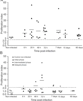
Initially, we observed a progressive increase of parasite load in the spleen of hamsters infected with
L. (L.) chagasi (data not shown). To better understand the increase of parasite burden and progression of the disease in this model, we investigated the proliferative response of spleen cells to a mitogen and a Leishmania
antigen. When the spleen cells in culture were stimu-lated with Con A, the response was preserved during all
periods of infection (Figure 1A), but a specific response
to Leishmania antigen was only detected at 48 and 72 h post-infection (pi) (Figure 1B). No proliferation in response to the Leishmania antigen was observed from 7 to 60 days
pi. When spleen cells from hamsters immunized with total
L. (L.) chagasi antigenwere stimulated with Leishmania
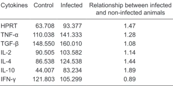
antigen and presented a proliferative response, they were used as a positive control for antigen proliferation. Using RT-PCR to detect cytokine mRNA in spleen cells from infected hamsters, we observed the presence of mRNA
of all cytokines evaluated, i.e., TNF-α, TGF-β, IL-2, IL-4, IL-10, and IFN-γ during all periods of infection evaluated
(Figure 2), as well as in non-infected cells. As a control of RT-PCR, we detected hamster HPRT. No qualitative and quantitative differences in cytokine mRNA produced by spleen cells were observed between infected and non-infected hamsters (Table 1).
Discussion
Hamsters infected with Leishmania develop a progres-sive and severe disease, as is also observed in humans
Figure 1. Proliferation of spleen cells of hamsters infected by
Leishmania (Leishmania) chagasi. Spleen cells (2 x 10⁷ cells/mL)
at initial (6, 20, 48, and 72 h), intermediate (7 and 15 days) and late (60 days) time of infection were stimulated with concanavalin
A (0.5 μg/mL) (A) or total antigen of LLC (5 x 10⁷ parasites/mL)
(B). The results are reported as proliferation index (count per min of stimulated cells/count per min of non-stimulated cells) with a
cut-off (dashed line) of 2.0. *P < 0.05 vs non-infected; #P < 0.05
vs 48 h (Kruskal-Wallis test).
Figure 2. Detection of hamster cytokine mRNA. The mRNA of spleen cells (2 x 10⁷ cells/mL) was analyzed by reverse transcriptase
polymerase chain reaction (RT-PCR) for lane 1 = HPRT (420 bp), lane 2 = TNF-α (260 bp), lane 3 = TGF-β (270 bp), lane 4 = IL-2 (400 bp), lane 5 = IL-4 (290 bp), lane 6 = IL-10 (280 bp), and lane 7 = IFN-γ (320 bp) expression both in non-infected and infected-hamsters
Immunosuppression in the experimental visceral leishmaniasis 669
with active VL (12-15). In the present study, we observed a progressive time-dependent increase of parasite burden in
the spleen of hamsters, thus confirming that these animals
are good models for the development of active VL and for the study of immunosuppression. For a better
understand-ing of VL progression in hamsters, we first evaluated the
proliferative response of T cells to a mitogen and to a
Leishmania antigen. We observed specific immunosuppres -sion in response to Leishmania antigen, which, however, was limited to the initial phase of infection (48 and 72 h
pi). We have observed a Con A-induced lymphoprolifera-tive response during all experimental periods but a total absence of a Leishmania antigen-induced response in the late phase (3). According to the literature, the Leishmania
antigen-induced response was suppressed (13), but there is a disagreement about the Con A-induced response. Some studies showed a response preserved throughout the experi-ment (13) while others did not observe it after 42 days of infection (14). Some factors, such as route of inoculation, may affect these results. Intracardiac inoculation produces an important impairment of the T-cell proliferative response to both a mitogen and a Leishmania antigen (13), while subcutaneous inoculation induces an immune response to both mitogen and Leishmania antigen (15,16). In the present study, we used the intraperitoneal route of inoculation, which promotes progressive infection and development of disease, indicating the occurrence of progressive immunosuppres-sion during active VL similar to that observed in natural infection. Although hamsters are considered to provide a good model of visceral leishmaniasis, the almost complete absence of markers for T cells, macrophages and cytokines impairs a better understanding of the occurrence of the immune response in this model. However, after cytokine cloning from hamsters (6) there was an improvement in the understanding of the immune response in this experimental
model. The profile of Th2 cytokines is directly responsible
for suppression, but other factors such as adherent cells (17) and macrophage-mediated suppression have been reported to lead to increasing parasite growth and also to be linked to defective antigen presentation (18). The Th1
cytokine IFN-γ plays a key role in the control of infection with
many intracellular pathogens, including Leishmania spp, and is the cytokine primarily responsible for macrophage activation and killing of intracellular parasites (19,20). In hamsters infected with L. (L.) donovani, adherent spleen cells have been shown to be important lymphoproliferative suppressors and to play a role in defective antigen
pre-sentation (13). Furthermore, TGF-β produced by adherent
antigen-presenting cells from infected hamsters has been
implicated in immunosuppression since a high level of TGF-β
was observed in the cell culture supernatant when the
Leishmania antigen-induced lymphoproliferative response was inhibited (7,8). In the present study, we investigated
the cytokine profile produced during the course of VL by
spleen cells in culture upon stimulation with Con A and
Leishmania antigen using RT-PCR and we did not observe a qualitative or quantitative change in cytokine expression by non-infected and infected hamsters during any period of infection studied, in agreement with results obtained by Melby et al. (6). These results are similar to those observed in human VL but contrast with the dominant Th2 responses observed in progressive L. major infection in BALB/c mice. The immunosuppression mechanism is not related to the
cytokine profile, although the change of a nucleotide in the final portion of sequence of mRNA from IFN-γ has been
observed in hamsters (6), possibly contributing to a reduc-tion of the biological effect of this cytokine and indicating the absence of power to control the progression of infection.
Interestingly, we observed a strong TGF-β band during all
periods of infection evaluated. We emphasize the
impor-tance of TGF-β in susceptibility and immunosuppression,
a fact that has been demonstrated by the restoration of the proliferative response of non-adherent cells from infected
hamsters treated with anti-TGF-β (8). It is attractive to
speculate about the possible participation of CD4+CD25+
-regulatory cells in immunosuppression during active VL in hamsters (9). According to our data, the immunosuppression observed in hamsters during active visceral leishmaniasis
is an antigen-specific response since the initial phase of
infection (after 72 h of infection), as determined by the absence of a lymphoproliferative response to Leishmania
antigen and a preserved response to Con A. Interestingly, based on our results, the production of cytokines does not contribute to the development of immunosuppression in this experimental model.
Acknowledgments
Research supported by CAPES, FAPESP, and CNPq.
Table 1. Quantitation of cytokine mRNA in infected and
non-in-fected control hamsters.
Cytokines Control Infected Relationship between infected and non-infected animals
HPRT 63.708 93.377 1.47
TNF-α 110.038 141.333 1.28
TGF-β 148.550 160.010 1.08
IL-2 90.505 103.582 1.14
IL-4 86.538 124.538 1.44
IL-10 44.007 83.234 1.89
IFN-γ 121.803 105.299 0.89
Cytokine mRNA was measured using the Image Lab 1D L340 software. Data are reported as DNA band intensity and are rep-resentative of only 1 animal from each group. The relationship between infected and non-infected animals was measured by the
C. Fazzani was the recipient of CAPES and FAPESP fel-lowships. P.A. Guedes and A. Senna were recipients of
FAPESP fellowships. H. Goto was the recipient of a CNPq fellowship.
References
1. Carvalho EM, Teixeira RS, Johnson WD Jr. Cell-mediated immunity in American visceral leishmaniasis: reversible im-munosuppression during acute infection. Infect Immun 1981; 33: 498-500.
2. Nickol AD, Bonventre PF. Immunosuppression associated with visceral leishmaniasis of hamsters. Parasite Immunol 1985; 7: 439-449.
3. Goto H, Lindoso JA. Immunity and immunosuppression in experimental visceral leishmaniasis. Braz J Med Biol Res 2004; 37: 615-623.
4. Bunn-Moreno MM, Madeira ED, Miller K, Menezes JA, Campos-Neto A. Hypergammaglobulinaemia in Leishmania donovani-infected hamsters: possible association with a polyclonal activator of B cells and with suppression of T cell function. Clin Exp Immunol 1985; 59: 427-434.
5. Dasgupta S, Mookerjee A, Chowdhury SK, Ghose AC. Im-munosuppression in hamsters with progressive visceral leishmaniasis: an evaluation of the role of nitric oxide toward impairment of the lymphoproliferative response. Parasitol Res 1999; 85: 594-596.
6. Melby PC, Chandrasekar B, Zhao W, Coe JE. The hamster as a model of human visceral leishmaniasis: progressive disease and impaired generation of nitric oxide in the face of a prominent Th1-like cytokine response. J Immunol 2001; 166: 1912-1920.
7. Rodrigues V Jr, Santana da Silva J, Campos-Neto A. Transforming growth factor beta and immunosuppression in experimental visceral leishmaniasis. Infect Immun 1998; 66: 1233-1236.
8. Mookerjee A, Sen PC, Ghose AC. Immunosuppression in hamsters with progressive visceral leishmaniasis is associ-ated with an impairment of protein kinase C activity in their lymphocytes that can be partially reversed by okadaic acid or anti-transforming growth factor beta antibody. Infect Im-mun 2003; 71: 2439-2446.
9. Rodrigues OR, Marques C, Soares-Clemente M, Ferronha
MH, Santos-Gomes GM. Identification of regulatory T cells
during experimental Leishmania infantum infection. Immu-nobiology 2009; 214: 101-111.
10. Melby PC, Tryon VV, Chandrasekar B, Freeman GL. Cloning of Syrian hamster (Mesocricetus auratus) cytokine cDNAs and analysis of cytokine mRNA expression in experimental visceral leishmaniasis. Infect Immun 1998; 66: 2135-2142. 11. Dwyer DM. Antibody-induced modulation of Leishmania
donovani surface membrane antigens. J Immunol 1976; 117: 2081-2091.
12. Barbosa Junior AA, Andrade ZA, Reed SG. The pathology of experimental visceral leishmaniasis in resistant and sus-ceptible lines of inbred mice. Braz J Med Biol Res 1987; 20: 63-72.
13. Rodrigues Junior V, Da Silva JS, Campos-Neto A. Selective inability of spleen antigen presenting cells from Leishmania donovani infected hamsters to mediate specific T cell pro -liferation to parasite antigens. Parasite Immunol 1992; 14: 49-58.
14. Vasconcellos RC, Urago KP, Bunn-Moreno MM, Madeira ED. Suppressor activity in Leishmania donovani-infected hamster serum: reversion by delipidated bovine serum albumin and role in cell cycle events. Braz J Med Biol Res 1996; 29: 615-622.
15. Gifawesen C, Farrell JP. Comparison of T-cell responses in self-limiting versus progressive visceral Leishmania dono-vani infections in golden hamsters. Infect Immun 1989; 57: 3091-3096.
16. Streit JA, Recker TJ, Filho FG, Beverley SM, Wilson ME. Protective immunity against the protozoan Leishmania chagasi is induced by subclinical cutaneous infection with virulent but not avirulent organisms. J Immunol 2001; 166: 1921-1929.
17. Basak SK, Saha B, Bhattacharya A, Roy S. Immunobio-logical studies on experimental visceral leishmaniasis. II. Adherent cell-mediated down-regulation of delayed-type hypersensitivity response and up-regulation of B cell activa-tion. Eur J Immunol 1992; 22: 2041-2045.
18. Reiner NE, Ng W, McMaster WR. Parasite-accessory cell in-teractions in murine leishmaniasis. II. Leishmania donovani suppresses macrophage expression of class I and class II major histocompatibility complex gene products. J Immunol 1987; 138: 1926-1932.
19. Heinzel FP, Sadick MD, Holaday BJ, Coffman RL, Lock-sley RM. Reciprocal expression of interferon gamma or interleukin 4 during the resolution or progression of murine leishmaniasis. Evidence for expansion of distinct helper T cell subsets. J Exp Med 1989; 169: 59-72.