Effects of diet and exercise training on
neurovascular control during mental
stress in obese women
1Instituto do Coração, Hospital das Clínicas, 2Disciplina de Endocrinologia,
Faculdade de Medicina,
3Escola de Educação Física e Esporte,
Universidade de São Paulo, São Paulo, SP, Brasil A.C. Tonacio1, I.C. Trombetta1,
M.U.P.B. Rondon1,
L.T. Batalha2, F.H.S. Kuniyoshi1,
M.C. Laterza1, P.H. Suzuki1,3,
M.M.G. Gowdak1,
A.C.P. Barretto1, A. Halpern2,
S.M.F. Villares2 and
C.E. Negrão1,3
Abstract
Since neurovascular control is altered in obese subjects, we hypoth-esized that weight loss by diet (D) or diet plus exercise training (D + ET) would improve neurovascular control during mental stress in obese women. In a study with a dietary reduction of 600 kcal/day with or without exercise training for 4 months, 53 obese women were subdivided in D (N = 22, 33 ± 1 years, BMI 34 ± 1 kg/m2), D + ET (N
= 22, 33 ± 1 years, BMI 33 ± 1 kg/m2), and nonadherent (NA, N = 9,
35 ± 2 years, BMI 33 ± 1 kg/m2) groups. Muscle sympathetic nerve
activity (MSNA) was measured by microneurography and forearm blood flow by venous occlusion plethysmography. Mental stress was elicited by a 3-min Stroop color word test. Weight loss was similar between D and D + ET groups (87 ± 2 vs 79 ± 2 and 85 ± 2 vs 76 ± 2 kg, respectively, P < 0.05) with a significant reduction in MSNA during mental stress (58 ± 2 vs 50 ± 2, P = 0.0001, and 59 ± 3 vs 50 ± 2 bursts/100 beats, P = 0.0001, respectively), although the magnitude of the response was unchanged. Forearm vascular conductance during mental stress was significantly increased only in D + ET (2.74 ± 0.22 vs 3.52 ± 0.19 units, P = 0.02). Weight loss reduces MSNA during mental stress in obese women. The increase in forearm vascular conductance after weight loss provides convincing evidence for D + ET interventions as a nonpharmacologic therapy of human obesity.
Correspondence
C.E. Negrão
Unidade de Reabilitação Cardiovascular e Fisiologia do Exercício, InCor-HC FMUSP Av. Dr. Enéas C. Aguiar, 44 05403-000 São Paulo, SP Brasil
Fax: +55-11-3069-5043 E-mail: [email protected] Research supported by FAPESP (No. 1998/15983-8) and by Fundação Zerbini. A.C. Tonacio was the recipient of a CNPq-PIBIC fellowship.
Received January 24, 2005 Accepted August 11, 2005
Key words •Vasodilation
•Sympathetic nervous system •Weight loss
Introduction
Neurovascular alteration in human obe-sity has been recently described (1). Muscle sympathetic nerve activity (MSNA) and fore-arm vascular resistance are increased, whereas forearm vascular conductance is significantly reduced in obese subjects (1). This scenario suggests an increased risk for
cardiovascular disorders in obese persons (2,3).
ques-tions emerge: 1) is it possible to improve the neurovascular control during mental stress in obese subjects? 2) If so, which is the best strategy to reverse this neurovascular abnor-mality? In a recent study, a hypocaloric diet plus exercise training, in contrast to a hypocaloric diet alone, significantly im-proved the vasodilatory responses during mental stress in obese children (7). How-ever, that study provided no information regarding sympathetic activation in obese children.
In the present study, we report the impact of body weight reduction by a hypocaloric diet and hypocaloric diet associated with exercise training on MSNA and forearm blood flow during mental stress in obese women.
Our hypothesis was that weight loss would reduce MSNA and would increase forearm blood flow during mental stress in obese women. In addition, this improvement in neurovascular control would be increased if the hypocaloric diet were associated with exercise training.
Subjects and Methods
Study population
Fifty-three consecutive out-patient sed-entary obese women from the Women Obe-sity Ambulatory of the Endocrinology De-partment, University of São Paulo Medical School, were invited to participate in the study. They were randomly divided into a group receiving a hypocaloric diet (N = 26) and a group receiving a hypocaloric diet associated with exercise training (N = 27) for 4 months. Nine obese women did not want to take part in the study, although they agreed to be studied before and after 4 months. Thus, our final sample consisted of three subgroups: 1) hypocaloric diet (N = 22, age 33 ± 1 years), 2) hypocaloric diet associated with exercise training (N = 22, age 34 ± 1 years), and 3) nonadherent to any of the two interventions (N = 9, age 35 ± 2
years). None of the individuals were taking medication or contraceptives and all were non-smokers, with no evidence of metabolic or cardiovascular disease at the time of the study. The hypocaloric diet, diet plus exer-cise training, and nonadherent groups were similar regarding total cholesterol (170 ± 7, 169 ± 6, and 166 ± 11 mg/dL, respectively, P = 0.94), HDL-cholesterol (39 ± 2, 39 ± 2, and 42 ± 4 mg/dL, respectively, P = 0.74), LDL-cholesterol (105 ± 6, 108 ± 6, and 105 ± 10 mg/dL, respectively, P = 0.91), triglyc-erides (129 ± 18, 116 ± 13, and 107 ± 6 mg/ dL, respectively, P = 0.69), glucose (80 ± 2, 86 ± 3, and 85 ± 4 mg/dL, P = 0.11, respec-tively), and insulin levels (15.4 ± 2, 13.3 ± 1.4, and 12.5 ± 2.2 µIU/mL, P = 0.60, respec-tively). All subjects were in the pre-hyper-tensive stage according to the Seventh Re-port of the Joint National Committee on Detection, Evaluation, and Treatment of High Blood Pressure (8). Since hormonal varia-bility during the regular menstrual cycle may affect blood flow and vascular resistance (9), all women were studied between the first and the fifth day after the onset of menstruation. The study protocol was ap-proved by the Human Subject Protection Committees of the Heart Institute (InCor) and University Hospital, University of São Paulo Medical School, and written informed consent was given by each subject that par-ticipated in the study.
Muscle sympathetic nerve activity.
established and described criteria. Muscle sympathetic bursts were identified by visual inspection by a single investigator (C.E.N.), and were reported as burst frequency (bursts per minute) and bursts per 100 heart beats.
Forearm blood flow. Forearm blood flow was measured by venous occlusion plethys-mography (1). The nondominant arm was elevated above heart level to ensure adequate venous drainage. A mercury-filled silastic tube attached to a low-pressure transducer was placed around the forearm and con-nected to a plethysmograph (Hokanson, Bellevue, WA, USA). Sphygmomanometer cuffs were placed around the wrist and upper arm. At 15-s intervals, the upper cuff was inflated above venous pressure for 7 to 8 s. Forearm blood flow (mL
.
100 mL tissue-1.
min-1) was determined on the basis of a
minimum of four separate readings. Fore-arm vascular conductance was calculated as forearm blood flow/mean arterial pressure x 100, and is reported as ‘units’ [100 mL(dL tissue)-1
.
min-1.
mmHg-1].Blood pressure and heart rate. Blood pressure was monitored noninvasively with a finger photoplethysmograph device (Fina-Press 2300; Ohmeda, Englewood, CO, USA) on a beat-to-beat basis (AT/CODAS soft-ware) at a frequency of 500 Hz. Heart rate was monitored continuously through lead II of the ECG.
Mental stress. In humans, the defense reaction can be elicited by mental challenge (11). In the present study, as well as in others, mental stress was induced by a 3-min period of the Stroop color word test (10-12). During this test, subjects were shown on a panel a series of names of colors written with ink of a different color from the color speci-fied. The subject was asked to identify the color of the ink, not to read the word. The difficulty of the task was tailored to the ability of the subject. The subject was urged to proceed as rapidly as possible and was gently chastised for incorrect responses. The subject was asked to try her best, but the
number of correct answers was not quanti-fied. Each subject was asked to assess task difficulty on completion of the protocol us-ing a standard five-point scale: 0, not stress-ful; 1, somewhat stressstress-ful; 2, stressstress-ful; 3, very stressful; 4, extremely stressful.
Hypocaloric diet
The hypocaloric diet was based on the basal energy requirements estimated by the FAO/WHO/UNU Expert Committee (13). Because under normal circumstances physi-cal activity accounts for 15 to 30% of a person’s total daily energy expenditure, the basal metabolic rate must be multiplied by 1.15 or 1.3 to calculate the daily energy expenditure (13). A 10% reduction in body weight leads to a substantial reduction in cardiovascular risk (14). In general, reduc-tions of 500 to 1,000 kcal/day are used to reduce body weight by 0.5 to 1.0 kg per week. Because the planned reductions were of 8 to 10 kg over a period of 16 weeks, energy intake was reduced by 600 kcal/day. The diet consisted of 50-70% carbohydrate, 10-15% protein and 15-30% fat. On alter-nate weeks, every patient visited the clinical nutritionist for a regular check-up. On the occasion of each visit, the subjects were weighed and encouraged to record their in-take to ensure adherence to the dietary pro-tocol.
Exercise training
point obtained in a progressive cardiopul-monary exercise test. The anaerobic thresh-old was considered to occur at the point where there was a loss of linearity between oxygen uptake and carbon dioxide produc-tion (15), or at the point where the ventila-tory equivalent for the oxygen and end-tidal oxygen partial pressure curves reached their respective minimum values and then began to rise during the progressive exercise test (16). The respiratory compensation point was determined as the point where the venti-latory equivalent for carbon dioxide was lowest before a systematic increase or where end-tidal carbon dioxide partial pressure reached a maximum value and began to decrease (17). The peak oxygen uptake was considered to occur at the end of the bicycle cardiopulmonary exercise test (ramp proto-col with a 10- to 15-W increment every minute up to exhaustion), when the subject no longer maintained the bicycle velocity at 60 rpm. Compliance was assessed as per-centage of exercise sessions attended. Com-pliance with the exercise program was very good, with the subjects attending more than 80% of the exercise sessions.
Experimental protocol
After a sleep of at least 7 h, no physical exercise for 24 h prior the study, and a light meal without caffeine, between 7:00 and 8:00 am, the subject lay down in a comfort-able position on a laboratory bed. The leg was positioned for microneurography and an adequate nerve recording site was ob-tained. Baseline MSNA, forearm blood flow, blood pressure, and heart rate were recorded for 3 min. The Stroop color word test was performed for 4 min and MSNA and heart rate were recorded continuously during the test. Blood pressure was measured on a beat-to-beat basis. Forearm blood flow was meas-ured each 15 s. The exercise-trained women were submitted to the experimental protocol two days after the last training section. This
experimental protocol was repeated after 4 months of intervention (diet alone or diet plus exercise training).
Statistical analysis
Data are reported as means ± SEM. The similarity in demographics among the three groups was tested by one-way analysis of variance. Two-way analysis of variance with repeated measures was performed to test differences (pre- versus post-intervention) within each group at rest and during mental stress. When significance was detected,
Scheffé post hoc comparison was performed.
Probability values of <0.05 were considered to be statistically significant.
Results
Baseline measurements
Baseline body weight and body weight mass were similar for the diet, diet plus exercise training, and nonadherent groups (87 ± 2, 85 ± 2, and 87 ± 3 kg, P = 0.66, respectively, and 34 ± 1, 33 ± 1, and 33 ± 1
kg/m2, P = 0.14, respectively). Similarly,
2, 72 ± 2, and 72 ± 3 bpm, P = 0.46). Regarding forearm measurements, there were no significant differences in baseline fore-arm blood flow or forefore-arm vascular conduc-tance among the diet group, diet plus exer-cise training group, and nonadherent group (2.17 ± 0.22, 1.88 ± 0.08, and 1.82 ± 0.20 mL
.
100 mL tissue-1.
min-1, P = 0.34,respectively, and 2.25 ± 0.23, 2.02 ± 0.11, and 1.89 ± 0.24 units, P = 0.48, respective-ly). Peak oxygen uptake was also similar for all groups (17.7 ± 0.8, 18.5 ± 0.6, and 18.4 ± 1.0 mL
.
kg-1.
min-1, P = 0.69, respectively).Effects of diet and exercise training
Diet and diet associated with exercise training significantly reduced body weight (87 ± 2 vs 79 ± 2 and 85 ± 2 vs 76 ± 2 kg, respectively, P < 0.05). Body weight was unchanged in the nonadherent group (86 ± 3
vs 85 ± 3 kg). Similarly, diet and diet plus
exercise training significantly reduced body mass index (34.4 ± 0.5 vs 31.0 ± 0.6 and 32.9
± 0.4 vs 29.4 ± 0.5 kg/m2, respectively).
Body mass index was unchanged in the non-adherent group (33.2 ± 0.9 vs 32.6 ± 0.8 kg/
m2). Diet associated with exercise training
significantly increased peak oxygen uptake (18.8 ± 0.6 vs 22.2 ± 0.7 mL
.
kg-1.
min-1, P< 0.05). In the diet and nonadherent groups, peak oxygen uptake was unchanged (17.9 ± 0.7 vs 18.7 ± 0.7 and 18.3 ± 1.0 vs 18.9 ± 1.2 mL
.
kg-1.
min-1, respectively).Regarding task difficulty on completion of the protocol of mental stress, there were no significant differences among the diet, diet plus exercise training, and nonadherent groups pre- and post-intervention (pre = 1.3 ± 0.2, 1.5 ± 0.2, and 1.3 ± 0.3 scores, and post = 1.3 ± 0.2, 1.6 ± 0.1, and 1.3 ± 0.4 scores, P = 0.90, respectively).
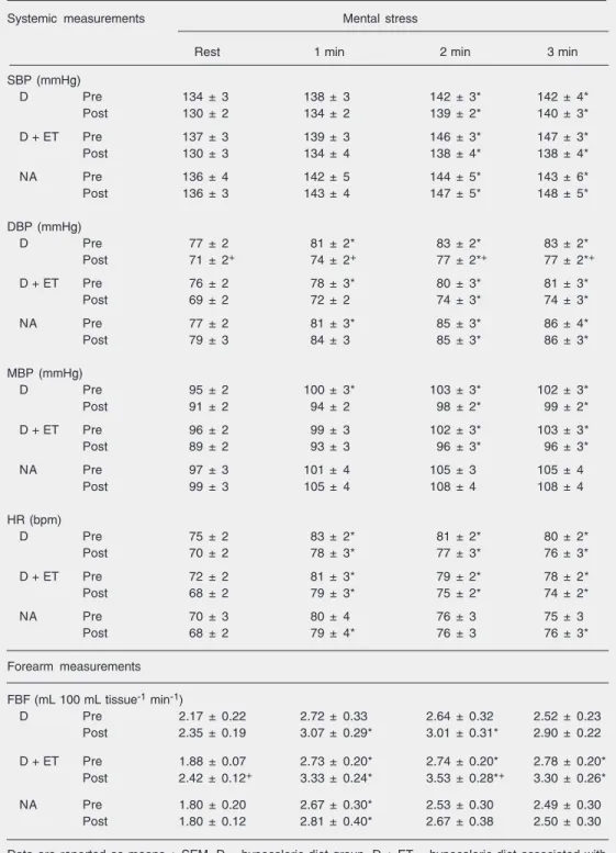
Diet and diet associated with exercise training slightly decreased systolic and mean blood pressure levels at rest and during men-tal stress (Table 1). Diet significantly re-duced diastolic blood pressure (P = 0.02),
whereas diet associated with exercise train-ing only tended to reduce diastolic blood pressure (P = 0.06). Both interventions, diet and diet associated with exercise training, slightly decreased heart rate levels at rest and during mental stress (Table 1). Systolic, diastolic, mean blood pressure, and heart rate levels were unchanged in the nonadher-ent group at rest or during mnonadher-ental stress.
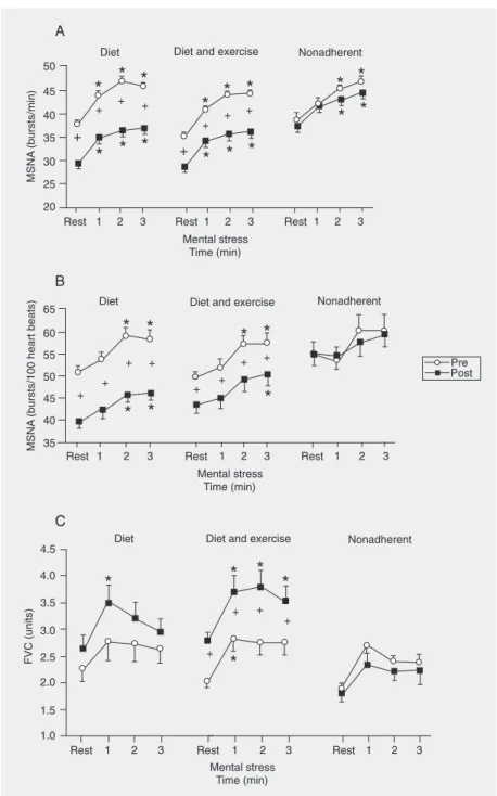
Diet and diet associated with exercise training significantly reduced MSNA burst frequency (bursts/min) at rest and during mental stress, although the magnitude of the responses, analyzed by the interaction of phase and time, showed no significant dif-ference between pre-intervention and post-intervention (Figure 1A). Similar findings were observed in pulse synchronous sympa-thetic activity (bursts/100 heart beats). Diet and diet associated with exercise training significantly reduced pulse synchronous sympathetic activity at rest and during men-tal stress, but caused no change in the mag-nitude of the responses of pulse synchro-nous sympathetic activity (Figure 1B). MSNA burst frequency and pulse synchro-nous sympathetic activity were unchanged in the nonadherent group.
Table 1. Systemic and vascular measurements during mental stress before and after interventions in obese women.
Systemic measurements Mental stress
Rest 1 min 2 min 3 min
SBP (mmHg)
D Pre 134 ± 3 138 ± 3 142 ± 3* 142 ± 4*
Post 130 ± 2 134 ± 2 139 ± 2* 140 ± 3*
D + ET Pre 137 ± 3 139 ± 3 146 ± 3* 147 ± 3*
Post 130 ± 3 134 ± 4 138 ± 4* 138 ± 4*
NA Pre 136 ± 4 142 ± 5 144 ± 5* 143 ± 6*
Post 136 ± 3 143 ± 4 147 ± 5* 148 ± 5*
DBP (mmHg)
D Pre 77 ± 2 81 ± 2* 83 ± 2* 83 ± 2*
Post 71 ± 2+ 74 ± 2+ 77 ± 2*+ 77 ± 2*+
D + ET Pre 76 ± 2 78 ± 3* 80 ± 3* 81 ± 3*
Post 69 ± 2 72 ± 2 74 ± 3* 74 ± 3*
NA Pre 77 ± 2 81 ± 3* 85 ± 3* 86 ± 4*
Post 79 ± 3 84 ± 3 85 ± 3* 86 ± 3*
MBP (mmHg)
D Pre 95 ± 2 100 ± 3* 103 ± 3* 102 ± 3*
Post 91 ± 2 94 ± 2 98 ± 2* 99 ± 2*
D + ET Pre 96 ± 2 99 ± 3 102 ± 3* 103 ± 3*
Post 89 ± 2 93 ± 3 96 ± 3* 96 ± 3*
NA Pre 97 ± 3 101 ± 4 105 ± 3 105 ± 4
Post 99 ± 3 105 ± 4 108 ± 4 108 ± 4
HR (bpm)
D Pre 75 ± 2 83 ± 2* 81 ± 2* 80 ± 2*
Post 70 ± 2 78 ± 3* 77 ± 3* 76 ± 3*
D + ET Pre 72 ± 2 81 ± 3* 79 ± 2* 78 ± 2*
Post 68 ± 2 79 ± 3* 75 ± 2* 74 ± 2*
NA Pre 70 ± 3 80 ± 4 76 ± 3 75 ± 3
Post 68 ± 2 79 ± 4* 76 ± 3 76 ± 3*
Forearm measurements
FBF (mL 100 mL tissue-1 min-1)
D Pre 2.17 ± 0.22 2.72 ± 0.33 2.64 ± 0.32 2.52 ± 0.23
Post 2.35 ± 0.19 3.07 ± 0.29* 3.01 ± 0.31* 2.90 ± 0.22
D + ET Pre 1.88 ± 0.07 2.73 ± 0.20* 2.74 ± 0.20* 2.78 ± 0.20* Post 2.42 ± 0.12+ 3.33 ± 0.24* 3.53 ± 0.28*+ 3.30 ± 0.26*
NA Pre 1.80 ± 0.20 2.67 ± 0.30* 2.53 ± 0.30 2.49 ± 0.30
Post 1.80 ± 0.12 2.81 ± 0.40* 2.67 ± 0.38 2.50 ± 0.30
Data are reported as means ± SEM. D = hypocaloric diet group, D + ET = hypocaloric diet associated with exercise training group, NA = nonadherent group, SBP = systolic blood pressure, DBP = diastolic blood pressure, MBP = mean blood pressure, HR = heart rate, FBF = forearm blood flow.
*P < 0.05 compared to rest; +P < 0.05 compared to pre-intervention (two-way ANOVA for repeated
Discussion
The novelty of the present study is that weight loss improves neurovascular control during mental stress in obesity. We found that weight loss decreases MSNA levels dur-ing mental stress in obese subjects. In addi-tion, weight loss by diet associated with exercise training, in contrast to weight loss by diet alone, increased forearm vascular conductance during mental stress.
Reduction in MSNA after weight loss has been previously reported during moder-ate exercise (18) when sympathoexcitation is mediated by a central command, by mecha-noreceptors and by chemoreceptors. The present study, however, brings about a new understanding of human obesity. Mental stress isolates the central command and may precipitate cardiac events, especially in obese subjects, in whom neurovascular alteration is the rule (6). Our results show that the central neural outflow during a stressful con-dition, directly measured by microneurogra-phy, was markedly reduced after weight loss. In addition, the unchanged magnitude of the increase in MSNA levels during mental stress after the interventions suggests that the low-ered sympathetic activation during this physi-ological maneuver is a consequence of a reduction in baseline MSNA. Although the mechanisms underlying the reduction in cen-tral neural outflow are beyond the scope of the present investigation, the improvement in baroreflex control is a potential mechan-ism for explaining the decrease in the cen-trally mediated MSNA after weight loss (19). The decreased plasma leptin levels and the increased insulin sensitivity after weight loss can also be involved in the reduction of MSNA, although this physiological link still needs to be demonstrated in humans (18). Alternatively, the decrease in free fatty acids after weight loss (20) is a suggestive candi-date to explain the reduction in MSNA in human obesity. Because free fatty acids com-ing from visceral adipose tissue or the portal
vein can sensitize liver afferent nervous fi-bers which, in turn, centrally modulate sym-pathetic activity (21), it is conceivable that the reduction in plasma free fatty acid
con-Figure 1. Muscle sympathetic burst frequency (MSNA, Panel A), pulse synchronous sym-pathetic activity (MSNA, Panel B), and forearm vascular conductance (FVC, Panel C) at rest and during mental stress in obese women after intervention by diet or diet associated with exercise training, and in obese women with nonadherence to either intervention. *P < 0.05 compared to rest; +P < 0.05 compared to pre-intervention (two-way ANOVA for
centration after weight loss can attenuate central neural outflow in humans.
The tendency towards a decrease in heart rate during mental stress suggests that sym-pathoinhibition also took place at the cardiac level. Although we have not measured sym-pathetic activity on the heart, MSNA is highly correlated with cardiac noradrenaline spill-over at rest, and changes in MSNA and cardiac noradrenaline spillover during men-tal stress are qualitatively similar (22). There is also the possibility that weight loss pro-voked an increase in the vagal activity that controls heart rate. A previous study (18) has shown that a diet and exercise regimen caused a significant reduction in heart rate during mild and moderate exercise, although no information on sympathetic or vagal func-tioning was provided in that study.
Even though we were not dealing with hypertensive women, but rather with pre-hypertensive women (8), diet and diet asso-ciated with exercise training tended to re-duce diastolic blood pressure at rest and during mental stress. The effect of weight loss on blood pressure during physiological maneuvers has been recently demonstrated in obese children (7). The systolic and dia-stolic blood pressure levels during mental stress and exercise were significantly low-ered after weight loss by diet and exercise training (7). Because high blood pressure is consistent with sympathetic activation, the question may be raised that the changes in neurovascular control changes found here were sue, at least in part, to the reduction in blood pressure.
As anticipated, weight loss increased fore-arm vascular conductance during mental stress. However, this vascular adaptation is only obtained when diet is associated with exercise training. These findings confer an important role on exercise in the treatment of human obesity. It is unlikely that this exercise effect reflects the decline in sympa-thetic activation because exercise training and diet, and diet alone equally reduced
MSNA. It seems more reasonable that exer-cise improved endothelium function, increas-ing the bioavailability of nitric oxide. In fact, the benefits of exercise on endothelially mediated blood flow have been reported in adult obese subjects (23), and, more recently, in obese children submitted to diet associ-ated with exercise training. In obese chil-dren, a hypocaloric diet plus exercise train-ing restored muscle endothelium-mediated vasodilatory responses during mental stress and handgrip exercise (7). The effects of exercise training on endothelial function are not limited to obese individuals and skeletal muscle. A previous study has demonstrated that exercise training improves coronary flow in response to adenosine infusion in patients with ischemic heart failure (24).
The mechanisms involved in the improve-ment of endothelial function after exercise training are not fully understood. However, we can hypothesize that the reduction in pro-inflammatory mediators plays a role in this
matter. It is well known that TNF-α and
interleukin-6 enhance reactive oxygen spe-cies, which, in turn, increase nitric oxide degradation. Thus, exercise training, by
re-ducing plasma and muscle TNF-α and
inter-leukin-6 as previously demonstrated (25,26), would increase nitric oxide production in obese individuals, in whom these pro-in-flammatory mediators are augmented (27, 28). Alternatively, we suggest that exercise training improves endothelial function by increasing endothelial nitric oxide synthase. A previous study demonstrated that exercise training increased endothelial nitric oxide synthase expression in dogs with heart fail-ure (29).
Study limitations
that more prominent benefits would be seen if our diet and exercise regimen were pro-longed. Our sampling was restricted to women. This strategy avoided gender con-founding, but at the same time reduced the generality of our findings. The non-adherent group included individuals who did not ad-here to the diet plus exercise training or to the diet. Thus, the nonadherent group as a control group should be viewed with cau-tion. One may argue that the decreased muscle sympathetic nerve activity in obese women after weight loss is due to the per-ceived exertion. This interpretation is un-likely, because the task difficulty on comple-tion of the protocol using a five-point scale
was similar across the three groups studied (data not shown).
The present study extends our knowl-edge by showing that weight loss reduces MSNA levels during mental stress in obese subjects as a consequence of the decrease in baseline MSNA. The improvement in muscle vascular conductance during a mental chal-lenge depends on the weight loss strategy chosen. The increase in muscle vascular con-ductance during a stressful condition is achieved if a hypocaloric diet is associated with exercise training. These findings dem-onstrate that these two interventions should be the mandatory strategy for the nonphar-macologic treatment of human obesity.
References
1. Negrao CE, Trombetta IC, Batalha LT et al. (2001). Muscle metaboreflex control is diminished in normotensive obese women. American Journal of Physiology, 281: H469-H475.
2. Masuo K, Mikami H, Ogihara T et al. (2000). Weight gain induced blood pressure elevation. Hypertension, 35: 1135-1140.
3. Krauss RM, Winston M, Fletcher BJ et al. (1998). Obesity: Impact on cardiovascular disease. Circulation, 98: 1472-1476.
4. Hilton SM (1982). The defense-arousal system and its relevance for circulation and respiratory control. Journal of Experimental Biology, 100: 159-174.
5. Hjemdahl P, Freyschuss U, Juhlin-Dannfelt A et al. (1984). Differen-tiated sympathetic activation during mental stress evoked by Stroop test. Acta Physiologica Scandinavica, 527: 25-29.
6. Kuniyoshi FH, Trombetta IC, Batalha LT et al. (2003). Abnormal neurovascular control during sympathoexcitation in obesity. Obesity Research, 11: 1411-1419.
7. Ribeiro MM, Silva AG, Santos NS et al. (2005). Diet and exercise training restore blood pressure and vasodilatory responses during physiological maneuvers in obese children. Circulation, 111: 1915-1923.
8. Chobanian AV, Bakris GL, Black HR et al. (2003). Seventh report of the Joint National Committee on Prevention, Detection, Evaluation, and Treatment of High Blood Pressure. Journal of the American Medical Association, 289: 2560-2572.
9. Bartelink ML, Wollersheim H, Theeuwes A et al. (1990). Changes in skin blood flow during the menstrual cycle: the influence of the menstrual cycle on the peripheral circulation in healthy female vol-unteers. Clinical Science, 78: 527-532.
10. MiddLekauff HR, Nguyen AH, Negrao CE et al. (1997). Impact of acute mental stress on sympathetic nerve activity and regional blood flow in advanced heart failure: implications for ‘triggering’ adverse cardiac events. Circulation, 96: 1835-1842.
11. Steptoe A & Vogele C (1991). Methodology of mental stress testing in cardiovascular research. Circulation, 83 (Suppl II): 14-24.
12. Trombetta IC, Batalha LT, Rondon MUPB et al. (2005). Gly16 + Glu27 ß2-adrenoceptor polymorphisms cause increased forearm blood flow responses to mental stress and handgrip in humans. Journal of Applied Physiology, 98: 787-794.
13. WHO (1985). Energy and Protein Requirements. Report of a Joint FAO/WHO/UNU Expert Consultation. World Health Organization Technical Report Series, 724: 1-206.
14. Yamashita T, Sasahara T, Pomeroy SE et al. (1998). Arterial compli-ance, blood pressure, plasma leptin and plasma lipids in women are improved with weight reduction equally with a meat-based diet and a plant-based diet. Metabolism, 47: 1308-1314.
15. Beaver WL, Wasserman K & Whipp BJ (1986). A new method for detecting the anaerobic threshold by gas exchange. Journal of Applied Physiology, 60: 2020-2027.
16. Wasserman K, Whipp BJ, Koyal SN et al. (1973). Anaerobic thresh-old and respiratory gas exchange during exercise. Journal of Ap-plied Physiology, 33: 236-243.
17. Wasserman K, Hansen JE, Sue DY et al. (1986). Measurement of the physiological response to exercise. In: Wasserman K, Hansen JE, Sue DY et al. (Editors), Principles of Exercise Testing and Interpretation. Lea & Febiger, Philadelphia, PA, USA, 27-46. 18. Trombetta IC, Batalha LT, Rondon MU et al. (2003). Weight loss
improves neurovascular and muscle metaboreflex control in obe-sity. American Journal of Physiology, 285: H974-H982.
19. Grassi G, Seravalle G, Colombo M et al. (1998). Body weight reduction, sympathetic nerve traffic, and arterial baroreflex in obese normotensive humans. Circulation, 97: 2037-2042.
20. Keim NL, Barbieri TF & Van Loan M (1991). Physiological and biochemical variables associated with body fat loss in overweight women. International Journal of Obesity, 15: 283-293.
21. Rao SV, Donahue M, Pi-Sunyer X et al. (2001). Obesity as a risk factor in coronary artery disease. American Heart Journal, 142: 1102-1107.
meas-urements of cardiac noradrenaline spillover and sympathetic out-flow to skeletal muscle in humans. Journal of Physiology, 453: 45-58.
23. Sciacqua A, Candigliota M, Ceravolo R et al. (2003). Weight loss in combination with physical activity improves endothelial dysfunction in human obesity. Diabetes Care, 26: 1673-1678.
24. Hambrecht R, Wolf A, Gielen S et al. (2000). Effects of exercise on coronary endothelial function in patients with coronary artery dis-ease. New England Journal of Medicine, 342: 454-460.
25. Adamopoulos S, Parissis J, Karatzas D et al. (2002). Physical training modulates proinflammatory cytokines and the soluble FAS/ soluble FAS ligand system in patients with chronic heart failure. Journal of the American College of Cardiology, 39: 653-663. 26. Gielen S, Adams V, Mobius-Winkler S et al. (2003).
Anti-inflamma-tory effects of exercise training in the skeletal muscle of patients with
chronic heart failure. Journal of the American College of Cardiology, 42: 861-868.
27. Kirchgessner TG, Uysal T, Wiesbrock SM et al. (1997). Tumor necrosis factor-alpha contributes to obesity-related hyperleptinemia by regulating leptin release from adipocytes. Journal of Clinical Investigation, 100: 2777-2782.
28. Mantzoros CS, Moschos S, Avramopoulos I et al. (1997). Leptin concentrations in relation to body mass index and the tumor necro-sis factor-α system in humans. Journal of Clinical Endocrinology
and Metabolism, 82: 3408-3413.